Papilledema
Historical document, translated for reference. It reflects medical knowledge of the 1920s–30s and is not medical advice.
Summary
Papilledema is a disease of the optic nerve characterized by swelling of the optic disc protruding above the retinal level, most commonly caused by increased intracranial pressure. The concept was introduced by A. Graefe in 1860, who first described its clinical picture and connection to increased intracranial pressure.
Encyclopedia article (1928–1936)
PAPILLEDEMA (papilla oedematosa, Stauungspapille), a disease of the optic nerve, the most characteristic feature of which is edema of the disc with protrusion above the level of the retina, and the cause of which in the vast majority of cases is increased intracranial pressure. The concept of P. was introduced by A. Graefe in 1860, who also described for the first time the clinical picture of P. and indicated its connection with increased intracranial pressure. Subsequently, due to different interpretations of the nature of the process, various designations were applied. Thus, Leber, considering P. one of the forms of inflammation of the optic nerve, called it papillitis—a name now abandoned as not corresponding to the understanding of the origin of P.; Adamyuk called P. neuritis oedematosa, but the majority accepts the name papilla oedematosa (Stauungspapille) as a concept that clearly distinguishes this form of optic disc lesion from its inflammation. Etiology. The causes of P. are extremely diverse, but the most common are brain tumors. In addition to brain tumors, P. can be caused by gummas, solitary tubercles, tumors of the meninges, bones of the skull, abscesses, various forms of meningitis, especially serous, the so-called pseudotumor, acute and chronic hydrocephalus, aneurysms and hemorrhages, cysticerci and echinococci, deformation of the skull (tower skull), severe skull injuries, sinus thrombosis; it is also encountered in individual cases with multiple sclerosis, with blood diseases (chlorosis, anemia, polycythemia, leukemia), during pregnancy, with nephritis, with lead poisoning. All these various conditions causing P. are accompanied in the vast majority of cases by increased intracranial pressure, one of the main symptoms of which is P. The nature and size of brain tumors do not play a special role in the development of P., and the greatest importance is only the location of the tumor and the speed of its growth. At present, it is believed that tumors located in the cerebellum, in the region of the quadrigeminal bodies, the fourth ventricle most frequently and early cause P.; more rarely—tumors of the occipital, temporal, and frontal lobes; very rarely—tumors of the base, infundibulo-hypophyseal, and especially rarely tumors of the meninges. In addition to intracranial causes, P. is sometimes caused by processes in the orbit (tumors of the orbit, optic nerve, etc.). Finally, P. is also encountered with purely intraocular diseases, as for example with penetrating injuries to the eye, when there is a prolonged decrease in intraocular pressure. Pathological anatomy. On macroscopic examination of typical forms of P., a mushroom-like protrusion of the disc is noted, often descending steeply onto the retina. The protrusion of the disc sometimes reaches 1-2 mm or more. Then, macroscopically in the majority of cases (60-70%, according to Hippel), stretching of the inter-
4 6
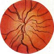
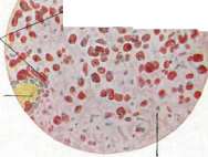
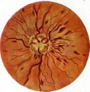
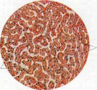
Figure 1. Multiple serous and blood cysts of the corpus luteum. Figure 2. Corpus luteum: a. - layer of lutein cells; b - islands of large cells of theca interna; c - blood clot; d - fibrin. (From Sobotta). Figure 3. Papilledema (pure edema in fresh cases). Figure 4. Papilledema in later stages. Figure 5. Granular bodies (a) in the brain in softening of the brain; b - vessels; c - fatty degenerated substance (stained with Sudan). Figure 6. Fatty degeneration of the liver in chloroform poisoning (fat droplets stained with Sudan in yellow-red color): a - fatty-degenerated liver trabeculae; b - capillaries of the liver lobule. (Figures 1, 5 and 6 from the author's preparations). To the articles: Corpus luteum, Fatty degeneration, Papilledema, Granular bodies. Papilledema is an ampulliform dilation of the optic nerve sheath spaces. On microscopic examination, edema of the optic nerve's non-myelinated segment is found, with protrusion of the axial cylinder forward, and edema of the retina around the papilla. The edema displaces the nerve fibers and the interstitial tissue, causing the entire papilla to take on a spongy appearance [see separate table (p. 475-476), figure 4]. With pronounced edema, the nerve fibers soon show signs of degeneration. In the segment of the optic nerve directly behind the lamina cribrosa, edematous changes are also present, with the edema noted not only around the septa of the optic nerve but also within them (Behr). All these phenomena in the initial stages of papilledema occur without any signs of inflammation. In late cases, inflammatory phenomena are found both in the area of the papilla and in the trunk and sheath of the optic nerve (Elschnig and others), but they are not characteristic of papilledema and are undoubtedly of secondary order. In the initial stages of papilledema, atrophy of the nerve fibers is observed with extremely pronounced proliferation of glial tissue (Abelsdorff). Pathogenesis. Explanations of the pathogenesis of papilledema are still contradictory to this day. Two main theories opposed each other in the past - Leber's inflammatory theory and the mechanical, or circulatory, theory of Schmidt-Rimppler-Mantz. Leber's theory explains papilledema as the formation of inflammatory products in brain tumors, which penetrate along with the cerebrospinal fluid into the subarachnoid space of the optic nerve and cause edema of the papilla as an inflammatory reaction to the penetrating harmful substances. However, this theory is now rejected primarily because inflammatory phenomena are absent in fresh cases of papilledema. The mechanical theory is based on the observations of Schwalbe, who proved that the subarachnoid space of the optic nerve communicates with the subdural and subarachnoid spaces of the brain; based on this, Schmidt-Rimppler and Manz explain papilledema by the accumulation of cerebrospinal fluid in the blind end of the subarachnoid space of the optic nerve when intracranial pressure increases. The optic nerve with its vessels is compressed, and the papilla becomes saturated with stagnant fluid. This theory received further development in two new theories - those of Schieck and Behr, each of which has its followers among modern ophthalmologists. According to Schieck's theory, based on observations by Levinsohn, when intracranial pressure increases, lymph flowing from the vitreous body along the perivascular spaces of the axial cylinder of the optic nerve encounters an obstacle to its outflow due to the increased pressure of cerebrospinal fluid flowing from the subdural space into the sheaths of the optic nerve; lymph stasis, compression of vessels, and venous stasis occur. In addition to increased intracranial pressure, according to Schieck, an increase in cerebrospinal fluid is also necessary. Behr in 1911 presented his theory of papilledema, which is based on his comprehensive study of the nutrition of the optic nerve fibers and the role of glia in this process. According to his view, parenchymal tissue fluid flows inside the nerve fibers of the optic nerve along the system of glial fibers in the direction from the papilla to the brain, and this flow of fluid can be disrupted at any point of the optic nerve. In particular, when intracranial pressure increases, a reverse flow of tissue juices into the optic nerve inevitably occurs, and subsequently their stasis, edema of nerve tissue, and impaired circulation, phenomena that manifest themselves most easily and earliest on the papilla of the optic nerve. To explain the mechanism of the effect of increased intracranial pressure on papilledema, Behr points to the easy possibility of compression of the optic nerve at its entrance into the bony canal, which is covered here by a duplication of the dura mater. From the standpoint of his theory, Behr explains all types of papilledema, considering that in some cases it is a passive lymph stasis (with increased intracranial pressure), while in others it is an active lymph stasis (with increased lymph excretion through functionally altered vessel walls in blood diseases, etc.). Symptomatology and course of papilledema. The ophthalmoscopic picture of papilledema at its height of development is very characteristic: the papilla mushrooms out, sometimes protruding very significantly above the surface of the retina, the size of the papilla increases, its boundaries are indistinct, the color is grayish-red, sometimes the papilla, especially initially, takes on a vitreous luster (see separate table, fig. 3), the central veins are dilated, the arteries are narrowed, when passing onto the protruding papilla the vessels make a distinct bend; often radial striation is noticed on and near the papilla, and hemorrhages in the form of streaks and spots are often visible both on the papilla and near it. In late and pronounced cases, the appearance of white streaks and plaques near the papilla, sometimes in the area of the macula, is noted (see separate table, fig. 4). Having reached its development, papilledema can pass into the atrophic stage - it becomes paler, the protrusion decreases, sometimes disappears, the arteries narrow, while the veins remain wide and tortuous, the boundaries of the papilla are irregular (so-called inflammatory atrophy of the papilla); after a long period, papilledema sometimes gives a picture of simple atrophy. Functional disorders of the organ of vision in papilledema are usually absent not only initially but also for a long time with a fully developed ophthalmoscopic picture, which is characteristic of papilledema in contrast to inflammations of the optic nerve. Relatively early, an increase in the blind spot is observed, especially clearly established by perimetry according to Bjerrum. To a certain extent, even in the early stages of papilledema, periodic blurring of vision, sometimes repeating several times within a day, is pathognomonic. Over time, however, with papilledema, a decrease in central vision and narrowing of the field of vision in various forms, most often concentric, also appear; in far-advanced cases, vision is completely lost. In the vast majority of cases, papilledema with increased intracranial pressure is bilateral, with a difference often observed in the degree of papilla edema and other phenomena on one side and the other. Diagnosis and prognosis. The diagnosis of papilledema is difficult at the beginning of the process when there are no pronounced signs of papilla edema. According to Behr, the first signs of edema are noted, especially with binocular examination using the Gullstrand ophthalmoscope, at the upper edge of the papilla, less often at the lower, while the other parts remain unaffected for a long time. When the picture of edema has already developed, for diagnosis the degree of papilla protrusion was previously used, and according to Uhthoff, a protrusion of 2/3 mm or more (a difference in refraction of 2 D or more) was considered characteristic of papilledema. At present, it is taken into account that, on the one hand, the degree of papilla protrusion of 2 D and above can also occur in inflammations of the optic nerve (in so-called neurorecurrences, in myelitis transversa, etc., according to Hippel), and on the other hand, papilla edema can be less than 2 D but be accompanied by such local and general symptoms that fully justify the diagnosis of papilledema. Thus, in the diagnosis of papilledema, one should be guided not only by ophthalmoscopic data and examination of eye functions but also by general examination, primarily neurological, as well as by observation of the course of the process. Sometimes lumbar puncture serves for diagnosis (its use is dangerous in tumors of the posterior fossa). The prognosis in papilledema depends on its cause and the degree of impairment of eye functions. In cases of inoperable causes, the prognosis is most often poor, but as long as eye functions have not changed sharply, the prognosis for vision with surgical intervention is favorable. However, if visual acuity is significantly reduced (0.1 and below), especially if regressive changes have already occurred, the prognosis is always doubtful; when blindness has occurred, the prognosis for vision is hopeless except in cases where blindness arose quickly and recently. Treatment. Drug therapy in papilledema is successful only in some cases of syphilitic diseases that caused papilledema. The greatest importance is attached to surgical intervention provided it is timely.
In establishing the localization of a brain tumor, radical surgery is indicated, while in other cases, various palliative operations are applied with the aim of reducing intracranial pressure, of which trepanation of the skull is foremost. When performed in a timely manner, i.e., early, when there is sufficient visual acuity and field of vision, this operation in most cases leads to the reverse development of papilledema with preservation of vision or even improvement of it. Furthermore, in papilledema, so-called puncture of the pons (Balkenstich) is performed in order to create communication between the ventricle and the subdural space. More recently, the so-called suboccipital puncture has been proposed, which aims to drain the fourth ventricle. In addition, there are attempts at surgical intervention directly on the optic nerve, in the form of Müller's orbital operation, in which the dura mater of the optic nerve is excised in order to eliminate edema of the optic nerve, but this operation is technically difficult and yields unreliable results. One or another decompressive operation is applied depending on the nature and localization of the process that caused the increase in intracranial pressure and the development of papilledema. In some cases, careful repeated lumbar punctures prove to be beneficial. In addition to surgical treatment, in recent years, radiotherapy has been applied in papilledema, however, the best results have been achieved with it only when it was preceded by a decompressive operation (Hippel).
Related articles
Mentioned in
Cite this page
“Papilledema.” Soviet Medical Encyclopedia. English translation of Bolshaya Meditsinskaya Entsiklopediya, 1st ed. (Moscow, 1928–1936), ed. N. A. Semashko. https://sovietmedicalencyclopedia.pages.dev/article/papilledema/