Optic Nerve
Historical document, translated for reference. It reflects medical knowledge of the 1920s–30s and is not medical advice.
Summary
The optic nerve (II cranial nerve) originates from ganglion cells in the retina's deep layers and consists of fibers extending to the brain and some returning to the retina. The article describes its embryological development, anatomical structure, divisions, and surrounding tissues.
Encyclopedia article (1928–1936)
OPTIC NERVE, visual nerve (II cranial nerve), originates from the ganglion cells of the retina, located in its deep layers. Through the mediation of bipolar cells, the ganglion cells come into contact with the visual cells, whose protoplasmic processes—rods and cones—receive visual stimulation (see Retina). In addition to nerve fibers extending from the retina to the brain—to the visual nerve centers—the optic nerve contains a small number of fibers that go to the retina, beginning from large cells of the superficial layer of the anterior colliculus and ending in the inner granular layer of the retina. According to some data, part of the fibers going to the retina comes from the sympathetic nerve; they are probably vasoconstrictors of the retina. Embryol
in
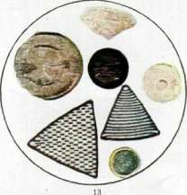
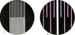
Figs. 1 and 2G Optical spectra of a medium with a rare system of thin lines and a dense system of thick lines. All continuous spectra are closed, showing the primary (absolute) maximum—the grating is not visible. Figs. 5 and 6
Passed through the first.......jh (absolute) maximum and the empty spectra of the rare system—showing the system of rare lines: in place of the dense system—a narrow band. Figs. 7 and 8. The 1st and 2nd spectra of the rare system and the 1st spectrum of the dense system of lines are passed; both systems are visible. Figs. 9 and 10. The 3rd spectrum of the rare system and the 1st spectrum of the dense system are passed; strengthening of the lines in the rare system. Fig. 11. Radial lines at weak magnification, "suspended" by a narrow non-diffracting beam. Due to diffraction and depending on thickness and density of lines, different zones are colored in different spectral colors1 not visible with wide open diaphragm. Fig. 12. The same place covered at the same magnification. The central diaphragm excludes the primary minimum; the result is a dark field of vision, in which a bright spot of diffraction rings stands out. Diffraction pictures, depending on the change in the path of light rays, others. Fig. 13. False structures in diaphragms—triangular—Triceratii..... fairns) with a slit diaphragm over the objective, excluding the appearance of secondary spectra. To the article. Microscope. Description of the histological structure of the optic nerve and retina. They are formed from an outgrowth of the intermediate brain, which at a certain stage of development takes the form of a double-walled cup, connected by a hollow stalk with the base of the intermediate brain. The wall of the optic cup transforms into the retina with its pigment epithelium, and the stalk becomes the optic nerve. The transformation of the stalk of the optic cup into the optic nerve passes through the following sequentially developing processes: 1) differentiation of the cells forming the stalk into glial cells and fibers; 2) growth of visual fibers in a centrifugal direction; 3) penetration from the outside of the mesoderm containing vessels. The entry of a. centralis into the nerve occurs when the embryonic fissure closes, initially it lies peripherically and only after some time penetrates into the center of the optic nerve. The optic nerve extends from the retina to the chiasm, where its crossing occurs; it has an average length of about 5 cm; it is divided into four parts; the first part, intraocular or bulbar, consists of the optic disc (papilla n. optici) with non-myelinated nerve fibers, the lamina cribrosa, and the scleral part containing myelinated fibers of the optic nerve; the second part, orbital or retro-bulbar, represents the part of the optic nerve lying between the eyeball and the optic foramen (for. opticum), with an average length of 3 cm; it in turn is divided into two segments—the anterior 10-20 mm long, containing the central vessels (a. et v. centralis retinae), and the posterior, not containing central vessels; the third part, intra-canalicular, lies along the bony optic canal, and finally the fourth part, intracranial, extends from foramen opticum to the chiasm. From the eyeball, the optic nerve exits through an opening in the vascular layer, through the lamina cribrosa inside from the posterior pole, at an average distance from it of 2.5-3 mm and 1 mm below the horizontal meridian of the eye. In the retrobulbar part, the optic nerve has an arcuate course in the shape of the letter S, so that all movements of the eyeball are not accompanied by tension of the optic nerve. In this part, the optic nerve is surrounded by adipose tissue and has relations with various nerves and vessels: on the outer surface of the nerve lies the ciliary ganglion; ciliary nerves first lie on the nerve and then depart from it; the upper branch of the III nerve and the ramus nasalis (branch of the V nerve) go over the upper surface of the optic nerve; of the vessels, the ophthalmic artery, central artery of the retina, and posterior ciliary arteries have relations to the optic nerve; when exiting the orbit, the optic nerve is surrounded by the apex of the muscular cone formed by the straight muscles of the eye (see Eye). In the intracranial part, the optic nerve lies with its lower surface on the base of the skull; here, on the outer side of the nerve lies the internal carotid artery, above lies the olfactory nerve; further posteriorly, the upper part of the nerve is adjacent to the substantia perforata anterior, forming with it a groove—sulcus opticus. Over a considerable extent, the optic nerve has several sheaths. The entire retrobulbar part of the optic nerve has three sheaths, which are continuations of the meninges. The outermost is the dura (tunica dura), corresponding to the dura mater, consisting of dense fibrous tissue; at the posterior pole of the eye it merges with the sclera (see figure), and at the entry of the optic nerve into the
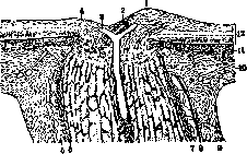
Optic nerve in longitudinal section: 1- nasal part of the optic nerve disc; 2- a. centralis retinae; 3- v. centralis; 4- temporal part of the disc; 5- intravaginal space; 6 and 7- pia mater; 8- arachnoidea; 9- dura mater; 10- sclera; 11- choroid; 12- retina.
The bony canal forms the periosteum of the canal wall. From the inner side, adjacent to the dura mater, is the thin, cell-rich arachnoid membrane (tunica arachnoidea), and the third is the pia mater (tunica pia), closely connected to the trunk of the optic nerve itself. These membranes delimit the so-called intravaginal space, filled with cerebrospinal fluid, which is divided into the subdural and subarachnoid spaces, having a connection with the spaces of the same name in the brain and ending at the sclera of the eye as a blind sac. The intracranial part of the optic nerve has only one pia mater. The blood supply to the optic nerve is provided mainly by the central artery—a branch of the a. ophthalmica. The central artery of the retina, accompanied by a vein, enters the optic nerve at a distance of 10-20 mm behind the eyeball. Proceeding along the axis of the optic nerve forward to the retina, the central artery gives off branches supplying the optic nerve in the anterior segment, and at the same time sends backward a large branch supplying the posterior parts of the optic nerve. In the orbital part, the a. ophthalmica sends small branches to the dura mater and pia mater of the optic nerve. The vascular network of the membranes is connected with each other, and the vessels of the pia mater give off numerous branches into the trunk of the optic nerve, participating together with the central vessels in the blood supply of the nerve. In the intracranial part of the optic nerve, the supply is through the vessels of the pia mater from the a. ophthalmica. The trunk of the optic nerve, upon microscopic examination, appears to consist of a mass of individual fibers, separated by connective tissue septa extending into the optic nerve from the pia mater. The nerve fibers, non-myelinated in the region of the optic disc, behind the lamina cribrosa have a myelin sheath; their caliber varies: most are thin, 2-4 μ in diameter, others are thicker—5-10 μ in cross-section. The thin fibers are attributed to the transmission of visual perceptions, while the thicker ones participate in the pupillary reflex.--The fibers of the optic nerve are divided into macular and peripheral; the former originate from the central parts of the retina—from the macula lutea—and are related to central vision; the latter begin from the peripheral parts of the retina and serve for peripheral vision (see Retina). In addition, both types participate in the decussation, and therefore are divided into direct and crossed (see Visual pathways, centers). Between the nerve fibers of the optic nerve lie cells and fibers of neuroglia.--In recent years, much has been done in the study of the morphology of the optic nerve glia, and its very complex functions have been clarified. The latest methods (mainly by Rio del Hortega) allow in the optic nerve, as in the central nervous system, to isolate three types of glial cells—astrocytes, oligodendroglia, and microglia (Marchesani, Trubin G. A., and others), to which various functions are attributed—both as supporting tissue and as tissue participating in metabolism, nutrition. The glial fibers lie most densely at the periphery of the bundles of nerve fibers and penetrate into the bundles, forming a dense fibrous network, through which the nerve fibers pass. The pathology of the optic nerve is diverse and concerns both the membranes of the optic nerve and the trunk or both together. Congenital defects of the optic nerve. In congenital anophthalmia, the optic nerve is completely absent (aplasia n. optici) and instead only a connective tissue strand can be found. More commonly, coloboma of the optic nerve is observed, which upon clinical examination should more correctly be called 'coloboma at the entrance of the optic nerve' ('Koloboma am Sehnerveneintritt'—German authors). Coloboma of the optic nerve is determined by ophthalmoscopic examination as follows: at the site of the optic disc, a large depression with steep edges is visible, and the diameter of this depression exceeds that of a normal optic disc by 2-4 times. The color of this depression is white with a bluish tint, sometimes the depression is surrounded by pigment, and vessels approach the depression most often from below, but sometimes from all sides. (The origin of colobomas—see Coloboma.) Clinically defined as colobomas of the optic nerve—anatomically they do not always represent such. More often instead of a true coloboma of the optic nerve, colobomas of the choroid at the lower border of the optic nerve disc are found, often with cystic protrusion of the sclera. Furthermore, to the developmental anomalies of the optic nerve disc belong various deviations in its shape (presence of a cone, especially downward, etc.), the presence of an embryonic a. hyaloidea, usually in the form of a strand extending from the optic disc to the vitreous body and either ending freely or extending to the posterior capsule of the lens (a. hyaloidea persistens); then it should be noted the sometimes observed anomaly in the color of the optic disc, which appears significantly redder than normal, and at the same time its borders are indistinct, giving the impression of hyperemia of the optic disc and even its inflammation. The frequent combination with anomalies of refraction—high hypermetropia, astigmatism—absence of functional changes and the stationary state of the ophthalmoscopic picture allow considering this phenomenon an anomaly of the optic disc and to call it pseudoneuritis. Inflammation of the optic nerve—neuritis n. optici—occupies the most important place in the pathology of the optic nerve. It can manifest in various forms and depends on extremely diverse causes. Despite the great diversity in the etiology of the process and the same clinical picture in many cases, the classification of inflammations of the optic nerve is very difficult. The most satisfying practical needs will be the division of inflammations of the optic nerve from the point of view of the ophthalmoscopic picture, taking into account functional disorders in the organ of vision and the patho-anatomical essence of the disease. Based on such signs, inflammations of the optic nerve are divided into two groups—inflammation in the parts of the optic nerve visible in ophthalmoscopy, i.e., inflammation of the optic disc—papillitis (Entzundungspapille according to Schröder) or neuritis optica simplex (E. V. Adamyuk), contrasting this condition with the edematous optic disc—neuritis oedematosa (Staungspapille) (see Congestive optic disc). The second group of inflammations of the optic nerve is characterized clinically by the absence of visible signs of inflammation of the optic disc throughout the process or at least at the beginning of it. This inflammation is localized in the posterior—retrobulbar—parts of the optic nerve, and can involve either only the periphery of the trunk of the optic nerve—the so-called neuritis peripherica, s. perineuritis—or its central, axial part—neuritis centralis, s. axialis—or finally the entire cross-section of the trunk—neuritis transversa totalis. Neuritis of the optic nerve is also divided according to its spread: either from the brain to the periphery—descending neuritis, neuritis descendens—or from the periphery, from the retina or optic disc toward the brain—ascending neuritis, neuritis ascendens. Etiology and pathogenesis of inflammation of the optic nerve. Most often, neuritis of the optic nerve arises as a result of the spread of inflammation from the brain or its membranes, from the side of the orbitgfrom the side of the nasal sinuses, or can develop independently—by the direct action of the causative agent on the optic nerve. The cause of inflammation is primarily various infections, among which syphilis, tuberculosis, typhus, especially typhus, influenza, epidemic cerebrospinal meningitis, scarlet fever, measles, erysipelas, etc., are in the first place. Besides infections, neuritis of the optic nerve can be caused by various types of poisoning; of particular importance are lead, alcohol, tobacco, carbon disulfide, arsenic, quinine, etc. Furthermore, neuritis of the optic nerve is observed in various conditions, such as nephritis, diabetes, sometimes in disorders of menstruation, during pregnancy, lactation. A number of nervous disorders (e.g., multiple sclerosis, myelitis, encephalitis, meningitis, lues cerebri) are also accompanied in some cases by neuritis of the optic nerve. Local processes in the eye—inflammation of the retina, choroid—often also involve the optic nerve; eye injuries, especially penetrating ones, purulent processes in the anterior part and especially general ones, also cause neuritis of the optic nerve. The patho-anatomy of inflammation of the optic nerve has much in common with inflammation occurring in the white matter of the brain, which is understandable in view of the embryological connection of the optic nerve with the brain. In accordance with the difference in histological structure between the optic disc and its trunk, the patho-anatomical picture of inflammation of these parts is different. In papillitis, there is edema of the optic disc tissue with displacement of nerve and glial fibers, increase in glial cells; phenomena of severe hyperemia—dilation of vessels, changes in their walls, often hemorrhages; at a certain stage of inflammation—new formation of vessels; then, depending on the intensity of the process, exudative phenomena of different extent and localization are observed. The nerve fibers, their axis cylinders undergo a series of changes, mainly consisting of swelling, thickening and subsequent disintegration.--In inflammation of the trunk of the optic nerve, vascular changes, inflammatory infiltration are localized either diffusely or in foci in one or another part of the optic nerve. If the process originates from the membranes of the optic nerve, the infiltration is located mainly in them, spreading to the peripheral parts of the optic nerve along the connective tissue septa and giving a picture of neuritis peripherica. Cellular infiltration is especially abundant around the vessels. The nerve tissue itself, when inflammatory phenomena are localized mainly in the connective tissue septa, may be involved in the process secondarily, but undoubtedly it is also affected independently.
In the nerve fibers, degeneration occurs—first, there is a breakdown of myelin, partial or widespread, and then involvement of the axis cylinders, their swelling, spindle-shaped expansion, and disintegration. The glia participates significantly in the inflammation—both progressive and regressive phenomena are noted in it. In the nerve bundles, hypertrophy, proliferation of glial cells, and in places an increase in the number of glial fibers can be seen. The deeper and more widespread the inflammation, affecting the nerve tissue, the more noticeable are the phenomena of replacement of the dead tissue by glia. The glia also participates in the absorption and removal of the products of disintegration of nerve tissue—hence the frequent appearance in the foci of inflammation of glial granular cells. When the inflammation is localized in the retrobulbar portions of the optic nerve, the inflammatory process observed in the papillomacular central bundle of the optic nerve is of particular importance from a pathohistological side, which gives the picture of neuritis axialis. However, clinically designated as axial, retrobulbar neuritis is not always histologically an expression of inflammation of the papillomacular bundle; there are processes (alcohol, tobacco intoxication) that occur with the symptoms of retrobulbar neuritis, and histologically represent predominantly a degenerative process in the optic nerve. Symptoms and course of inflammation. Objective symptoms of inflammation of the optic nerve are most clear when the inflammatory process is localized in the area of the optic nerve head (papillitis, s. neuritis optica simplex). Ophthalmoscopically, in such cases, the optic nerve head appears hyperemic due to the dilation of its numerous small vessels, it is redder than normal, sometimes merging in color with the surrounding fundus of the eye. The large vessels—the central veins—are dilated, tortuous, the arteries are usually somewhat narrowed; hemorrhages are often visible near and on the nerve head. The boundaries of the nerve head are blurred and indistinct due to the presence of exudative phenomena; the same reason explains the cloudiness and thickening of the nerve head tissue, due to which the convergence of vessels on the nerve head is unclear, the course of the vessels both on the nerve head and in the surrounding retina often cannot be traced throughout its entire length; the physiological excavation of the nerve head is obscured, smoothed out. The inflammatory exudate, accompanied by edema of the nerve head tissue, can in some cases be very significant, and then the nerve head appears protruding above the surface of the fundus, becoming noticeably larger in its diameter (see separate table, figure 5). Such an ophthalmoscopic picture can be close to the picture of a congested nerve head, and only the examination of visual functions and the general condition of the patient allow for a correct diagnosis (see Congested nerve head). The pronounced involvement of the retina in the inflammation of the optic nerve—its edema, appearance of hemorrhages, white spots—gives grounds to consider such cases as neuroretinitis. In retrobulbar inflammation of the optic nerve, the ophthalmoscopic picture can remain unchanged for a long time, and sometimes throughout the entire course of the process, and only in those cases when the inflammation is localized near the eye, the process can involve the optic nerve head; ophthalmoscopically this is expressed in the picture of hyperemia of the nerve head, its inflammation. Most often, retrobulbar inflammation in the form of neuritis axialis is recognized in the ophthalmoscope by the pallor of the temporal part of the nerve head, when the process has already caused degenerative changes in the visual fibers of the papillomacular bundle, located in the area of the nerve head on the temporal side (see separate table, fig. 6). Subjective symptoms, which accompany the inflammation of the optic nerve in its various forms, are mainly reduced to a decrease in vision. The degree of decrease in visual acuity, both in strength and in time, is extremely diverse depending on the intensity of the process and its localization. All possible transitions are observed from a slight, slowly increasing decrease to sudden complete blindness. An important symptom for diagnosis is the change in the visual field, and particularly characteristic is the visual field in perineuritis (concentric narrowing) and retrobulbar axial neuritis (central scotoma). Usually, the visual field for colors suffers first, especially green and red. When evaluating the significance of functional disorders in inflammation of the optic nerve, it is necessary to keep in mind the often observed discrepancy between the clinical manifestations of inflammation of the optic nerve and the disorder of functions, and not only in retrobulbar localization of inflammation, but also in lesions of the optic nerve head. Sometimes a sharply expressed ophthalmoscopic picture is accompanied by a relatively weak disturbance of functions and vice versa. The course of neuritis of the optic nerve can be both acute and chronic; the latter is observed more often and usually lasts for months. The resolution of the process occurs with a noticeably changing ophthalmoscopic picture: the hyperemia of the nerve head becomes weaker over time, its redness gradually diminishes, starting from the center, the cloudiness of the nerve head decreases, its edema, and the boundaries gradually become clear. In favorable cases, the functions of the visual organ are completely restored, but very often the inflammatory process, in accordance with pathoanatomical changes, passes into an atrophic one. The nerve head from hyperemic red slowly changes to pale, in some cases partially, most often in the temporal half of the nerve head, or it becomes pale entirely, so that a picture of inflammatory atrophy of the optic nerve is obtained, when the pale nerve head also has irregular boundaries, indicating a former exudative process in the area of the nerve head and around it. Separate forms of inflammation of the optic nerve. Despite the diversity of etiology of neuritis of the optic nerve, certain forms can be distinguished as the most frequent and typical in their clinical picture and course. In this respect, syphilitic neuritis of the optic nerve (neuritis optica syphilitica) must be mentioned first. Syphilis is one of the most frequent causes of inflammation of the optic nerve, causing neuritis in both early and late periods of its course. Inflammation of the optic nerve in syphilis arises either independently or the process on the optic nerve extends from the meninges or the walls of the orbit (especially in the place where the optic nerve passes through the bony canal). The frequency of syphilitic neuritis of the optic nerve can be illustrated by some statistical data. Thus, Badal had 57 cases of inflammation of the optic nerve out of 631 cases of syphilitic lesions of the eye (38 neuritis optica, 16 neuroretinitis, 3 neuritis oedematosa), i.e., in 9% of cases of syphilis of the eye. According to the data of the Kazan Eye Clinic, out of 1,529 cases of syphilis of the eye, inflammation of the optic nerve was observed in 84 cases, i.e., in 5.5% (Protopopov). In recent years, attention has repeatedly been drawn to the possibility of inflammation of the optic nerve in the fresh period of syphilis, to which little attention has been paid until now. Indeed, both in foreign and Russian literature there are great contradictions in this respect—some find syphilitic inflammation in the form of perineuritis extremely often (up to 90% according to Kazas and Shafra), others rarely (Gurvich—1.33%), but in any case it must be assumed that previous statistical data do not fully reflect the frequency of inflammation of the optic nerve in syphilis. Inflammation of the optic nerve in syphilis manifests in different forms—here perineuritis, papillitis, and neuroretinitis are encountered. The so-called neurorecurrences—inflammation of the optic nerve due to insufficient treatment of syphilis, which was especially often observed when salvarsan was introduced into the therapy of syphilis with insufficient doses—are worthy of attention. Of other forms of inflammation of the optic nerve, neuritis caused by general infections, such as typhus, especially typhus fever, then cerebrospinal meningitis, are further common. Typhus fever, as recent epidemics have shown, is relatively often complicated by neuritis, usually in the form of severe papillitis. Neuritis occurs in both mild and severe cases of typhus, usually is bilateral, and often ends with atrophic changes of the nerve head. Neuritis of the optic nerve in typhus fever mostly represents the spread of the process from the brain and its meninges, but in a number of cases they can also be an independent disease of the optic nerve of bacterial or toxic origin. Neuritis is detected both at the height of the general process and during the period of recovery, in typhoid fever, more often on the 3rd-4th week of the disease. Further, some epidemics of influenza are complicated by inflammation of the optic nerve. Cerebrospinal meningitis also often causes inflammations of the optic nerve, sometimes with severe outcomes up to complete atrophy. Retrobulbar neuritis occupies a separate place both clinically and etiologically, especially in its typical form—p. axialis. The causes of such neuritis can be different, but retrobulbar neuritis on the basis of disseminated sclerosis, myelitis, and the so-called rhinogenic neuritis deserves special attention. Retrobulbar neuritis in disseminated sclerosis can be one of the earliest signs of this process, sometimes many years preceding the main manifestations of sclerosis. A characteristic form for disseminated sclerosis is retrobulbar axial neuritis, both of acute and chronic course. The ophthalmoscopic picture, in accordance with the localization of the process, does not show special changes, or partial pallor of the nerve head appears—pallor of the temporal part.
The outcome of the process in its acute form is often favorable from a clinical point of view—the acute disturbance of vision in the form of central scotoma in axial neuritis or even complete blindness in transverse neuritis of the optic nerve trunk may resolve with restoration of vision, but chronic forms usually lead to persistent atrophic changes of the optic nerve. Patho-anatomical changes in the optic nerve in disseminated sclerosis consist in the development of sclerosis plaques in the optic nerve of the same structure as in the foci located in various parts of the nervous system in this process. In myelitis, especially in its acute form, inflammation of the optic nerve (neuromyelitis optica, resp. opticomyelitis) is sometimes also observed in the form of retrobulbar neuritis, which clinically and anatomically has much in common with retrobulbar neuritis in disseminated sclerosis. The process usually affects both optic nerves and often precedes the general symptoms of myelitis. Ophthalmoscopically, in a number of cases, no changes in the fundus are found, but more often the picture of neuritis optica is observed, and in rare cases even a choked disk. Patho-anatomically, a widespread lesion of the optic nerve is usually determined throughout its entire length including the optic tract, and the inflammation may involve the entire cross-section of the optic nerve or occur in scattered foci. Basically, the histological changes consist in the breakdown of nerve fibers with early death of the myelin sheaths, in the proliferation of glial cells with noticeable infiltration of the connective tissue septa of the optic nerve. The histological picture in many cases is so similar to that found in the optic nerve in sclerosis disseminata that great difficulties arise for differential diagnosis (Wilbrand, Saenger, Abelsdorff, etc.). Of great practical importance is also rhinogenic retrobulbar neuritis, i.e., arising secondarily as a result of lesions of the paranasal sinuses. The anatomical proximity of the optic nerve to the sinuses, the commonality of blood circulation and lymph circulation, especially of the posterior sinuses and the optic nerve in its intracanalicular part, make this particular part of the retrobulbar section of the optic nerve especially vulnerable, but often the optic nerve is involved in inflammation in lesions of the anterior sinuses—the maxillary and frontal. It is important to note that retrobulbar neuritis often occurs in the absence of severe, purulent sinusitis; sometimes only a catarrhal process in the sinus mucosa causes a lesion of the optic nerve, and most frequently and earliest the papillomacular bundle of the optic nerve suffers as the most sensitive part of the optic nerve to various harmful influences. Hence also the most frequent clinical form of rhinogenic neuritis—axial neuritis with its typical central scotoma, but in severe cases the matter is not limited to axial neuritis alone, but the entire trunk of the optic nerve is involved. Among the early signs of rhinogenic neuritis, especially depending on the lesion of the posterior sinuses, the so-called Huette symptom (v. Hoeve)—early enlargement of the blind spot, often preceding the appearance of central scotoma—was recently included, but the diagnostic significance of this symptom at the present time is considerably shaken by observations of enlargement of the blind spot in various diseases of the nose, nasopharynx and anterior sinuses. The outcome of rhinogenic retrobulbar neuritis with timely measures taken (often surgical) can be favorable, widespread, persistent atrophies of the optic nerve are relatively rare. To retrobulbar neuritis in terms of clinical symptoms also belong lesions of the macular bundle of the optic nerve in poisonings, of which the most well-known are alcoholic and nicotine (so-called alcoholic and tobacco amblyopia). Chronic poisoning with ethyl alcohol and nicotine with abuse of tobacco, often both poisons together, sometimes causes the appearance of symptoms of axial neuritis of the optic nerve (central scotoma, pallor of the temporal part of the disk). Patho-anatomically, the process is evaluated differently—some see in it 'interstitial neuritis,' others—degeneration of nerve fibers in connection with the primary lesion of the ganglion cells of the retina. Abelsdorff considers that although the primary process is degenerative, but along with it some inflammatory phenomena are also observed. Poisoning with methyl alcohol causes more dangerous lesions of the optic nerve, which is understandable in view of the significantly greater toxicity of it compared to wine alcohol. Although the process in mild cases manifests itself in the symptoms of axial neuritis, usually the lesion is not limited to the macular bundle alone, but involves the entire optic nerve, often causing complete blindness. Patho-anatomically, the matter is a widespread toxic degeneration of the nerve fibers of the optic nerve and ganglion cells of the retina as a result of the primary action of methyl alcohol on nervous tissue. The treatment of neuritis of the optic nerve aims to eliminate the main causes of inflammation and naturally due to the variety of causes it is different. Specific treatment in the most energetic form in syphilis is usually successful, other specific and symptomatic means are less reliable in various infections causing neuritis of the optic nerve. Retrobulbar neuritis of rhinogenic origin require timely surgical treatment, sometimes locally applied prolonged anemia of the nasal mucosa by introducing tampons with cocaine and adrenaline is also beneficial. In alcoholic and tobacco amblyopias, it is important to completely eliminate the poisons causing the lesion. In neuritis of various origins in a certain period of the process, the use of resorbing agents (iodine) is recommended. Locally, bloodletting in the form of artificial leeches by Heurteloup has some importance. Protection of the eyes from light irritation, prohibition of work, etc., is necessary. In addition to inflammations, atrophies of the optic nerve (atrophia n. optici) occupy a significant place in the pathology of the optic nerve, which are divided by origin into two groups: inflammatory atrophy (atrophia inflammatoria) and genuine, or simple atrophy (atrophia genuina). Inflammatory atrophy arises as a result of past inflammation of the optic nerve or after a choked disk, which caused degeneration of nerve fibers. Simple atrophy is caused by various processes, and in some cases the degeneration of the optic nerve is due to a lesion of the ganglion cells of the retina, and as a result ascending atrophy develops, or the process occurs along the course of the optic nerve in its retrobulbar or intracerebral part up to and including the subcortical centers, causing descending atrophy. The causes of simple atrophy can be extremely varied, but the most characteristic are atrophy in tabes dorsalis, in progressive paralysis, disseminated sclerosis, myelitis; then simple atrophy can be the result of interruption of conductivity of the fibers of the optic nerve along its course, whether it is a direct traumatic injury in the retrobulbar part or compression, rupture in fracture of the base of the skull with violation of the integrity of the bones of the orbit, compression by hemorrhages, tumors in one or another part of the optic nerve or compression by tumors inside the skull; tumors of the hypophysis are particularly important in this respect; sclerotic vessels (a. carotis int. or a. ophthalmica) have the same effect. Simple atrophy further develops after a number of poisonings (methyl alcohol, quinine, optochin, atoxyl, etc.), after abundant loss of blood. Finally, there are forms of atrophy of hereditary origin: such is familial hereditary atrophy, or Leber's disease. Patho-anatomically, atrophy of the optic nerve appears different depending on its origin. Atrophy as an outcome of neuritis has been mentioned above. In simple, genuine atrophy of the optic nerve, especially in tabes dorsalis, degeneration of nerve fibers is found in the optic nerve, and this process is accompanied by proliferation of glial cells and especially in-
OPTIC
connective tissue framework of the optic nerve in late stages of atrophy undergoes thickening, homogenization of the sclerosed connective tissue septa occurs. The pathogenesis of tabetic atrophy still remains an unresolved question. The former view that tabetic atrophy is primarily a degenerative process, after a series of studies, especially by Stargardt, is being reconsidered, and many believe that degeneration is associated with an inflammatory process localized mainly in the meninges of the optic nerve, and in nature the atrophy is not ascending, as has been claimed until now, but descending. Symptoms and course. Atrophy of the optic nerve is confirmed by means of an ophthalmoscope when it reaches the optic disc or is localized in the disc. Every atrophy is characterized by pallor of the optic disc, and in complete atrophies the disc is pale throughout its entire length, in partial atrophies on separate areas, pallor of the temporal half of the disc is often observed due to atrophy of the papillomacular bundle of the optic nerve. Inflammatory atrophy is characterized by indistinct, sometimes irregular borders of the discs (see separate table, fig. 7). In simple atrophy, on the contrary, the borders of the disc are clear and regular. Often the color of the disc in simple atrophy is noticeably gray, hence the gray atrophy of the discs in tabes dorsalis (atrophia grisea tabetica) (see separate table, fig. 8). The central vessels and especially the arteries appear narrowed. The main symptom of atrophy is decreased vision, which in complete atrophy reaches the loss of light perception. The course in many cases is chronic—in this respect an example is tabetic atrophy, in which the atrophic process can progress over a number of years. On the other hand, there are processes, such as poisonings, where in a short time the atrophic process reaches its full development. In progressive atrophies, the impairment of function, decrease of central and peripheral vision gradually increase. For tabetic atrophy, for example, early loss of the visual field for colors, sector-shaped narrowing of the peripheral visual field for white color is characteristic. The prognosis in atrophies is always serious and especially poor in progressive atrophies—usually the condition ends in blindness. Treatment is of little hope and only timely treatment directed at the cause of the suffering can stop the process. As symptomatic treatment, subcutaneous injections of strychnine, galvanization are used; in recent years, retrobulbar injections of atropine and others have been tried without noticeable success.
Tumors of the optic nerve occur relatively rarely and develop either primarily in the optic nerve itself or secondarily due to spread from the eye or orbit. Primary tumors of the optic nerve are divided into extradural and subdural (Golovin). The former arise from the outer sheaths of the optic nerve and spread mainly into the orbital fat tissue. They usually have a malignant course, belonging to endotheliomas and sarcomas. True tumors of the optic nerve or subdural ones involve the trunk of the optic nerve and usually form a pear-shaped thickening of the optic nerve in its retrobulbar part. These tumors are currently characterized as gliomas (or gliomatosis of the optic nerve), identical to gliomas of the brain with their various forms (Fleischer, Oberling et Nordmann, Odintsov and others). These tumors are benign in their course, do not grow beyond the dura mater of the optic nerve, do not give metastases, but become dangerous when they involve the entire optic nerve, spreading to its intracranial part. A tumor of the optic nerve is diagnosed by the appearance of exophthalmos with good mobility of the eye in all directions, with signs of early decreased vision. Ophthalmoscopically—at first, signs of stagnant disc are often observed, and then atrophy. Treatment is surgical: removal of the tumor usually by means of the Krenlein operation in Golovin's modification.
Injuries to the optic nerve are observed under various conditions—direct injury through the soft tissues of the orbit, through the bony walls, often bilateral (bullet wounds), usually accompanied by death of the optic nerve—its atrophy. Fractures of the base, bruises of the supraorbital arch, accompanied by violation of the bony walls of the apex of the orbit, also give injuries to the optic nerve, ending in its atrophy. Complete avulsions of the optic nerve from the eyeball (avulsio n. optici) have been described many times in trauma.
Related articles
Mentioned in
Cite this page
“Optic Nerve.” Soviet Medical Encyclopedia. English translation of Bolshaya Meditsinskaya Entsiklopediya, 1st ed. (Moscow, 1928–1936), ed. N. A. Semashko. https://sovietmedicalencyclopedia.pages.dev/article/optic-nerve/