Scotoma
Historical document, translated for reference. It reflects medical knowledge of the 1920s–30s and is not medical advice.
Summary
Scotoma refers to any defect in the field of vision, classified as positive (subjective) or negative (objective), and absolute or relative. The article describes physiological scotomas like the blind spot and pathological forms including central, ring-shaped, and isolated defects.
Encyclopedia article (1928–1936)
SCOTOMA. Some authors call any defect in the field of vision a scotoma, while others only refer to delimited defects not connected with the periphery. Defects connected with the periphery are described as various types of constriction of the visual field (see). Scotomas are divided into positive (subjective), perceived by the person being examined, and negative (objective), not subjectively perceived. According to the degree of expression, scotomas are distinguished as absolute and relative. An absolute scotoma for white and black color refers to defects in which a white or black object is not perceived at all, while relative scotomas are such losses in which a white or black object is visible but not sufficiently clearly, appearing rather gray. Figure 1. Configuration of the scotomatous spot according to Helmholtz. Figure 2. The same according to Bjerru. PKTPr./'t,G1ЯpHтTX oP7Tp_. Figure 3. The same according to Igersheimer.
Absolute scotoma for red, green, blue) for a particular color refers to defects in which the color of the object is not recognized, even if the object itself is perceived as colorless, gray, while relative scotomas are those in which the color of the object appears only less saturated than in the areas free of scotoma. Thus, a relative scotoma for white color can at the same time be absolute for other colors. Even in the visual field of a normal eye, a number of defects not connected with the periphery are noted-physiological scotomas. These scotomas are: 1) the blind, or Mariotte's, spot, 2) the vascular defects originating from it-angioscotomas, and 3) physiological central scotoma. 1) The place of exit from the eye of the optic nerve (papilla nervi optici) as lacking light-sensitive neuro-epithelial elements is blind. The defect in the visual field corresponding to the projection into space of the optic nerve head is the blind spot, discovered and described for the first time by the English physicist Mariotte in 1668. Under ordinary conditions, it is not subjectively perceived and therefore belongs to the category of objective, or negative, scotomas. Corresponding to the anatomical form of the optic nerve head, the blind spot has the shape of an oval somewhat elongated in the vertical diameter, the center of which is in the horizontal meridian at a distance of approx. 15 arc degrees from the fixation point outward, and in the vertical meridian-approx. 2-3°. The greatest size of the horizontal diameter of the blind spot fluctuates around 5°, of the vertical-around 8-9°.
2) The branches of the central artery and vein of the retina, nourishing the inner layers of the latter, lie in front of the light-sensitive layer of the retina and therefore cast a shadow on this layer. If the visual field is carefully examined using a thin technique (according to Bjerrum's method, on Lloyd's stereoperimeter, according to Igersheimer's, Evans's methods, etc.) under a sufficiently small angle of view, the image of the object falling on the blood vessel during such examination of very small size may turn out to be smaller than the cross-section of the vessel and is not perceived by the eye. Thus, vascular scotomas-angioscotomas-are discovered. The latter are always necessarily connected with the blind spot. The evolution of our concepts about the configuration of the blind spot and the angioscotomas originating from it is vividly illustrated in fig. 1-5. Of particular interest in this respect are the studies that appeared in 1926-27 by Evans, who with the help of a very thin technique managed to obtain almost a complete negative image of the vascular tree of the retina (fig. 5). 3) Defects in the area of the fixation point-central scotomas-are observed in the visual field of a normal eye only under special conditions of adaptation of the eye to reduced brightness of illumination-dark adaptation of the eye.
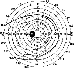
Pathological scotomas not connected with the periphery can be schematically reduced to the following forms: 1) Scotomas originating from or connected with the blind spot. 2) Central scotomas, 3) Ring-shaped scotomas, 4) Isolated island-like defects. 1) The blind spot can change in relation to its size and shape under the influence of the most diverse causes. Among congenital anomalies, myelin fibers of the optic nerve, which obstruct due to their opacity the path of light rays to the light-sensitive layer of the retina, are accompanied by an increase in size and change in configuration of the blind spot. The same can be observed in myopia accompanied by peripapillary atrophy of the vascular coat. Particularly characteristic is an increase in the blind spot in all directions with a stagnant optic disc (fig. 6). This symptom is assigned certain importance in the differential diagnosis between a stagnant optic disc and inflammation of the optic nerve. In 1909-10, van der Hoeve described an increase in the blind spot as an early symptom of
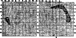
Figure 6. Enlargement of the blind spot with a stagnant optic disc. 5 20 15 10 5 4=0,7? H 1.5 D 5 10 15 20 25 25 20 15 10 5 Tension 20 mmHg т-0.7; H 1,5D Figure 7. Bjerrum's scotoma. 5 10 15 20 25. Teusio.45mmHg
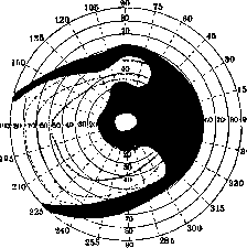
Figure 8. A ring-shaped scotoma merged with the periphery, Bjerrum's.
of the optic nerve in diseases of the paranasal sinuses, known since then under the name of van der Hoeve's symptom. According to this author's observations, this symptom occurs in the vast majority of cases only in diseases of the posterior sinuses (sphenoid and ethmoid) and precedes the appearance of central scotomas, which are so characteristic of optic nerve lesions of rhinogenic origin. In general, the data of v. d. Hoeve were subsequently confirmed by numerous studies. Nevertheless, a number of researchers do not consider it possible to attribute significant clinical value to the symptom of v. d. Hoeve. In this connection, it was found that this symptom is observed not only in posterior but also in anterior sinusitis, as well as in some other diseases of the nose and nasopharynx, primarily in adenoids of the nasopharynx, that it does not necessarily accompany rhinogenic retrobulbar neuritis and therefore has only facultative value. Of undoubted clinical importance is the characteristic enlargement of the blind spot in glaucoma, described by Bjerrum. It consists in the appearance of scotomas originating from the blind spot, arc-shaped surrounding the fixation point and called Bjerrum's scotomas (Fig. 7). Sometimes originating from the upper and lower poles of the blind spot, they can form a closed ring around the fixation point. In far-advanced cases of glaucoma, these ring-shaped scotomas merge with peripheral defects (Fig. 8). Although not pathognomonic for glaucoma, Bjerrum's scotomas are nevertheless a very characteristic phenomenon in glaucoma, contributing to the early diagnosis of the process. They also have a certain prognostic significance. 2) Central scotomas have exceptionally important clinical significance, since in many cases only on the basis of their detection is the correct diagnosis of the disease established. They may be the result of lesions of the retina and choroid in the area of the yellow spot or maculo-papillary (axial) bundle of fibers of the optic nerve, selectively and exclusively sensitive to certain harmful influences. They may be unilateral, but more often they are bilateral and are usually accompanied by more or less sharp decrease in visual acuity. Since color vision is the most delicate function of the eye and suffers first of all under the influence of various harmful influences, central scotomas arise first and foremost as scotomas for green, red, and blue colors. The presence of a unilateral central scotoma, generally speaking, forces one to look for its cause mainly in local suffering of the eye and the areas surrounding it, whereas a bilateral scotoma is in the vast majority of cases the result of a general disease. The causes of unilateral scotomas are diverse. They may be observed in the eye in the absence of any visible anatomical changes in congenital or acquired in early youth amblyopia. Congenital developmental defects of the yellow spot (coloboma maculare congenita), inflammatory processes - central tuberculous, syphilitic and other chorioretinitis, hemorrhages developing in the area of the yellow spot can be the cause of central scotomas. For inflammatory processes, it is characteristic that the appearance of scotomas is preceded or accompanied by subjective phenomena of metamorphopsia, macro- and micropsia. Among the causes of unilateral central scotomas arising on the basis of lesions of the maculo-papillary bundle in retrobulbar (axial) neuritis, first place is taken by disseminated sclerosis (sclerosis disseminata) and inflammatory diseases of the paranasal sinuses, especially the ethmoid and sphenoid. Disseminated sclerosis often causes bilateral eye lesions (Fig. 9), whereas in sinusitis this is observed but is not characteristic. Bilateral central scotomas in the vast majority of cases occur in acute and chronic retrobulbar (axial) neuritis on the basis of exo- or endogenous intoxications. Among exogenous intoxications, first place is taken by chronic poisoning of the organism by alcohol; (methyl) and tobacco. Starting with small relative, and later absolute, scotomas for green and red color, they can within several weeks turn into extensive central scotomas for all colors, accompanied then by a sharp decrease in vision and pallor of the temporal halves of the optic nerve heads. For them, their form of a horizontal oval is characteristic, sometimes merging with the area of the blind spot (Fig. 10). In poisoning with much more toxic methyl alcohol, the changes often do not limit themselves to central scotomas but have a more extensive character, often leading to complete blindness. Among other exogenous intoxications also damaging the maculo-papillary bundle, mention should be made of poisoning with carbon disulfide (in the rubber industry), lead, veronal, dinitrobenzene, arsenic, especially atoxyl, iodoform, chloral, thyroidin, etc. Among auto-intoxications, mention should be made of cancerous cachexia and diabetes mellitus. The latter can give central scotomas due to lesions of the macular regions of the retina, especially in the form of retinitis punctata centralis diabetica. Single, sometimes difficult to establish ophthalmoscopically, small white shiny foci of retinal degeneration in the area of the yellow spot can clinically manifest as a clearly expressed central scotoma. But more often diabetes leads to bilateral, and also unilateral, central scotomas due to axial neuritis, which greatly aggravates the condition. A special form of lesions of the optic nerves with central scotomas is represented by the familial-hereditary Leber's form of atrophy of the optic nerves, affecting mainly men in the period of puberty. Symmetrical lesion of the area of the yellow spot with central scotomas is further observed in senile degeneration of the yellow spot in old age, in central chorioretinitis and degenerations of the yellow spot on the basis of high myopia, as well as in a special form of its familial-hereditary degeneration, sometimes transmitted from generation to generation and manifesting itself most often in the period of puberty. Transient, and sometimes persistent, central scotomas can occur in retinitis due to blinding of the eyes by bright light, especially direct sunlight. The above-mentioned causes of unilateral central scotomas can obviously also lead to bilateral scotomas and vice versa. Sometimes bilateral scotomas have a hemianopic character (Fig. 11). 3) Ring-shaped scotomas (Fig. 12), leaving the periphery and center free, surround the latter in the form of a completely or incompletely closed ring. Often they merge with the periphery
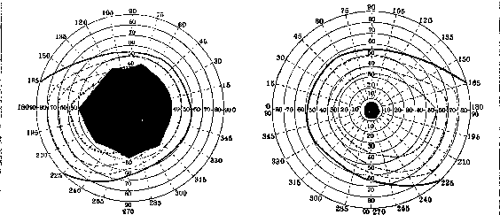
Figure 9. Central scotomas in sclerosis disseminata.
with difficulty ophthalmoscopically ascertainable small white shiny foci of retinal degeneration in the area of the yellow spot can clinically manifest as a clearly expressed central scotoma. But more often diabetes leads to bilateral, and also unilateral, central scotomas due to axial neuritis, which greatly aggravates the condition. A special form of lesions of the optic nerves with central scotomas is represented by the familial-hereditary Leber's form of atrophy of the optic nerves, affecting mainly men in the period of puberty. Symmetrical lesion of the area of the yellow spot with central scotomas is further observed in senile degeneration of the yellow spot in old age, in central chorioretinitis and degenerations of the yellow spot on the basis of high myopia, as well as in a special form of its familial-hereditary degeneration, sometimes transmitted from generation to generation and manifesting itself most often in the period of puberty. Transient, and sometimes persistent, central scotomas can occur in retinitis due to blinding of the eyes by bright light, especially direct sunlight. The above-mentioned causes of unilateral central scotomas can obviously also lead to bilateral scotomas and vice versa. Sometimes bilateral scotomas have a hemianopic character (Fig. 11). 3) Ring-shaped scotomas (Fig. 12), leaving the periphery and center free, surround the latter in the form of a completely or incompletely closed ring. Often they merge with the periphery
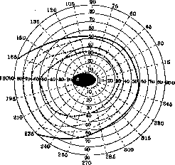
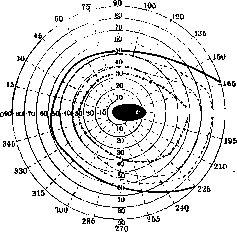
Figure 10. Central scotomas for red and green color in alcoholic and tobacco intoxication.
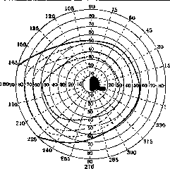
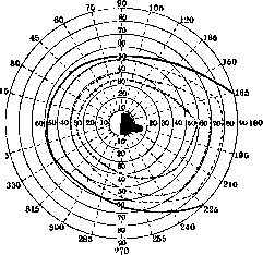
Figure 11. Hemianopic central scotomas from gunshot wound of the occipital region. f23 lice, diabetes and other ailments. The aforementioned retinitis from blinding by sunlight often accompanies central scotoma with a ring-shaped one. As a mass phenomenon, this was described by Jess in persons who observed a solar eclipse with the naked eye. 345' 370 Figure 12. Ring-shaped scotoma. Sometimes a central scotoma of this origin, resolving, transforms into a ring-shaped one. The latter usually undergoes regression. Since pilots often have to be blinded by direct sunlight, ring-shaped scotoma is characteristic for ma scintillans. It usually arises suddenly with the following subjective phenomena: in the field of vision of both eyes, more often near the fixation point, there appears a peculiar release of flickering light spots, bordered by a lighter zigzag line; these spots more or less rapidly move to the periphery of the field in the same direction for both eyes; then the flickering ceases, and in the same parts of the field relative or absolute scotomas of homonymous hemianopic type arise. After lasting V4-u2 hours, the scotomas disappear, after which pains arise in the half of the head opposite the scotoma. These migraines can be of varying intensity and sometimes are combined with more severe general phenomena: nausea, vomiting, hemianesthesias and paresthesias, even speech disorders (with right-sided hemianopia) and hemipareses. Since the sensations of flickering are associated with a very characteristic appearance of zigzag lines in the field of vision, this peculiar functional condition of the eye received the name teichoscopy, or teichopsia (teichos- fortress wall). The described phenomena usually repeat at various intervals. Their basis lies in circulatory disorders in the subcortical centers of vision and the Gracile bundle with alternation of phenomena of irritation (teichopsia) and inhibition (scotoma) on the basis of angiopathies of cerebral vessels. In some cases, they are early symptoms of such severe diseases as lues cerebro-spinalis, tabes dorsalis, paralysis progressiva, but can also have a purely functional character.-All the above forms of scotoma can be combined and combined
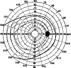
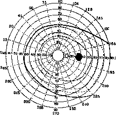
270 Figure 13. Cortical island-like hemianopia. (After Wilbrand.)
this profession. Prevention-wearing appropriate protective glasses.-^Isolated island-like defects can have the most diverse form and be observed in the most diverse ailments of the eye, associated with the presence of scattered focal pathological changes in the vascular membrane and retina (hemorrhages, tub. foci, foci of degeneration, chorioretinites of the most diverse etiology, etc.). They are very often and in typical form observed in chorioiditis disseminata. They can also have a hemianopic character (fig. 13). To the category of transient scotomas of hemianopic character also belongs the flickering scotoma.-scoto- in the most diverse ways, presenting in such cases very great difficulties for analysis.
Related articles
Mentioned in
Cite this page
“Scotoma.” Soviet Medical Encyclopedia. English translation of Bolshaya Meditsinskaya Entsiklopediya, 1st ed. (Moscow, 1928–1936), ed. N. A. Semashko. https://sovietmedicalencyclopedia.pages.dev/article/scotoma/