Perimetry
Historical document, translated for reference. It reflects medical knowledge of the 1920s–30s and is not medical advice.
Summary
Perimetry is a method for examining peripheral vision based on projecting the spherical surface of the retina onto a concentric spherical surface. The technique was first conceptualized by Purkinje in 1825 and later developed into practical instruments by Aubert and Forster in 1857.
Encyclopedia article (1928–1936)
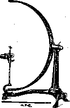
PERIMETRY (from Greek peri - around and metron - measure), one of the methods for examining peripheral vision, based on the projection of the spherical surface of the retina onto a similarly spherical and concentric external surface; in practice, this method is carried out with the help of a special instrument called a perimeter. The idea of the method itself belongs to Purkinje and was expressed by him as early as 1825, but practically in the form of a special instrument it was only realized in 1857 by Aubert and Forster, of whom the latter introduced a number of improvements to the original model and introduced the instrument into general use under the name perimeter, which is why it is usually named after him. Landolt (1872) significantly simplified and improved the Forster perimeter. Although over time the instrument underwent numerous modifications and improvements introduced by various authors, the basic principle of Forster and Landolt remained unchanged. The main part of the perimeter in modern models (Figure 1) is an arc made of a blackened metal strip 5 cm wide, with dimensions of half or a quarter of a circle described by a radius of 30 cm. Degree divisions are marked on the inner or outer surface of the arc, starting from its center, which is taken as 0 and at the same time serves as the fixation point for the subject, which is why it is marked with a white circle. With its apex, the arc is attached to a vertical stand on a massive cast iron base in such a way that at the point of attachment it can rotate around its horizontal axis. The inclination that the arc takes during these rotations with respect to the vertical, in other words, the position of the meridians, is indicated on a special disk mounted on the same stand, behind it, with the help of a special pointer that rotates together with the arc. A. N. Maklakov (1884) proposed taking two arcs instead of one, intersecting at the center at a right angle, so that the field could be examined in two meridians at once. As a test object for P. serves a small white square, which is moved along the inner surface of the arc or simply by the researcher's hand at the end of a thin black stick or with the help of a special mechanism with a rope transmission on a special device of the sled type. The latter is a combination of three disks, of which the lower, rotating one, is divided into a number of sectors colored in the primary colors (for white and color P.), the second above it, stationary,
Fig. 1. Forster's perimeter; modern model. with a quadrangular cutout on the front edge of 20 mm, and the third between the first two, also rotating, with a whole series of square cutouts on the edge from 2 to 20 mm, regulating the size of the test object similar to a diaphragm. In view of the fact that the nodal point of the subject's eye must coincide exactly with the center of the arc, which in turn requires the subject's head to remain stationary, on the opposite end of the cast iron base in front of the arc there is a special column with a horizontal chin rest, which can be set to any height by means of a special screw, and in addition in front of the rest there rises a vertical rod with a small platform at the upper end, to which the subject presses with the lower edge of the orbit.
Figure 2. Perimeter in the form of a hemisphere; model of Scherk.
From the numerous variants of the perimeter, more or significantly different from the model just described, the following deserve mention: 1. Hollow peri-meters (for example Scherk's), in which the arc is replaced by a hemisphere (Fig. 2); the inner surface of the latter is blackened, and meridians and parallel circles are marked with red paint; in the vertical meridian the hemisphere is divided into two halves connected by hinges, so that if necessary either half can be swung aside. The test object at the end of a thin black stick is moved along the inner surface of the hemisphere by the researcher's hand, and the results are immediately marked with chalk. The advantages of this model are, firstly, the elimination of impressions from extraneous objects that distract the subject's attention, and secondly, the free movement of the test object not only along meridional, but also in all other directions.
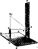
2. Perimeters without an arc (e.g. Helmbold's), an ingenious (Fig. 3) and very simple in concept model that can be easily constructed with the most primitive means. As a test object, a small white square is taken,
Fig. 3. Perimeter without an arc; model of Helmbold. fixed on a long, thin stick, with a miniature loop at the very end. If, by hooking the loop onto a hook, one pulls the cord so that the test object describes a 90° arc, a weight on the free end of the cord is raised to a certain height, and the point on the back board opposite which it is positioned is marked with the number 90, and the entire distance between 0 and 90 is divided into 9 equal parts, each corresponding to 10°. To determine the boundaries of the visual field according to this scale, it is sufficient to mark those divisions opposite which the weight will stop when the test object moves in different directions or meridians, the deviations of which the researcher determines approximately.
3. Portable perimeters. From
Figure 4. Hand perimeter; model of Schweigger.
the numerous models, the most common is the hand perimeter of Schweigger (Schweigger), which in appearance resembles a regular perimeter in miniature (Fig. 4), with an arc of 15-20 cm radius; the entire instrument is held by the subject in their hands by a special handle, and the test object is moved along the arc by hand. Elschnig somewhat modified this model, making it foldable.
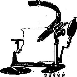
4. Self-recording perimeters (for example, Hardy, Fig. 5). The advantages of self-recording perimeters lie in that they eliminate the need to specially mark the boundaries of the visual field on the arc and to draw these boundaries on paper or a diagram, and also eliminate any possibility of error during this manipulation, since the registration of the visual field is carried out automatically. This is achieved by the fact that all movements of the test object by means of a cord and toothed wheels are transmitted to a special recording system located behind the arc and equipped with a special pointed stylus marking the position of the object with point punctures on the opposite diagram. The entire system rotates together with the arc, and in addition the stylus also performs lateral movements.
5. Lighted perimeters (e.g. Michel's, Fig. 6), intended for examination in the dark, for some reason it is undesirable or impossible to use daylight or artificial lighting, as well as for those cases when due to
Fig. 6. Lighted perimeter; model of Michel.
clouding of transparent media, for example the lens, the subject cannot distinguish




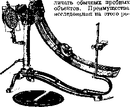
The advantages of such perimeters also include that the examination is always conducted under identical lighting conditions, and the subject's attention is not distracted by extraneous objects. Externally, these instruments resemble the self-recording perimeter just described, with the only difference being that on the inner surface of the arc, painted gray, there is a deep groove along which a small box containing a small electric lamp instead of the test object moves; the box is equipped with a series of diaphragms of varying widths and a set of colored glass plates in case color perimetry in the dark is necessary. The fixation point is a small red lamp at the center of the arc. In addition to those listed, there are still a whole range of perimeter models of various types that are not widely used. It should also be kept in mind that the perimeter is used not only for the study of peripheral vision but also for other purposes, such as determining the angle of strabismus, the position of a cysticercus in the eye, etc. The study of peripheral vision generally amounts to a comprehensive examination of the so-called visual field (see). For practical purposes, the most important aspects are: 1. Determination of the outer boundaries of the visual field, which under normal conditions depend on the configuration of the facial structures surrounding the eye (eyelids, bridge of the nose, nose, edges of the orbit), further on the position of the eye itself in the orbit, on the size of the pupil, etc. 2. Determination of partial defects or scotomas in the visual field, which develop under the influence of focal diseases of the visual apparatus. 3. Determination of color sensation on the periphery of the retina. The actual technique of perimetry is as follows: the patient sits with their back to the light, places their chin on a special stand of the instrument, and with the eye being tested, fixes the zero division at the center of the arc; the other eye is covered during this time. The doctor, positioned opposite the patient, must carefully ensure that the patient's eye does not move away from the designated mark throughout the examination. It is preferable to conduct the examination in diffused daylight, placing the instrument between two windows, set back somewhat from the wall, or even better under similar conditions but in a corner of the room. If necessary, artificial lighting can also be used, which should also be sufficiently strong and diffused. When determining the boundaries of the visual field for white color, with which perimetric research usually begins, the test object is slowly and silently moved along the perimeter arc from the periphery toward the center until the patient, while continuing to fixate on the central mark, notices the movement of 'something white' and reports this to the examiner. The division of the arc at which the test object first becomes visible to the subject corresponds to the outer boundary of the visual field at that location. By sequentially rotating the perimeter arc around its center by 45°, this type of manipulation is performed in at least 4 meridians, namely: the vertical, horizontal, and two intermediate oblique ones, starting at least with the vertical, and in each meridian, 2 points at opposite ends are determined. The 8 points found are plotted on the corresponding degree divisions of special diagrams (see Visual field), and by connecting them, the outline of the boundaries of the visual field of that eye is obtained. To determine suspected scotomas, the test object is moved along a series of meridians, 10-15° apart, throughout the entire length of the arc, starting from the outer boundaries of the visual field to the fixation point itself, and as soon as the object enters such a defect area, it will immediately disappear from the subject's view and reappear upon leaving this area. The degree divisions of the arc at which the object disappears and reappears are transferred to the diagram, and by connecting them, the outline of the scotoma itself is obtained. The examination of color sensation on the periphery of the retina is carried out on the same principle as the determination of peripheral perception of white color, with the only difference that instead of white objects, corresponding colored objects of the same size as the white ones are used, and the subject should not in advance know what color the object is, and the boundary of the visual field for a given color is considered to be the place where it is correctly recognized by the subject. For clinical purposes, it is usually sufficient to determine the boundaries of the visual field for blue, red, and green colors. Color perimetry is also a very important and precise method of research, as it often reveals pathological changes where the white object does not yet show them.
D. strupov.
Related articles
Mentioned in
Cite this page
“Perimetry.” Soviet Medical Encyclopedia. English translation of Bolshaya Meditsinskaya Entsiklopediya, 1st ed. (Moscow, 1928–1936), ed. N. A. Semashko. https://sovietmedicalencyclopedia.pages.dev/article/perimetry/