Strabismus
Historical document, translated for reference. It reflects medical knowledge of the 1920s–30s and is not medical advice.
Summary
Strabismus is a condition in which the visual axis of only one eye is directed toward an object of attention, while the visual axis of the other eye assumes a different direction. It is classified into concomitant and paralytic types based on whether the deviation remains constant or varies with gaze direction.
Encyclopedia article (1928–1936)
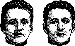
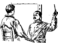
Strabismus (from Latin strabo- crooked), or loxophthalmos (from Greek loxos- crooked and ophthalmos- eye), is a condition in which the visual axis of only one eye is directed toward an object of attention, while the visual axis of the other eye assumes a different direction. The angle of strabismus is the angle between the direction that the visual axis of the deviating eye actually occupies and the direction it should occupy with the eye properly fixed on the point of fixation. Strabismus divides into two large groups. One includes all cases in which the deviation of the deviating eye remains the same in all directions of gaze, while the other group includes cases in which the deviation of the deviating eye increases in certain directions. In cases of the first group, we are dealing with an anomaly of position with healthy muscles, in cases of the second group-with an anomaly of movement as a result of paralytic condition of the muscles. In cases of the first group, the deviating eye by association repeats all movements of the fixing eye in full. In cases of the second group, the movements of the deviating eye in the sphere of action of the paralyzed muscles lag behind the movements of the fixing eye (Fig. 1). The second group of strabismus is called paralytic, the first group-concomitant, or concomitant strabismus (strabismus concomitans). If in strabismus the fixing eye is covered with a screen and the deviating eye is asked to fixate, the latter establishes its visual axis on the fixation point. For this it must make an adjustment movement of a certain volume in the direction opposite to the former deviation. The covered eye, by association with the adjusting eye, also makes a movement in the same direction as the adjusting eye. According to the law of uniform distribution of nerve impulse between both eyes, the volume of movements of the adjusting eye and the secondarily deviating eye
Figure 1. On the left-primary position, on the right-lag of the right eye (paresis of the right external muscle).
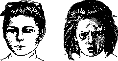
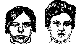
in concomitant strabismus will exactly correspond to each other. According to the nature of the deviation, the most common are convergent strabismus (strab. convergens; Fig. 2) and divergent (strab. divergens; Fig. 3). In convergent strabismus, the visual axis of the deviating eye deviates from the fixation point toward the midline of the body; in divergent strabismus it moves away from the midline. When the visual axis deviates from the fixation point upward, it is called strab. sursum vergens, when it deviates downward-strab. deorsum vergens. - Deviation of the visual axis from
Figure 2.
the fixation point in some cases may be only apparent (pseudostabismus). Pseudostabismus is determined by the size and position of the angle formed by the visual axis with the line connecting the posterior pole of the eye with the center of the cornea (Z?). The angle is more often open inward from the center of the cornea (positive), but can also be open outward from it (negative). If the angle is positive (predominantly in hypermetropes), then with its significant size, for the deviated inward from the center of the cornea visual axis to coincide with the fixation point, the anterior segment of the eyeball must move outward, while with a negative angle-inward (predominantly in myopes) (Fig. 4). Unlike true strabismus, in pseudostabismus covering the non-deviating eye is not accompanied by an adjustment movement of the deviating eye. And if it is known that the apparently deviating eye has good visual acuity and is capable of central fixation, then the diagnosis of pseudostabismus is established. Concomitant strabismus can be
Figure 3
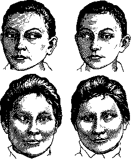
constant or periodic (strab. periodicus and intermittens). Concomitant strabismus can be limited to only one side in all circumstances (strab. unilateralis) or shift from one eye to another (strab. alternans; Fig. 5).
Figure 4.
Concomitant strabismus can at all times remain unchanged (strab. constans) or appear only when fixing at a certain distance or when fatigued (strab. facultativus). Strabismus that is immediately noticeable is called manifest (strab. manifestus). To determine the degree of deviation of the deviating eye, there are several methods. This includes the method of determining the magnitude of deviation in linear measure using the Lawrence strabometer. This method is very inaccurate and applicable only when central vision exists in the deviating eye. The Lawrence strabometer is a curved plate corresponding to the shape of the lower eyelid, divided along its upper edge into millimeters. The divisions go in both directions from the zero located in the center. When measuring, the patient's non-deviating eye is covered and they are asked to fixate a distant object with the deviating eye. At the same time, the strabometer is applied to their lower eyelid so that the zero division corresponds to the center (or outer edge) of the cornea. Then they are asked to fixate the same object with the previously covered eye and look at which division the center (or outer edge) of the cornea of the deviating eye, which has returned to its usual position, now corresponds to. The number of millimeters found determines the linear magnitude of the deviation. It is considered that each millimeter approximately corresponds to 5°. More accurate methods include angular measurement using a perimeter and Maddox's tangent scale. When measuring with a perimeter, the deviating eye is placed in the center of the perimeter arc, and the fixing eye-on an object located 5-6 m along the radius passing through the zero division of the perimeter arc. The observer looks over the arc and moves a candle flame along its inner surface until they see the reflection of the flame in the center of the pupil of the deviating eye. The number on
Figure 5. Above-strabismus divergens alternans, below-convergens alternans.
the perimeter arc where the flame is located at that moment indicates in degrees the magnitude of the angle of strabismus together with the angle kappa. Maddox's scale (Fig. 6) consists of two cardboard strips placed crosswise. On the horizontal one there are two rows of numbers-large and small. The large numbers correspond in degrees to angles for which tangents have been calculated at a distance of 5 m, and the small numbers-angles for which tangents have been calculated at a distance of 1 m. In the center of the horizontal strip is placed a light source, which the patient fixes. The distance between the light source and the point corresponding to which the visual axis of the deviating eye rests against the scale is the tangent of the angle of strabismus. This point is considered found when, with a known rotation of the fixing eye, the image of the light source coincides with the center of the pupil of the deviating eye. In practice, for initial orientation, one can use the following guidelines by Hirschberg: if the reflection from the light source with an average pupil width (3.5 mm) is at its edge, then the angle of deviation is 15-20°; if the reflection is placed on the limbus, then the deviation is almost 45°. The position of the visual axes and consequently of the eyeballs is determined by anatomical relationships. Since ideal symmetry in the structure of the orbits and their contents on both sides is rarely encountered, it becomes necessary to have a corrective effect from the sensory-motor apparatus. This corrective effect can manifest itself timely only when control exists. Such control is performed by the "aversion" to double vision inherent in normal people-diplophobia. The eyes of people deprived of diplophobia are left exclusively to static influences that result from anatomical relationships. The static strabismus developing under their influence is usually insignificant and causes patients no inconvenience, as diplopia does not bother them, and cosmetically strabismus becomes inconvenient only at higher degrees. Persons to whom diplophobia is characteristic seek to get rid of double images and achieve this in two ways: either they achieve fusion of images or they suppress one of them. These people in turn can be subdivided into those with a tendency to fusion and those without it. The former, through muscular tension, can overcome not too significant abnormalities of position and thus get rid of diplopia. The latter try to eliminate diplopia by excluding the image of the deviating eye. For this purpose, they try to give this eye such a position that its image can be suppressed with the greatest ease. The farther from the center the image is located on the retina, the easier it is to exclude it from sensation. Therefore, the deviating eye, as it were, runs before its extrafoveal image, it deviates from the symmetrical position with the other eye more than
10987654 321012 3 456789 10|
40
Figure 6.
this is required by anatomical relationships, and thus to static strabismus is added 'escape strabismus'. This combined strabismus almost always becomes concomitant. The constancy of the angle of deviation, characteristic of concomitant strabismus, ensures the constancy of the area of the retina onto which the extrafoveal image falls, and thereby facilitates its suppression. There exists a great dependence of strabismus on refractive anomalies. This dependence is based on the innate connection between accommodation and convergence. In hypermetropes, as a result of this connection, strabismus convergens can develop, in myopes-strabismus divergens. Of course, strabismus convergens is also found in emmetropes and even in myopes, since besides refractive anomalies, strabismus is caused by other reasons, which at the same time can overcome the influence of refractive anomalies. If a hypermetrope is forced to converge excessively, he faces diplopia. In those cases when the extensibility of the connection between accommodation and convergence is exhausted, he can only suppress the extrafoveal image. In search of the most convenient position for the squinting eye for this purpose, he does everything that was described in static strabismus with diplophobia. As a result, his final strabismus consists of static (since it is at different times common to all people), refractive, and escape strabismus. The refractive anomaly in most cases determines only the direction of strabismus, but not its degree. At the beginning of its development, refractive strabismus can be completely eliminated by correcting the refractive anomaly (Fig. 7).
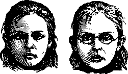
Figure 7. Disappearance of convergent strabismus under the influence of correction with glasses.
But as soon as the tissues adapt to the incorrect position, this is no longer possible: refractive strabismus is partially or completely transformed into static strabismus. Even in these cases, it is sometimes still possible with the help of glasses to eliminate part of the deviation by changing the impulse that accommodation gives to convergence. Since strabismus is only a symptom that can be caused by various causes, determining the cause for each case is the first and most important task. Determining the etiological factor is particularly difficult in cases of convergent strabismus. The difficulties here are primarily related to the early period of life, during which strabismus convergens develops in most cases. However, with some patience, it is possible to determine refraction by means of skiascopy during the first year of life and thus obtain a basis for therapeutic measures. If hypermetropia exists, it is necessary to prescribe as early as possible for constant wear approximately full correction. For very small children, the glasses should be fastened with ties at the back of the head.-In early childhood, one more condition can be fulfilled. Experience leaves no doubt that in many cases amblyopia of the squinting eye develops only after strabismus sets in, and the younger the child, the sooner this happens. According to the data of Worth, if treatment is started early enough, the squinting eye never loses the ability to fixate centrally; without treatment, in cases where strabismus develops in the first year of life, a decrease in vision may occur as early as after 6-8 weeks. For treatment, it is first necessary to determine whether the child has alternating or unilateral strabismus. Since it is impossible to test the visual acuity of a child, it is necessary at least to test whether the squinting eye is capable of central fixation (of a flame or a shining object). The constantly or predominantly deviated eye must be brought into use for vision. For this, the fixing eye is either bandaged or atropinized for a prolonged period. In this connection, it should be remembered that prolonged exclusion of the fixing eye may also lead to the development of amblyopia in it.-There is no doubt that abnormal excitations of the convergence center as a cause of convergent strabismus can also arise reflexively from distant organs (e.g., from the intestinal canal in the case of worms). These reflex influences should be attributed, for example, to cases of intermittent strabismus, as well as to the much more frequent cases in which strabismus convergens, although permanent, varies periodically in degree.-Cases of static strabismus, in which the only or most significant cause for strabismus are anatomical (mechanical) relationships, have a very definite character. As long as the tendency to fusion acts as a regulating factor, the anomaly of the position of rest may remain hidden. In fatigue, states of weakness and aimless vision, when images on the retina do not attract attention and thus do not influence the oculomotor apparatus, the abnormal state of rest becomes apparent. The encouragement that occurs spontaneously or under the influence of a call causes strabismus, along with the return of attention, to return to its hidden state (see Heterophoria).-Strabismus of hypermetropics, in turn, has characteristic features. It also occurs only at times (when carefully examining objects), but is precisely absent during aimless vision, i.e., when there is no interest in its distinctness. The periodicity of onset or fluctuations in the angle of strabismus is thus a common phenomenon for different types of strabismus and therefore requires careful observation in order to carry out treatment corresponding to the etiology. In some cases, an attempt is made to discover and eliminate the harmful factors acting on convergence reflexively (this group of cases ultimately also includes the correction of existing hypermetropia with glasses), in others (where there are indications of an anomaly of position at rest) one comes to the necessity of surgical intervention.-How and when to proceed with the operation depends on various other conditions: the age of the patient, subjective complaints (asthenopia), the size of the angle of strabismus, possible disturbances in the sphere of action of the internal and external rectus muscles of the eye. In periodic strabismus convergens during the first 6 years of life, surgery is performed only in exceptional cases. Prolonged waiting in periodic strabismus can never be harmful, as long as patients remain under systematic control and the tendency to fusion is strengthened by stereoscopic exercises. Also in permanent convergent strabismus, one should never operate before the completion of the 4th year of life. And even then only in the case that the treatment indicated above, despite its application for months, has had no effect on the size of the angle of strabismus, and the angle of strabismus exceeds 15°. The early start of treatment (prescription of glasses, bringing the squinting eye into use for vision) may be so important that the final, generally achievable result in this case does not suffer from postponing the time of operation. If through prolonged observation the constancy of the angle in strabismus convergens is proven and it is found that preliminary treatment has no effect on it, operative intervention thus becomes indicated. The operation may consist either in the backward displacement of the attachment of the tendon of the m. rectus internus (tenotomy), or in the forward displacement of the attachment of the tendon of the m. rectus externus (transplantation), or in a combination of both operations simultaneously. Some authors regard tenotomy negatively because of unexpected failures (artificial paresis of the m. rectus internus and strabismus divergens). These failures are due to incorrect indication and careless performance of the operation. An example of such incorrect indication may be those cases in which, with spontaneous or postoperative paralysis of one muscle, tenotomy of its antagonist is undertaken. The inevitable result is that the loss of mobility in the sphere of action of one muscle is joined by an equally great loss in the sphere of action of its antagonist. From the foregoing it follows that where strabismus depends on muscle weakness, transplantation of this muscle is absolutely indicated, not tenotomy of its antagonist. Tenotomy is permissible only in the case that the action of the corresponding muscle significantly exceeds the average norm, i.e., if in strabismus convergens a significant excess of action of the m. rectus internus can be proven. The latter usually occurs in long-standing strabismus. In such cases, the inner margin of the pupil at maximum contraction of the m. rectus int. passes across a line mentally drawn between the lacrimal points by a more or less considerable distance. This strengthening of the m. rectus int. does not always correspond to a weakening of the action of the m. rectus externus, and in such case tenotomy of the m. rectus int. as the first measure is fully indicated. But even in this case, one should always remember that the function of muscles can only be weakened to normal limits. In order not to risk too strong an effect in tenotomy, a thread is first placed on the tendon, which is secured with a loose loop to the conjunctiva. If immediately after operation an excessive weakening of the action of the m. rectus int. is found, the loop should be slightly tightened, regardless of the fact that strabismus will again become greater. If the immediate effect is not too great, the loop is left in place and the eye is bandaged. Excessive effect can be corrected by means of a more or less strong tightening of the knot on the following and third day after operation. If the immediate effect is not too great, the thread can be removed on the day following the operation. The final result of the operation cannot be judged before the second week. If the effect is insufficient and it is necessary to operate further, one should again investigate whether there is a significant excess of action of one or the other muscle which would justify a new tenotomy, or whether transplantation of the m. rectus ext. seems more appropriate. Under no circumstances should tenotomy be repeated on the same muscle if the first tenotomy has already brought the mobility in the sphere of its action within normal limits. Simultaneous tenotomy of both m. recti int. is permissible only in cases of the most long-standing strabismus. In this connection, it should always be borne in mind that the effect of a simple tenotomy in some cases may be accompanied by an extremely strong effect. Transplantation of the m. rectus ext. is indicated: 1) if at maximum turning of the eye outward the temporal margin of the cornea remains far from the outer canthus, 2) if an excess of mobility in the region of the m. rectus int. is absent or has already been eliminated by tenotomy, and 3) in long-standing strabismus to support the effect of tenotomy of the m. rectus int.
When performing transplantation, three options must be considered: 1) simple displacement of the muscle attachment closer to the corneal edge, 2) shortening of the muscle by excising a piece (or creating a fold) while preserving the natural attachment, and finally 3) transplantation of the shortened tendon. All these methods suffer from one drawback - the unreliability of the final result due to the impossibility of precise dosage. Since the effect of all transplantations weakens, it is always necessary to aim for an immediate hyper-effect, for which however there is no measure, as the weakening is also beyond any accounting. With a strabismus angle of 20° or more, unilateral transplantation (or resection) of the rectus ext. muscle is insufficient. One must either operate on both rectus ext. muscles simultaneously (which is practiced in cases of amblyopia of the squinting eye) or combine transplantation of the rectus ext. muscle with tenotomy of the rectus int. muscle. If the detached rectus int. muscle is not released from the hand with the aid of a loop, then despite uncontrollable and individually very different influences (muscle tone, tension of muscle fasciae, etc.), the effect of the transplantation can be to some degree dosed with the aid of a combined tenotomy. Even the temporary elimination of tension in the rectus int. muscle by simply detaching its tendon, which is held by a thread as close as possible to the site of the old attachment, gives the transplantation effect reliability and is without any hesitation applicable even in those cases in which the function of the rectus interni muscle does not permit its weakening. After transplantation, a binocular bandage is required for approximately five days to prevent eye movements. If the strabismus angle after surgery becomes less than 15°, then, especially in children, a prolonged interval dedicated to observation should follow. During this interval, attempts to combat amblyopia of the squinting eye should be resumed. If age is taken into account, then in young patients one should never aim for the complete elimination of convergence as an immediate result. This is particularly undesirable in those cases in which the possibility of restoring binocular vision is excluded from the very beginning. Permanent strab. diverg. always requires surgical treatment. Tenotomy of the rectus ext. muscle as the only measure is insufficient, especially if one does not forget the basic rule against excessive weakening of muscles. In most cases, it is necessary to perform transplantation of the rectus int. muscle. This intervention, depending on the size of the strabismus angle on one or both sides, should always be combined with temporary detachment of the tendon of the rectus ext. muscle. The immediate effect should always be substantially greater than that which is desirable as a permanent result. One should aim for convergence and to a greater degree the stronger the previous divergence was. If the function of the external rectus muscles is preserved, postoperative convergence (up to 10°) disappears after a few days after removal of the binocular bandage. After strabismus operations, if there is no large difference in vision of both eyes, diplopia often develops, which can be of two origins: either as a result of congenital fusion defects or as a result of the formation during strabismus of an anomalous correspondence (as if the formation of a second macula on the squinting eye). In the first case, the distance between the double images is close to the existing deviation at the moment, in the second it corresponds to the difference between the former squint position and that obtained after surgery. Prolonged and distressing diplopia can only occur with fusion defects, but even in such cases it usually eventually passes by itself. -The most important task of treatment after strabismus surgery is to stimulate and strengthen the tendency to fusion. For this purpose, stereoscopic exercises are used. This aims at the simultaneous perception of images belonging to both retinas, the fusion of related halves of the image, and the exercise of that innervation which counteracts the existing squint position. The treatment of obvious vertical deviations (strab. sursum et deorsum vergens) is almost always surgical. Vertical deviations are very often mixed with horizontal ones and specifically with high degrees of strab. converg. They rarely occur in isolation. In mixed deviations, the vertical ones can first be eliminated if there are indications that they are the main obstacle to fusion. Usually only the straight elevators and depressors are operated on. The operation should be performed on those muscles in whose sphere of action the maximum deviation exists. The latter circumstance must be clarified before the operation in the most careful manner. -One cannot, for example, with deviation of the left eye upward, immediately perform tenotomy of the m. recti superioris sinistri. This operation would be contraindicated if it were found that in the sphere of action of this muscle, i.e., with the eye turned to the left, the vertical divergence disappears, while when turned to the right (sphere of action of the oblique muscles of the left eye) it becomes maximum. In such a case, if one wishes to equalize the position by operating on the squinting eye itself, the only correct procedure would be tenotomy of the m. obliqui inf. sinistri. Instead, the goal is better achieved by tenotomy of the m. recti inf. dextri or transplantation of the rectus super, dextri or by a combination of both operations simultaneously. With this measure, the relative position of the visual lines (in the vertical direction) for turning to the right will be changed most strongly, for the median position less, and for turning to the left minimally.
L. Sergievsky. Paralytic strabismus (strab. paralyticus) arises as a result of paralysis of the muscles rotating the eyeballs, in other words, as a result of damage to the corresponding nerves: oculomotor (III), trochlear (IV), and abducens (VI), or their nuclei located in the brainstem. Paralytic strabismus therefore has great importance in the diagnosis of nervous diseases. Here, the same forms are found as in concomitant strabismus, i.e., strabismus convergens, divergens, sursum vergens and deorsum vergens, or forms where the deviation of the squinting eye occurs in a complex direction, for example, simultaneously inward and downward, etc. For the diagnosis of paralytic strabismus, a number of characteristic symptoms are taken into account, which distinguish this type of strabismus from concomitant strabismus. These are: a) deviation of the eye in the direction opposite to the paralyzed muscle, with the angle of secondary deviation being greater than the angle of primary deviation; b) limitation of eye mobility in the direction of movement of the paralyzed muscle; c) presence of double vision-diplopia, especially pronounced in acute cases, but detectable by special examination methods (see Diplopia), and in cases of long duration. By examining the form of strabismus and especially by analyzing the nature of the observed diplopia, it is possible to accurately determine which muscle is paralyzed, and from this to establish topological diagnosis of the lesion that caused the muscle paralysis.-For lesions of m. rect. int. (n. oculo-motorius), external strabismus, weakening of the eyeball's movement inward, and crossed diplopia (diplopia cruciata) are characteristic. In lesions of m. rect. ext., the opposite picture is observed: internal strabismus, weakening of the eyeball's movement outward, and homonymous diplopia (diplopia homonyma). In isolated lesions of other eye muscles, strabismus is much less clear, as compensation by synergists (m. rectus sup.-m. obliquus inf., m. rectus inf.-m. obliquus sup.) is possible here. Paralysis of m. recti sup. causes deviation of the eyeball outward when looking upward due to the contraction of the synergist of the affected muscle-m. obliqui inf. At the same time, crossed diplopia occurs. Paralysis of m. recti inf. causes deviation of the eye outward and crossed diplopia when looking downward (action of m. obliqui sup.). In paralysis of m. obliqui inf., displacement of the eye inward and homonymous diplopia when looking upward (m. rectus sup.). In paralysis of m. obliquus sup., slight deviation of the eye inward and homonymous diplopia when looking downward (m. rectus inf.). Of particular clinical importance are strabismus convergens in paralysis of the n. abducens and str. divergens in paralysis of the n. oculo-motorii. Peripheral paralysis of the n. oculomotorii most often manifests as total involvement of its functions, i.e., the presence of strabismus and ptosis, mydriasis, and paralysis of accommodation. In nuclear lesions, dissociation of paralysis is more common. To determine the location of the lesion in the brainstem (midbrain), the presence of other phenomena of loss is of decisive importance. Thus, with a lesion located at the base of the midbrain (pes pedunculi), the so-called Weber syndrome occurs: paralysis of the n. oculo-mot. on the side of the lesion and hemiplegia on the opposite side. With a lesion in the region of the tegmentum-Benedict's syndrome: paralysis of the n. oculomot. on the side of the lesion and tremor on the opposite side. With a lesion of the n. oculomot. at the point where it passes through the red nucleus, Claude's syndrome occurs: paralysis of the n. oculomot. on the side of the lesion and cerebellar phenomena on the opposite side. Lesion of the n. abducens (strabismus convergens) at the point where it extends from the nucleus to the base of the brain (caudal part of pontis Varoli) also gives an alternating symptom complex: paralysis of the n. abducens (str. convergens) on one side and hemiplegia on the other. Lesion of the nucleus of the n. abducens almost always, in addition to paralysis of the n. abducens, also causes paralysis of the n. facialis.-The etiology of paralytic strabismus is very diverse. Mainly in diagnosis, one must think about lues cerebri, sclerosis disseminata, encephalitis, and meningitis. Further, strabismus is often a focal or distant symptom of a brain tumor (especially str. convergens in tumors of the cerebellopontine angle). The diagnosis is made based on consideration of other symptoms and the course of the disease. Then, paralytic strabismus can depend on various diseases of the orbit (periostitis, tumors, phlegmon, etc.). The therapy and prognosis of paralytic strabismus are determined by the etiology of the process. If etiological therapy is unsuccessful in chronic cases of paralytic strabismus, the question of surgical intervention in the forms indicated in the treatment of concomitant STRABISMUS is sometimes raised.
Related articles
Mentioned in
Cite this page
“Strabismus.” Soviet Medical Encyclopedia. English translation of Bolshaya Meditsinskaya Entsiklopediya, 1st ed. (Moscow, 1928–1936), ed. N. A. Semashko. https://sovietmedicalencyclopedia.pages.dev/article/strabismus/