Eyelids
Historical document, translated for reference. It reflects medical knowledge of the 1920s–30s and is not medical advice.
Summary
The eyelids are folds of skin that develop in the embryo, forming the palpebral fissure and protecting the eye. They contain muscles, glands, and connective tissue that enable blinking, tear distribution, and eye protection.
Encyclopedia article (1928–1936)
Eyelids (palpebrae), by their origin, represent folds of skin, which begin to develop at the end of the second month of embryonic life, grow toward each other and fuse together, separating again only shortly before the birth of the child. The border of the upper eyelid is marked by the eyebrow, and that of the lower eyelid approximately by the lower bony margin of the orbit. Converging at their ends, the eyelids form the palpebral fissure, with the outer angle of this fissure being pointed and the inner angle being horseshoe-shaped, where the lacus lacrimalis (tear lake) is located with the caruncula lacrimalis at its bottom. The width of the palpebral fissure varies individually, but, on average, with a normal forward gaze, the area of the palpebral fissure, i.e., that part of the cornea and scleral conjunctiva not covered by the eyelids, encompasses the entire cornea, except for its upper part. The shape and width of the palpebral fissure greatly influence the expression of the eyes. The skin of the eyelids is extremely thin and very mobile, due to the loose and fat-poor subcutaneous tissue. Therefore, it easily folds into wrinkles when the eyelids are opened and just as easily stretches when they are closed. But, due to this same mobility of the skin, it is easily pulled to the sides by adjacent scars, causing ectropion of the eyelids, and under it edemas and ecchymoses easily form. On the skin of the eyelids there are fine hairs, sebaceous and sweat glands. When the anterior surface of the eyelids is bent, a anterior border is formed on the free edge of the eyelid, and when the free edge is bent toward the posterior surface of the eyelid, a posterior border is formed, between which remains the intermarginal, or intermarginal, space. On the slightly rounded anterior border sit eyelashes in 2-3 rows, numbering 100-150 on the upper eyelid and 50-70 on the lower, with small sebaceous glands at their roots (glands of Zeis), between which and behind which there are also several modified sweat glands (glands of Moll). Somewhat in front of the posterior sharp border, the openings of the Meibomian glands are visible as dots, and between them and the eyelashes a thin grayish line is visible, dividing the intermarginal space into posterior and anterior halves. At the inner angle of the palpebral fissure, where the edges of the eyelids begin to form a horseshoe-shaped notch, small elevations are visible on both eyelids at the posterior border - the lacrimal papillae, on the tops of which are the lacrimal puncta, the beginning of the lacrimal canals, which run horizontally from here in the thickness of the eyelids to the lacrimal sac. When the eyelids close, their edges come into close contact with each other. The movements of the eyelids are caused by muscles located under the skin. These are, first of all, the m. orbicularis, a flat striated muscle surrounding the palpebral fissure, consisting of two portions - the inner one, located in the eyelids themselves (pars palpebralis), and the outer one, lying peripherically, along the edge of the orbit and its circumference (pars orbitalis). The fibers of the inner portion begin from the medial palpebral ligament and, running arch-like over the surface of the cartilage, meet each other at the outer angle of the palpebral fissure, where they connect with each other by means of the inscription tendinea, under which lies the lateral palpebral ligament. That part of the pars palpebralis, which begins from the posterior limb of the medial palpebral ligament and partly from the medial wall of the orbit, is called the pars lacrimalis of the m. orbicularis, or the muscle of Horner. Several bundles of the m. orbicularis, located near the medial edge of the eyelid, partly in front, partly behind the excretory ducts of the Meibomian glands, are called the m. ciliaris of Riolani s. subtarsalis (see Figure 1).
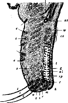
Figure 1. Vertical section through the upper eyelid: e-fine hairs of the skin; a-sweat gland; c-eyelashes; z-sebaceous gland; s-modified sweat gland of Moll; o-m. orbicularis palpebralis; r-m. ciliaris Riolani; m-excretory duct of the Meibomian gland; v-anterior border of the eyelid edge; h-posterior; d-Meibomian gland; t-pseudo-papillae, especially developed in the area of the upper edge of the cartilage (so); a-acinous glands of Waideyer; ai-arcus tarseus inferior; rp-arterial branch emerging from it; as-arcus tarseus superior; p-muscle of Heinrich Müller; (-levator palpebrae superioris; /-its bundles going to the skin (according to Fuchs).
The fibers of the outer portion attach to the bones of the medial edge of the orbit and, making a circular course, attach again to the bony edge or to the skin or interlace with other muscles. In the upper eyelid, the levator palpebrae superioris, a striated muscle, is also located, beginning at the edge of the optic canal, running along the upper wall of the orbit, attaching to the upper edge and anterior surface of the cartilage of the upper eyelid and lifting it upon contraction. This muscle, before reaching the upper edge of the orbit, forms a broad tendon, in which between two tendon layers lies a layer of smooth muscle fibers - the muscle of Heinrich Müller, which also attaches to the upper edge of the cartilage. On the lower eyelid, there is no muscle analogous to the levator, but there is the muscle of Heinrich Müller, located on the lower side of the m. rectus inferioris and attaching to the cartilage of the lower eyelid. Isolated contraction of the muscles of Heinrich Müller leads to a slight widening of the palpebral fissure. The dense foundation, the framework of the eyelids, is formed by the so-called tarsal plates - plates of dense fibrous connective tissue without cartilage cells. Internally, the fibers of the tarsal plate pass into the medial palpebral ligament, which attaches with one limb in front of the lacrimal sac to the frontal process of the maxilla, and the other - behind the lacrimal sac, to the crista lacrimalis posterior of the lacrimal bone. Externally, the fibers of the tarsal plate pass into the lateral palpebral ligament, which attaches to the lateral edge of the orbit. The remaining periphery of the tarsal plates connects with the edge of the orbit by means of a dense fascia - the f. tarso-orbitalis. The tarsal plates together with the fasciae form the septum orbitale, separating the contents of the orbit from the front. In the thickness of the tarsal plates lie the Meibomian glands - about 30 in the upper and fewer in the lower, consisting of a longitudinal excretory duct and short-stemmed alveoli emptying into it from both sides (see Figure 1). In their essence and structure, the Meibomian glands are modified sebaceous glands; their secretion, lubricating the edges of the eyelids, prevents tears from overflowing over the edge of the eyelids and protects the epithelium from maceration. At the upper edge of the cartilage, there are a few additional lacrimal glands, built on the type of lacrimal glands, the excretory duct of which opens into the fornix of the conjunctiva. The posterior surface of the eyelids is lined with a mucous membrane - the palpebral conjunctiva. Blood vessels of the eyelids. Arteries of the eyelids (see Figure 2) originate from two arches - arcus tarseus superior et inferior, running along the upper and lower edges of the tarsal plate, at a distance of 1-3 mm from them. On the lower eyelid, the arch at the orbital edge of the tarsal plate is sometimes absent. The veins of the eyelids, more numerous and wider, follow the same path and empty into the veins of the face and orbit. At the place where they pass through the m. orbicularis, the veins can be compressed, which accounts for the so often observed blepharospasm edema of the eyelids. The lymphatic vessels of the eyelids are very numerous and form two interconnected networks, lying on both surfaces of the tarsal plate. Lymph from them is carried away to the lymph gland located in front of the ear, which accounts for the swelling of the latter in severe inflammations of the eyelids. The sensory nerves of the eyelids receive from the n. trigemini: the upper eyelid - from the first branch and the lower - from the second; motor nerves: m. orbicularis - from the facial nerve, the levator - from the oculomotor nerve, and the muscle of Heinrich Müller - from the sympathetic. The eyelids are
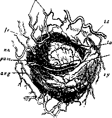
Figure 2. spo-a. supraorbitalis; fr-a. frontalis; pn-a. nasalis; ang-a. angularis, anastomosing with the previous one; mx-a. maxillaris externa; zy-a. zygomatica; la-terminal branches of a. lacrimalis; ts-a. temporalis superficialis; pal, pam-aa. palpebrales lateralis and medialis; ** arcus tarseus inferior formed by them (according to Megger). protection of the eye from external harmful influences; with any irritation they narrow the palpebral fissure, closing not only the entire upper half of the cornea but also its lower part. With constant movement during blinking, intensified with any irritation, they evenly moisten the eye and remove from its surface all the smallest particles. When the eyelids close during sleep ( or when squinting them when danger approaches the eye, with a reflex turning of the eye upward) the entire cornea is hidden under the upper eyelid. The muscle of Horner, due to its attachment to the medial palpebral ligament, pulling this ligament upward with each blink, and with it the walls of the lacrimal sac, expands the lacrimal sac and promotes the suction of tears into it through the lacrimal puncta. Corresponding to the structure of the eyelids, their diseases are reduced to diseases of the eyelid skin, common with skin diseases in general, diseases of the edges of the eyelids, diseases of the eyelid muscles and their glands.
Related articles
Mentioned in
Cite this page
“Eyelids.” Soviet Medical Encyclopedia. English translation of Bolshaya Meditsinskaya Entsiklopediya, 1st ed. (Moscow, 1928–1936), ed. N. A. Semashko. https://sovietmedicalencyclopedia.pages.dev/article/eyelids/