Conjunctiva
Historical document, translated for reference. It reflects medical knowledge of the 1920s–30s and is not medical advice.
Summary
The conjunctiva is a thin mucous membrane covering the posterior surface of the eyelids and the anterior part of the eyeball up to the cornea. It consists of three anatomical regions: the palpebral conjunctiva, the fornix, and the bulbar conjunctiva, each with distinct histological features and specialized structures.
Encyclopedia article (1928–1936)
CONJUNCTIVA, "conjunctiva (from Latin conjungere - to join), the membrane of the eye, covering the posterior surface of the eyelids and the anterior part of the eyeball up to and including the cornea. It consequently forms an open pouch forward, the anterior and posterior walls of which lie against each other. The conjunctival sac always contains a small amount of tear fluid, which continuously enters the sac from the lacrimal gland and, having moistened the eye, flows through the lacrimal passages into the nasal cavity. The conjunctiva is a very thin and delicate mucous membrane, containing in its deeper parts lymphoid elements and various kinds of glands, which although not numerous, are in cases of removal of the lacrimal gland still sufficient to maintain the necessary moisture in the conjunctival sac and on the anterior surface of the eyeball. -Anatomically three parts are distinguished in the conjunctiva: a) the conjunctiva of the eyelids (conjunctiva palpebrarum, s. tarsi), b) the conjunctiva of the fornix - the transitional fold (fornix, s. conjunctiva fornicis) and c) the conjunctiva of the eyeball, or sclera (conjunctiva bulbi, s. sclerae). The conjunctiva of the tarsus or cartilage appears moist, pale pink in color, but sufficiently transparent to allow one to see through it the pattern of the visible Meibomian glands located in the cartilage (see Eyelids). 11/2-2 mm above the edge of the eyelids and parallel to it on the tarsal conjunctiva there is a depression called the sulcus subtarsalis. The lower part of the tarsal conjunctiva is smooth. The upper part, on the contrary, forms conjunctival depressions, manifesting to a very varying degree and resembling on sections the picture of glands. Here the conjunctiva is smooth only when the adenoid layer is absent, i.e., in newborns. With the onset of mature age and the growth of adenoid tissue, folds of the conjunctiva are formed, leading to a series of depressions and elevations, the so-called Stieda grooves (Plateaux-und Rinnensystem Stieda); these grooves are observed mainly in the upper part of the tarsal conjunctiva and in the initial segment of conjunctivae mobilis, s. fornicis. The fornix is formed by the transitional fold connecting the tarsal conjunctiva with the bulbar conjunctiva; the upper fornix is deeper than the lower. The ability of the conjunctival fornix to fold and unfold allows the eyeball to maintain all its mobility. At the site of the transitional fold under the tunica propria there is subconjunctival tissue, consisting of loose connective tissue bundles, the presence of which explains the mobility of the conjunctiva. In the upper outer part of the upper fornix there are pinpoint openings - the excretory ducts of the lacrimal gland. In the conjunctiva of the eyeball, a movable part covering the eyeball itself and a part in the limbus area, fused with the underlying tissues, are distinguished. From the limbus the conjunctiva becomes completely transparent and passes onto the anterior surface of the cornea, forming its epithelial layer. In the inner corner of the eye the conjunctiva presents two formations: the lacrimal caruncle (caruncula lacrimalis) and the plica semilunaris (plica semilunaris).-The lacrimal caruncle, especially in the part of the lower eyelid, resembles skin in its structure (only the stratum corneum is missing); it contains small hairs, sebaceous and acinous glands. Somewhat outward from it is the semilunar fold, formed by the scleral conjunctiva: it is a rudimentary organ corresponding to the third eyelid in animals and birds. The rudimentary third eyelid (plica semilunaris) consists of connective tissue, multilayered epithelium or transitional epithelium. Mainly in the area of the conjunctival fornix the adenoid layer of the conjunctiva (covered in the normal state with a smooth layer of epithelium) on its surface presents a series of depressions and elevations, giving papilla-like formations called papillary bodies. The significance of these papillary bodies is important in view of the role they play in pathological processes of the conjunctiva. According to histological structure, two layers are distinguished in the conjunctiva: the superficial - epithelium and the deep - submucous layer (tunica conjunctivae propria). The epithelium is different in various parts of the conjunctiva. The epithelium of the eyelids (Fig. 1)


Figure 1. Conjunctiva of the eyelids. Cylindrical epithelium and submucous membrane.
Figure 2. Conjunctiva of the eyeball with goblet cells. consists of two layers. The superficial contains one layer of cylindrical cells; the deep - one, rarely two layers of smaller and more or less flat cells. The cylindrical cells are 10-25 microns in length. In addition, goblet cells are observed in the epithelium, which are also found on the transitional fold and in the mucous membrane of the eyeball (Fig. 2). On the transitional conjunctival fold we also encounter a layer of cylindrical cells, and the deep layer is thicker and consists of 2-3 rows of cells. The epithelium here is the same as on the palpebral part of the conjunctiva. At the level of the limbus, however, the epithelium is multilayered (Fig. 3), and in it three kinds of cells are distinguished: 1) a superficial layer of 1-2 rows of flat cells, 2) a middle layer consisting of polygonal cells arranged in several rows, 3) a deep layer consisting of one row of cylindrical or square cells with a large oval nucleus. -- The submucous layer is also divided into a superficial, or adenoid layer and a deep, or fibrous layer. The submucous layer of the conjunctiva is separated from the epithelium by a thin plate (basement membrane of some authors), which at the level of the limbus continues directly into Bowman's membrane. The adenoid layer consists of thin connective tissue with accumulations of lymphoid cells. This adenoid layer is richly supplied with vessels. Tunica propria consists of connective tissue and a small number of white blood cells; plasma cells are occasionally observed there. The fibrous layer consists of thick and dense connective tissue fibers. -The glands of the conjunctiva are as follows: Krause glands (Krause) acinous (Fig. 4), located on the upper and lower eyelids along a line corresponding to the area of the transitional fold. They number from 30 to 40 on the upper eyelid and only from 2 to 6 on the lower. Most of them are round or oval in shape and are located in the submucous layer. Each gland consists of a series of lobules and has a common excretory duct. Histologically ^'

Figure 4. Krause glands in the area of the upper eyelid
they have the same structure as the lacrimal gland and they indeed play the role of accessory lacrimal glands. Henle's glands, of tubular or tubular type, are located between the orbital edge of the cartilage and the transitional fold and are equipped with an excretory duct opening into the transverse grooves present in this part of the mucous membrane. The existence of Henle's glands is not recognized by all. The same is the case with Manz's glands, described by this author, in the vicinity of the cornea. Arteries supply the conjunctiva with a double network: on the one hand, palpebral branches supply the conjunctiva of the cartilage, the transitional fold, and part of the conjunctiva of the eyeball. On the other hand, the pericorneal area is supplied by branches of the art. ciliaris ant., closely connected with the ciliary body and iris, in which they end. This anatomical fact plays a major role in the pathology of the eye in the form of the so-called pericorneal (or ciliary) injection. The veins are arranged in the same way as the arteries; usually two veins accompany one artery. From the palpebral part, some veins pass into the v. ophthalmica, and others into branches going to the v. facialis et temporalis superf. The veins of the ciliary area (vv. ciliares anterior.) eventually pass into the vv. ophthalmicae. The study of the normal histology of the conjunctiva has been greatly facilitated by research with the slit lamp of Gullstrand, and a whole series of features, relating mainly to the study of the lymphatic pathways and nerves of the conjunctiva, is due to the microscopy of the living eye with the addition of methods of vital staining (see separate table, figure 5).-Lymphatic vessels are distributed in two layers: one, superficial, is located directly under the capillary layer, the other, deep,-in the submucous connective tissue and connects with the first by a whole series of anastomoses going in a vertical direction-some to the inner, others to the outer angle of the eye. The first pass-eventually into the submandibular lymph. node, and the second into the parotid lymph. node.- Nerves. Sensory branches come from several sources: for the outer part of the conjunctiva-from the n. lacrimalis, for the inner-from the n. naso-ciliaris. Branches originating from the n. lacrimalis and n. naso-ciliaris partly end in the epithelium, partly in the submucous layer of the connective tissue of the eye. The physiological significance of the conjunctiva lies in that it protects the eyeball from external harmful influences by moistening the surface of the eye with tears, thereby facilitating the movements of the eyeball. Finally, it protects the cornea from drying out and thus allows it to maintain moisture and transparency. Atmospheric dust falling on the eyeball is washed away by tears and removed by constant blinking movements from the conjunctiva into the tear drainage pathways. The same occurs with the removal of microorganisms from the conjunctival sac by tear fluid.- In comparative-anatomical terms, the most important part of the conjunctiva is the third eyelid. It is weakly developed in insectivores and rodents, large in size in carnivores and even larger in herbivores, and in birds and frogs it reaches such sizes that it covers the entire anterior surface of the eyeball; the third eyelid is thin and transparent in these animals, and in the frog it contains glands similar to skin glands. In the turtle, the third eyelid is a scaly formation. In monkeys, e.g. in the chimpanzee, it is reduced to a plate 10 mm long and 5 mm wide. Pathology of the conjunctiva (Inflammation of the conjunctiva. Conjunctivitis.) In different races, the degree of pigmentation varies. Pathological pigmentation of the conjunctiva of the sclera occurs in various general diseases of the body. The yellow color of the conjunctiva is a very typical symptom in jaundice; the yellow-brown, brown color is taken by the conjunctiva in Addison's disease; the appearance of dark brown spots in the conjunctiva of the sclera has been noted in alkaptonuria. But the most frequent change in the color of the conjunctiva to ashen, gray is observed in argyrosis (see Argyria). Rare cases of siderosis of the conjunctiva have also been described in the presence of a fragment of iron that has entered and remained for a long time in the tissue of the conjunctiva.- Among the conditions that disrupt the color of the conjunctiva, we should note the anemia of the conjunctiva. To properly evaluate this condition, it should be taken into account that only by the color of the conjunctiva of the eyelids, especially the area of the cartilage and transitional folds, can one form an idea of the presence of anemia, since the conjunctiva of the sclera, even in its physiologically normal state, appears white, as if bloodless; the area of the conjunctiva of the cartilage of the upper eyelid can incorrectly be recognized as anemic if the vessels of the eyelids are compressed by careless turning out of the eyelids. Therefore, it is best to judge the anemia of the conjunctiva by the condition of the conjunctiva of the lower eyelid, its transitional fold. Here, the diffuse pallor of the conjunctiva, its anemia, is noted especially sharply in malignant anemia, in cachexia, after large blood losses, as well as in fainting states, etc.- The opposite condition of the conjunctiva, hyperemia,-is a usual companion of inflammations, but it also occurs in non-inflammatory blood stagnation due to difficulty in blood outflow either in the eyeball itself (e.g. in increased pressure) or in the surrounding parts of the orbit (in orbital tumors, etc.). Hemorrhages not associated with inflammation of the conjunctiva also occur in it. They can occur from various causes, most often from injuries accompanied by a wound to the conjunctiva, contusion of the eyelids of the eye.- Recognition of hemorrhages of the conjunctiva presents no difficulties, especially with their extensive spread; they are sometimes so abundant that they raise the conjunctiva in the form of a ridge around the cornea. Hemorrhages usually resolve within a few days, even without any therapy. To accelerate resolution, dionin, compresses are useful.- Besides subconjunctival hemorrhages, hemorrhages on the surface of the conjunctiva are also encountered, in some cases being very dangerous. Thus, a number of cases of uncontrollable hemorrhage from the conjunctiva in hemophiliacs, ending in death, have been described. In rare cases, vicarious hemorrhages in the conjunctiva have been observed in violation of menstrual bleeding. Edema of the conjunctiva (chemosis conjunctivae)-accumulation of fluid under the conjunctiva of the eyeball, as well as sometimes under the conjunctiva of the transitional folds. Due to the loose structure of the submucosa of these parts of the conjunctiva, edema occurs very often under various conditions; in addition to inflammation of the conjunctiva or inflammation of the surrounding parts (hordeolum, dacryocystitis, panophthalmitis, etc.) it is observed in stagnation of blood and lymph in the orbital area, e.g. in orbital tumors, in protrusion of the eyeball. Edema of the conjunctiva is also observed with altered blood composition, e.g. in anemias, nephritis, and in the latter, edema of the conjunctiva sometimes appears for a short time and is repeated many times (the so-called chemosis fugax); then it is sometimes observed also in urticaria. Sometimes instillation of dionin into the conjunctiva is accompanied after a few minutes by a pronounced picture of edema. Minor degrees of edema of the conjunctiva are expressed by glassy-transparent swelling of the conjunctiva, especially noticeable in the area of the conjunctiva of the sclera, where through the transparent-edematous conjunctiva the deep layers of the conjunctiva and episclera can be well observed (examination with the slit lamp). With stronger degrees of edema of the conjunctiva, the appearance of a glassy ridge around the cornea is observed, sometimes with its edges hanging over the latter. Severe edema of the conjunctiva is sometimes accompanied by a slight clouding of the conjunctiva and a yellowish coloration. Subjectively, edema of the conjunctiva is noted only by a feeling of awkwardness in eye movements. It passes with the elimination of the causes that caused it.- Emphysema of the conjunctiva occurs simultaneously with emphysema of the eyelids, and sometimes with emphysema of the orbit. Emphysema of the conjunctiva develops in case of damage to the walls of the orbit, which creates conditions for the entry of air from the surrounding accessory cavities of the nose. The phenomena of emphysema of the conjunctiva are very characteristic: the tissue of the conjunctiva, having become edematous under the influence of air entry, easily yields to compression, and a peculiar crunch from the movement of air is heard. Emphysema of the conjunctiva disappears with the cessation of communication between the orbit and the nasal cavities, which is facilitated by the resting position of the eye, prohibition of deep, abrupt exhalations, nose-blowing, etc.- Lymphangiectasias of the conjunctiva,-a not infrequent phenomenon, especially in the area of the conjunctiva of the sclera. Usually this condition is characterized by the appearance under the epithelium of the conjunctiva of round or oval watery vesicles, the size of a pinhead. These vesicles are easily movable and often lie in rows in the form of beads in large numbers. Merging with each other, they sometimes give small translucent lymphatic cysts. Treatment in pronounced lymphangiectasias consists in their removal. The next group of lesions of the conjunctiva are various degenerative processes. Degenerative processes of the conjunctiva are due to both exogenous and endogenous harmful influences. A very common and at the same time harmless form of degeneration of the conjunctiva is pinguecula (the name is caused by the incorrect idea that it is a fatty degeneration of the tissue of the conjunctiva). Usually pinguecula develops in more or less elderly people, especially those frequently exposed to irritation by dust, etc., and represents a small yellowish-white area near the cornea, slightly elevated, of round or triangular shape, with the base of the triangle lying at the limbus and the apex directed toward the equator. Often pinguecula is located on both the inner and outer sides of the cornea in the area of the palpebral fissure.
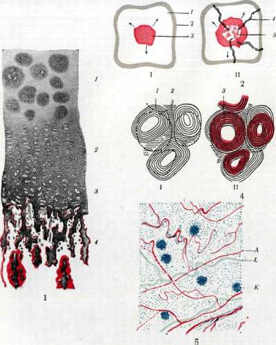
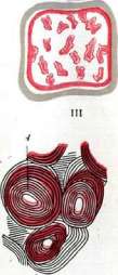
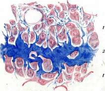
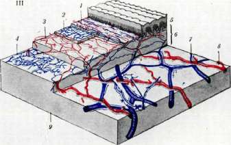
Patho-anatomically, in addition to changes in the epithelium (its keratinization) and thinning of the subepithelial tissue of the mucous membrane, the most significant change is the degeneration of the tissue itself, consisting of hyaline degeneration of collagen tissue and in various forms of prominent degeneration of elastic fibers. Hyaline degeneration of collagen tissue is accompanied by the formation of glassy-swelling fibers and the deposition of free hyaline in the tissue. Degeneration of elastic fibers begins with significant thickening and homogenization of them, and then leads to their granular disintegration and the formation of homogeneous masses. In addition, various transitional, not precisely definable products of tissue degeneration are also found in the tissue. Pinguecula is incapable of regression. The treatment measures - removal of pinguecula - may be prompted only by cosmetic considerations. A significantly more serious form of degeneration of the conjunctiva is its hyaline and amyloid degeneration (see Amyloid of the eye, Hyaline degeneration). T y-l o m a conjunctivae (callus of the conjunctiva) - a rare disease, first described by Hallenga, and then by Best. Tyloma manifests in the form of a sharply demarcated, rounded thickening of a matte luster and with somewhat dry surface. Figure 1. Endochondral (intracartilaginous) ossification: 1-field of normal cartilage with altered groups; 2-field of multiplication of cartilage cells arranged in columns; 3-field of swelling of cartilage cells and calcification of the ground substance; 4-field of growth of the perichondrium; remaining cartilage bars (gray) are covered with osteoid tissue (red) (according to Nicola). Figure 2. Phases of ossification of the cartilage bone rudiment: I-calcification of cartilage; II-vascularization; III-ossification of the entire rudiment; 1-cartilage; 2-perichondrium; 3-deposition of lime; 4-vessels; 5-island of ossification (according to Nicola). Figure 3. Island of ossification from the nasal bone of a cat embryo, stained according to Mallory: 1-osteoblasts (red); 2-osteoid substance (blue) (according to Petersen). Figure 4. Scheme of bone remodeling: I-initial stage; II-intermediate; III-final; 1-Haversian systems of the 1st generation; 2-interstitial systems; 3-Haversian systems of the 2nd generation; 4-Haversian systems of the 3rd generation. Dotted lines outline the areas undergoing resorption and being filled by systems of the next generation (according to Nicola). Figure 5. View of the conjunctiva of the eyeball in a binocular microscope with vital staining with Brillantkresylfclau: A-surface vessels; K-lymph nodes; L-lymphatic vessel. Figure 6. Scheme of distribution of blood vessels in the skin: 1-first venous network; 2-second venous network; 3-subpapillary arterial network; 4-third venous network; 5-epidermis; 6-true skin; 7-deep skin venous network; 8-deep skin arterial network; 9-subcutaneous tissue (according to Spalteholz). (For illustrations in the articles Skin, Conjunctiva, Bone).
See articles Skin, Conjunctiva, Bone.
the surface, and differs from the xerosis plaque in that it represents a plaque rising above the surface of the C. Tyloma conjunctivae develops in connection with chronic irritations of the C. Patho-anatomically, it is characterized by the formation of hyaline and amyloid degeneration of the tissue and pronounced keratinization of the epithelial covering in the affected area. The only possible treatment is surgical removal. Xerosis conjunctivae (dryness of the C.)-see Xerophthalmia. The pathology of pterygium-see Pterygium. Injuries to the C. can be caused by physical-chemical harmful agents or mechanical agents, and both can be of a professional nature. It should be noted here that chemical and thermal burns of the C. can be caused by acids and alkalis, as well as molten metal, etc., sometimes causing extremely severe and extensive damage to the C., often combined with damage to the cornea and eyelids. Immediately after injury, the surface of the C. in milder cases appears hyperemic, in severe cases it is necrotic, covered with a dirty-gray scab. While acids, due to the coagulation of tissue protein, form a well-detachable scab, in alkali burns, a process of tissue swelling and melting continues for a long time, destroying the C. to its deep layers. In this respect, burns with ammonia (resp. spirits of hartshorn) are particularly destructive, initially representing only a burn of the C., and then causing necrosis of the cornea, damage to the iris, lens, etc. Chemical injuries to the C. should include the often observed injuries to the C. from aniline dye-methyl violet, which is part of chemical pencils (see).-Very common burns of the C. are caused by lime, which gets into the eye in the form of slaked lime, lime milk, cement, etc. Lime burns are accompanied by extensive necrosis of the C. with subsequent extensive scarring; the cornea is also often involved in the process, not only due to direct burning, but also due to disruption of its nutrition in deep burns of the C. In connection with damage to the C. from lime, the so-called conjunctivitis petrificans should be mentioned-a lesion of the C., described by Leber (1895-1900), consisting of the presence of lime plaques in the C., most often in the lower transitional folds, which owe their origin not to a unique process of petrification of the C., as Leber assumed, but to the intentional introduction of lime into the C. and the damage caused by it.-Prevention of various chemical and thermal burns is especially important in professional eye injuries.-Treatment for various types of burns consists, on one hand, in the most rapid and complete removal of the damaging agent from the conjunctival sac, and on the other hand, in neutralizing its chemical action. Therefore, it is useful to use washing with acid solutions in case of alkali burns (boric acid solutions are commonly used), alkaline solutions in case of acid burns (soda, borax, etc.); in case of injury from aniline pencils-a solution of tannin (5-10%). In burns from spirits of hartshorn, transplantation of mucous membrane from the lip to the burn site has been successfully used in the fresh stages.-Wounds, tears of the C., which are common among injuries to the C., require the usual methods of diagnosis and treatment of wounds of mucous membranes; it should only be noted that due to the slight tendency of wounds to gape, there is rarely a need for suturing wounds of the C.-Foreign bodies in the C. are often encountered. These are usually small particles of metal, stone, wood, etc. Getting into the C. and preferably localizing in the area of the sulcus subtarsalis, they cause an extremely pronounced reaction of the eye-tearing, pain, inability to look. However, there are cases when foreign bodies, even of large size, remain in the C. without reaction for a long time. Removal of foreign bodies does not present difficulties due to their accessibility.-Tuberculosis of the C. usually occurs as a secondary process endogenously or as a result of spread from surrounding parts. Primary tuberculosis of the C. is an extremely rare phenomenon; at least the picture of the primary complex of the C. has been described only in individual cases. Clinically, tuberculosis of the C. manifests, according to Sattler (1891), in four forms. 1. Small, sometimes only millet-grain-sized ulcers of the C. with steep edges, with grayish-yellow uneven bottoms, with fresh miliary nodules around. This form is also characterized by frequent findings of tubercle bacilli. 2. In the C., scattered or grouped nodules are observed, resembling trachomatous follicles, gray or grayish-yellow in color with little tendency to break down. Here, bacilli are rare. 3. In this form, there are papillary proliferations, along with them, fatty-looking ulcers of the C. Bacilli are also very scarce here. 4. Lupus; on a strongly hyperemic, velvety C., there are more or less extensive ulcers with steep, thickened edges, with a clear tendency to scar formation. Eyre added a fifth form of tuberculosis of the C., characterized by proliferations in the C. in the form of papillomas or fibromas. Diagnosis, besides clinical examination, is made by bacteriological research, which is not always positive, by inoculation on animals, with the help of tuberculin tests, and in cases of suspicion of tuberculosis of the conjunctiva, ophthalmoreaction by Calmet is avoided. The course of tuberculosis of the C. is chronic, sometimes for years, with a tendency to relapses.-Therapy in the past was excision, curettage, cauterization (thermocautery, cauterization with 50% lactic acid), at present-tuberculin therapy, X-ray, radiotherapy, finzetherapy. Syphilis of the C. among other syphilitic diseases of the eye occupies one of the last places [according to the latest statistical data 0.2-0.5% (Batrachenko, Protopopov and others)]. Syphilis of the C. can manifest in all its stages. Naturally, primary syphilitic disease of the C. is rare-a hard chancre (although in Russian literature, such cases of extragenital syphilis infection have been noted in relatively large numbers-up to 50). In secondary syphilis, various forms of lesions of the C. are observed; some of them do not have specific features. The most common form are papules of the conjunctiva, observed in the C. of the eyeball as well as of the eyelids. Then a unique process in the C. is the so-called conjunctivitis granulosa specifica, s. syphilitica, first described in 1888 by Goldzieher. The lesion occurs in the secondary period in a form resembling trachoma: in the infiltrated C., follicle-like formations appear in the transitional folds of the C. as well as in the tarsus of the eyelids. Patho-anatomically, here too it is a matter of papules of the C. Sometimes the cartilage is also involved in the lesion (tarsitis syphilitica). Then Elsehnig described a gelatinous lesion of the C. of the sclera in secondary syphilis. Very rarely, in late syphilis, gummas of the conjunctiva are encountered. Tumors of the C. Among benign tumors, a congenital tumor of the C. is the dermoid or lipodermoid. Dermoid is usually localized in the outer or upper-outer edge of the cornea in the form of a hemispherical protrusion of pale pink or pale yellow color, firm consistency, with a dry surface, sometimes resembling skin. Located in the area of the limbus, the tumor often extends to the cornea. Dermoid can also be bilateral, often localized in symmetrical places of the eye, and in some cases is combined with other developmental defects-coloboma of the eyelids, iris, microphthalmia, etc. For lipodermoid, localization in the area of the equator of the eye is characteristic. Histologically, the structure of dermoid and lipodermoid of the C. is basically similar to dermoids of other localizations. In its course, lipodermoid differs from dermoid in that sometimes in the period of sexual maturity it shows signs of growth. Treatment for both types is surgical removal.-To congenital formations in the C. belong birthmarks of the C., which appear either in the form of dark pigmented spots of the C. (naevus pigmentosus) or non-pigmented-red-yellowish spots. Naevus pigmentosus is usually located in the area of the palpebral fissure, in the C. of the eyeball near the limbus and in most cases remains stationary and only sometimes (more often in the period of maturity) begins to grow, and the growth usually does not have a destructive character and only in rare cases can take the form of malignant growth of the type of melanoma. In view of the latter circumstance, early removal of pigmented birthmarks is recommended:-Besides typical birthmarks of the C., sometimes a very extensive pigmentation of the C. is encountered-i.e., melanosis conjunctivae. Cysts of the C. can be of different origins; besides congenital ones (such as dermoid cysts, palpebral cysts together with anophthalmia), acquired ones are most common: lymph cysts, which are one type of the above-mentioned lymphangiectasias of the C., and epithelial cysts of a retention type. The reason for the formation of the latter type of cyst is the retention of secretion of the Krause, Meibomian glands. Cysts develop both in the normal C. and especially after its inflammations (in particular often in trachoma).
Their usual localization is in the transitional folds; in size they range from a pinhead to a millet seed. Then cysts develop as a result of the proliferation of epithelium deep into the tissue during various inflammations of the conjunctiva, when the epithium protruding deep into the tissue forms gland-like structures; in some parts the epithium may die, which leads to the fusion of the surfaces exposed by the covering and the formation of cavities with subsequent development of the cyst. Furthermore, cysts can be of traumatic origin due to the formation of scar closures of the excretory ducts with the formation of cavities within them, etc. In some cases, when the conjunctiva is injured, a cyst may form from the introduction of epithelium deep into the tissue (Implantationscysten). Finally, false cysts - parasitic - can also form in the conjunctiva due to the settlement of parasites in it; here, although very rarely, a cysticercus blister is observed, simulating a cyst; in tropical countries and occasionally in Europe and America - pseudocysts from the introduction of Filaria loa, Rhinosporidium* Kinealyi. Angiomas (haemangioma, lymphangioma) occur as tumors that primarily arise in the conjunctiva or as tumors spreading from the eyelids onto the conjunctiva. Angiomas of the conjunctiva are observed either in the form of telangiectasias or as cavernomas. The former are mostly congenital, while cavernomas, developing from telangiectasias, appear in childhood or adolescence. Hemangiomas are predominantly located in the conjunctiva of the eyeball, more often at the inner angle. Lymphangiomas are encountered extremely rarely; sometimes they are associated with similar anomalies of the lymphatic vessels of the eyelids and facial skin. Removal of angiomas is indicated in some cases for cosmetic reasons, in others the tumor requires removal because its spread causes impairment of eye function and is occasionally accompanied by bleeding. Various methods exist for removing conjunctival angiomas: excision with preliminary ligation or infiltration of the vessels, or cauterization. Papillomas of the conjunctiva represent tumors that are generally benign but sometimes prone to recurrence and in some cases develop into malignant epitheliomas - carcinomas. They are most commonly found in the area of the palpebral fissure on the sclera in the region of the limbus, usually also involving the cornea; in addition to the limbus, these tumors are observed on the conjunctiva of the eyelids, not only singly but also multiply, and sometimes on both eyes. They are located with a broad base and a rough, uneven surface in the form of a cauliflower or mulberry; the consistency of the tumor is soft. Some fibromas observed on the conjunctiva of the eyelids also belong to papillomatous tumors. Histologically their structure is not complex; they, like papillomas, consist of papillary-like proliferations of connective tissue, but epithelial proliferation is observed to a lesser degree than in papillomas. Soft fibromas often bleed and are prone to recurrence; hard fibromas do not have these properties. Treatment of papillomas and fibromas is surgical - removal, which is especially important due to the possibility of papillomas transforming into epitheliomas. Malignant neoplasms of the conjunctiva. Sarcomas of the conjunctiva most often develop as a result of the spread of the tumor from the eyelids or from the uvea. Primary sarcoma of the conjunctiva is most commonly found on the sclera in the region of the limbus, from where it spreads to the conjunctiva of the sclera and the cornea. Clinically, sarcoma in the initial stages manifests as spots in the limbus area, which then proliferate into gray-red masses with a smooth surface on a broad base, situated in the conjunctiva. Leukosarcomas of the conjunctiva have been described in isolated cases. In their growth, sarcomas do not have a clear tendency to grow into the eye, but spread abundantly over the surface of the conjunctiva of the sclera and cornea, with the tumor in the cornea not forming a close connection with the tissue but only penetrating between the epithelium and Bowman's membrane. As they grow, the sarcoma gradually narrows the conjunctival sac and protrudes more and more above the surface of the eyeball in the form of a nodular tumor, which has a tendency to bleed with further growth. Sarcomas of the conjunctiva have a great tendency to local recurrence after surgery; but at the same time, in terms of their tendency to metastasize, sarcomas of the conjunctiva are considered less malignant than sarcomas of other locations. Melanosarcomas developing from naevus pigmentosus are particularly prone to recurrence, and they are also particularly prone to metastases. Sarcomas of the conjunctiva are usually observed in old age. As for the treatment of sarcomas of the conjunctiva, even the most thorough and possibly early removal often does not prevent recurrence; therefore, when recurrence occurs or when the tumor is more or less widespread in epibulbar sarcomas, enucleation is required, and when the sarcoma grows deep into the orbit - exenteration of the orbit. In the early stages of sarcomas, especially in naevus pigmentosus, favorable results have been noted with radiotherapy. Epitheliomas of the conjunctiva are localized like sarcomas most often in the limbus, i.e., where the character of the epithelium changes at the border of the conjunctival and corneal tissue, where the normal epithelium has a tendency to sink into the subepithelial tissue and where the conjunctiva is most often exposed to external influences. Patho-anatomically, epitheliomas of the conjunctiva in most cases represent squamous cell carcinoma; less commonly, tumors are found that stand at a lower stage of epithelial differentiation. As mentioned above, epitheliomas of the conjunctiva have a number of transitional forms to benign epithelial tumors, therefore some divide all epitheliomas into three groups - epithelioma papillomatosum, epith. carcinomatosum, also distinguishing an intermediate form - epith. initium, from which either a papilloma or a carcinoma can develop. Epitheliomas of the conjunctiva appear in the form of a grayish-red tumor, not particularly rich in blood vessels, with a nodular surface, sometimes with papillomatous proliferations, situated in the limbus area and usually spreading to the cornea and sclera. Epitheliomas of the conjunctiva have a tendency, as they grow, to encircle the conjunctiva around the entire eye, to penetrate inside the eye and into the orbit. Recurrences in epithelioma of the conjunctiva are very frequent. Treatment: in the early stages, radiant energy (X-ray, radium) can be tried, but mostly operations are indicated - thorough removal of the tumor followed by galvanocautery, and when the tumor has spread - enucleation, and when it grows into the orbit - exenteration of the orbit. Pemphigus conjunctivae is a rare disease that occurs either along with lesions of the skin and other mucous membranes or in isolation; it is characterized by the formation of blisters that rupture very quickly, leaving behind ulcers covered by membranes. Subsequently, extensive scar changes develop, leading to shrinkage of the conjunctival sac until its complete closure. The cornea is often involved in the process, and the condition ends in blindness. Treatment is usually unsuccessful - at least attempts to transplant mucous membrane to replace the scarred conjunctiva do not yield results in most cases. Symblepharon - scar adhesion of the sclera with the conjunctiva of the eyeball. Symblepharon occurs after a number of pathological processes of the conjunctiva accompanied by damage to it (injury, rupture, especially burns) or necrosis due to deep inflammations (diphtheria of the conjunctiva, smallpox, gonoblennorrhea, pemphigus), in short, in those processes when wound or ulcerative surfaces form on the opposing surfaces of the eyelid and eyeball conjunctiva, which subsequently fuse. If the adhesion of the eyelid and eyeball conjunctiva reaches the fornix of the conjunctiva, a so-called posterior symblepharon (symblepharon posterius) is formed; if the adhesion is a bridge and the fornix of the conjunctival sac remains free, it is called anterior symblepharon (symblepharon anterius). A special type in its origin is the posterior symblepharon, which forms due to extensive scarring of the conjunctiva with smoothing of the transitional folds and reduction of the entire sac due to trachoma. The degree of symblepharon varies depending on the cause that caused it - sometimes scar adhesions can lead to complete closure of the conjunctival sac with adhesion of the eyelids (ankyloblepharon). In rare cases, symblepharon can be congenital, combined with other developmental anomalies (cryptophthalmus, coloboma palpebrae). Treatment is surgical, usually plastic operations on the conjunctiva (see below); prevention of adhesions of the conjunctiva after its injury or ulcers is important - timely frequent retraction of the eyelid with abundant application of ointment into the conjunctival sac, separation of the exposed surfaces of the eyelid and eyeball conjunctiva by inserting temporary prostheses with an opening for the cornea, etc. Among surgical procedures on the conjunctiva, plastic operations hold a special place. Conjunctival plastic surgery. Various methods of conjunctival plastic surgery have received their modern development mainly in connection with the treatment of symblepharon; they are performed for functional and cosmetic indications, in the vast majority of cases under local anesthesia.
Plastic operations of the conjunctiva in general come down to: 1) simple incision of scar adhesions, 2) closing defects of the conjunctival sac by shifting the mucous membrane of adjacent sections, 3) closing defects with the help of mucous membrane flaps on a pedicle or with the help of a skin flap on a pedicle, 4) free transplantation of mucous membrane and 5) free transplantation of skin. Simple incision of scar adhesions is used in anterior symblepharon, not too extensive, in the form of a bridge extending across the transitional fold, where it is subsequently necessary by one means or another to prevent contact between the wound surfaces. In more extensive adhesions involving the transitional fold and the conjunctiva of the eyeball, the adhesions are separated from the eyeball, and the defects in the mucous membrane on the latter are closed with the shifted conjunctiva of the eyeball. The cut end of the symblepharon is secured with sutures in the depth of the transitional fold, and the symblepharon serves as material for closing the wound surface of the conjunctiva. Sometimes it is sufficient to incise the adhesions at the eyeball, pull the upper lip of the wound on the eyeball slightly to the side, and also shift the lower lip slightly to the side, but in the opposite direction, with the help of a suture in the area of the transitional fold. Operations by Arlt and others are based on these principles. If by shifting the symblepharon and tensioning the mucous membrane it is not possible to close the defect, recourse is had to closing the defect in the mucous membrane on the eyeball by cutting out a flap on a pedicle (one or two) from the surrounding healthy areas of the conjunctiva and sclera, if the latter has been preserved to a sufficient degree. Kellner proposed to replace defects of the conjunctiva of one eyelid with a flap on a pedicle from the conjunctiva of the other eyelid of the same eye. In case of insufficiently preserved mucous membrane of the conjunctival sac, recourse is had to replacing the defects in the mucous membrane with the help of a skin flap on a pedicle, where the flap is cut out from the surrounding skin coverings, directed with its base toward the nose, the temple, or downward, is passed through a through incision in the eyelid of larger or smaller sizes, applied with its wound surface to the wound surface in the conjunctiva, secured with sutures, and separated from its base after take. In very extensive scar changes of the conjunctiva, one has to resort to free autogenous transplantation of mucous membrane. This type of transplantation is a very unreliable method, since the flap transplanted to unfavorable scarred soil, with difficulty of its immobilization, often dies. Material for transplantation is taken from the mucous membrane of the lips, cheeks, conjunctiva of a healthy eye, and an important circumstance is the careful separation of the subepithelial elastic tissue, which contributes to the curling of the flap. Free transplantation of a skin flap is performed in the form of transplantation according to Lefort-Krause-Wolff or according to Thiersch. The success of the operation depends on proper fixation of the flap, especially in the area of the transitional fold. For this, various authors have proposed various methods of fixation: periosteal sutures at the edge of the orbit, various types of prostheses and semi-prostheses, lead, gutta-percha plates, special hollow silver 'fixators' proposed by Kuhnt, etc. At the same time, it is necessary to have further strong scar contraction and flattening of the transitional fold. Lit.: Averbakh M., Primary tubercle of the conjunctiva, Vestn. oft., vol. XXIII, No. 3, 1906 (lit.); Varshavsky Ya., On the question of plastic operations in the orbit and on the eyelids, Russ. oft. zhurn., vol. VIII, No. 6, 1928; Volkovich E., Experiments on the conditions of infection of the conjunctiva of the eye, diss., SPB, 1899; Lotin A., On some methods of plastic surgery of the conjunctival sac in the absence of the eyeball and its atrophy, Russ. oft. zh., vol. VII, No. 6, 1928; Pokrovsky A., On the doctrine of pigmented neoplasms of the eyelids and conjunctiva, Arkh. oft., vol. II, part 4, 1927 (lit.); Polev L., On the pathology of epithelial tumors of the cornea and limba - their classification according to histological principles, Russ. oft. zh., vol. VI, No. 2, 1927 (lit.); Protopopov, Syphilitic lesions of the eye, Arkh. oft., vol. VI, 1929; Rzhanitsyn F., Three cases of tuberculosis of the conjunctiva, Vestn. oft., vol. XXIX, No. 1, 1912 (lit. for 1906-1910); Chirkovsky V., On syphilitic lesions of the conjunctiva and cartilage of the eyelids, ibid., vol. XXXIII, No. 12, 1916; Augenarztliche Operationslehre, hrsg. v. A. Elschnig (Hndb. der gesamten Augenheilkunde, begr. v. A. Graefe u. Th. Saemisch. B. IV, Abt. 1, B., 1922); Bergmeister R., Die tuberkulosen Erkrankungen des Auges, B., 1927; Engelkind, Die Tuberkulose des Auges, Erg. d. allg. Pathologie, Jhrg. 21, 1920; Herrenschwand F., Die pathogenen Mikroorganismen des Auges, B.-Wien, 1927; Igersheimer J., Syphilis und Auge (Hndb. der Haut- u. Geschlechtskrankheiten, hrsg. v. J. Jadassohn, B. XVII, T. 2, B., 1928); Koeppe L., Die Mikroskopie des lebenden Auges, B. I, Lpz., 1920; Lohlein W., Bindehaut (Hndb. der speziellen pathologischen Anatomie u. Histologie, hrsg. v. F. Henke u. O. Lubarsch, B. XI, T. 1, B., 1928); Morax V., Maladies de la conjonctive (Encyclopedie francaise d'ophtalm., publ. sous la dir. de F. Lagrange et E. Valude, t. V, P., 1906); Saemisch Th., Die Krankheiten der Conjunctiva (Hndb. der ges. Augenheilkunde, begr. v. A. Graefe u. Th. Saemisch, B. V, T. 1, Lpz., 1904); Sattler H., Die bosartigen Geschwülste des Auges, Lpz., 1926. V. Chirkovsky.
Related articles
Mentioned in
Cite this page
“Conjunctiva.” Soviet Medical Encyclopedia. English translation of Bolshaya Meditsinskaya Entsiklopediya, 1st ed. (Moscow, 1928–1936), ed. N. A. Semashko. https://sovietmedicalencyclopedia.pages.dev/article/conjunctiva/