Conjunctivitis
Historical document, translated for reference. It reflects medical knowledge of the 1920s–30s and is not medical advice.
Summary
Conjunctivitis is an inflammation of the conjunctiva, a common eye disease caused by various infectious, chemical, physical, and other factors. The article details its etiology, clinical classification, pathological anatomy, and general symptoms.
Encyclopedia article (1928–1936)
CONJUNCTIVITIS, conjunctivitis, inflammation of the conjunctiva of the eye, a very frequent disease of the organ of vision and at the same time the most common form of lesion of the conjunctiva itself. The frequency of C. is explained on the one hand by the fact that the conjunctival sac, due to its anatomical position, is accessible to all sorts of external influences, and on the other hand, the conjunctiva, being a mucous membrane, is richly supplied with blood vessels and nerves and therefore easily responds to any irritation with a reactive inflammatory process. Etiology of C. Among the causes that give rise to C., infections occupy first place, in the vast majority of cases arising from the entry of an infectious agent from the outside - by the exogenous route - and relatively rarely - endogenously. The causative agents of exogenous infections are various microbial forms, many of which cause inflammatory infections of other mucous membranes or other parts of the body: such as gonococci, streptococci, staphylococci, the diphtheria bacillus, the intestinal bacillus, etc.; but there is a group of microbes that are specific causative agents of conjunctival inflammations; such are the Koch-Weeks bacillus (Bac. Koch-Weeks), the Morax-Axenfeld diplobacillus (Diplobac. Morax-Axenfeld) [see separate table (Vol. VII, p. 303-304), Fig. 2]; there are also a number of unquestionable conjunctival infections in which the causative agent is still unknown with precision; such infections include one of the most common among us - trachoma; in rare cases C. can be caused by fungal forms (Sporotrichon Beurmanni or Schencki). Besides exogenous, C. can also be due to endogenous influences, arising as bacteriotoxic C. during the circulation of microbes or their toxins in the blood (metastatic gonococcal C., conjunctivitis in general infections such as measles, scarlet fever, etc.). A frequent form of C. are the so-called scrofulous or phlyctenular C., which are considered by most as tuberculo-toxic. Then very often the cause of C. can be various physical and chemical agents; here various mechanical irritations and injuries play a role (entry of dust, shavings, etc.), very often associated with professional occupations. C. of the same category also include C. caused by various chemical substances, which, when acting on the conjunctiva in liquid or gaseous state, cause a sharp inflammatory reaction; such C. can be of accidental origin, but more often associated with various professional occupations (in chemical industries, laboratories, etc.). Such C. also arise from the action of various poisonous combat substances, such as asphyxiating (chlorine, chloropicrin), vesicant (especially mustard gas), and sneezing, lacrimatory (see Poisonous Combat Substances). Chemical action also reveals C. caused by a number of drugs commonly used in ophthalmology, such as eserine, atropine (see Atropine catarrh). Finally, the cause of C. can be the influence of physical agents, such as light, heat. Such are C. under the influence of light energy containing especially many ultraviolet rays - the so-called glacier catarrh (see), C. of workers in film production, in the electrical industry, in those exposed to X-ray, radium rays. In view of such a variety of causes of C., their classification according to the etiological principle encounters great difficulties, mainly because the same cause can be accompanied by a varied clinical picture; everything depends on the nature, intensity of action of the inflammatory agent, duration of action, etc. Therefore, the etiological classification can be carried out only in relation to certain groups of C., mainly infectious, and the old division according to the clinical picture with consideration of patho-anatomical changes remains in force in practical terms. Clinically. C. is divided into the following main forms: catarrhal, false-membranous, purulent, or blennorrhea, trachoma and follicular C., phlyctenular C. A number of forms, moreover, do not fit into this classification and require separate description (see Spring catarrh). Pathological anatomy of C. Despite the diversity of the clinical picture of C., in patho-anatomical terms they are characterized by a number of common symptoms. First of all, the hyperemia of the conjunctiva observed in every C. can involve the entire conjunctiva of both the eyelid and the eyeball or be limited to one or another of its parts; along with hyperemia, inflammatory edema and exudation appearing in various forms can spread to the entire conjunctiva or to its part; but usually these phenomena are most sharply observed in the conjunctiva of the transitional folds, in the conjunctiva of the eyeball. The exudate in acute forms of C., especially in infections, contains an abundant admixture of multinuclear leukocytes; they form large clusters around the vessels as well as in the subepithelial tissue of the conjunctiva. In the conjunctiva of the cartilage, cellular clusters are usually limited only to the area of the papillae, and only in individual forms does cellular infiltration spread to the entire area of the tarsal conjunctiva. Infiltration in later periods is characterized by the admixture of other elements - lymphocytes, eosinophils, plasma cells. Local eosinophilia is particularly characteristic for some forms (see Spring catarrh), and for chronic forms of C., lymphocytic, plasmacellular infiltration is characteristic. In addition to infiltration, proliferative and degenerative changes in the connective tissue elements are noted in the inflamed tissue of the conjunctiva. Thus, in some C., the formation of focal clusters of cellular elements in the form of follicles is observed; their presence, development and fate determine special forms of follicular lesions of the conjunctiva. For certain stages of the inflammatory process of the conjunctiva, and sometimes for its individual forms, the proliferation of papillary bodies is characteristic, due to the proliferation of connective tissue and the epithelial covering (chronic catarrhal C., chronic blennorrhea, etc.). Hypertrophy of papillae is observed in C. not only in the area of pre-existing papillae (upper edge of the cartilage, near the corneal limbus), but often also in those parts of the conjunctiva where normally there are no papillary bodies. In all forms of conjunctivitis, changes in the epithelium are observed in one form or another. The latter can also be a primary phenomenon, since external harmful agents (physical and chemical influences or infections) first encounter the epithelial covering when they act. More often it is a matter of secondary changes in the epithelius due to its penetration and loosening by the exudate. A characteristic change in the epithelium, especially in chronic C., is an increase in the number of so-called goblet cells. The epithelius undergoes deep widespread changes in severe C. with tissue necrosis, with the formation of a fibrinous exudate on the surface and in the tissue of the conjunctiva. Sometimes processes of proliferation are also observed in the epithelius, expressed in the fact that the epithelial covering thickens and grows into the depth of the tissue. In a number of cases of chronic C., the epithelius undergoes epidermization. In addition to all the mentioned changes, particularly characteristic is the exudation onto the surface of the conjunctiva, into the conjunctival sac. The character of the free exudate in C. varies: serous, sero-fibrinous, fibrinous, purulent. A purely watery exudate is a rare and rapidly passing phenomenon; usually in mild forms, a mucous exudate in the form of threads is observed, due to the secretion of mucin by the mass of mucous cells appearing in abundance in the epithelius of the conjunctiva during inflammation. In the so-called catarrhal C., in the secretion besides mucus there is a large admixture of purulent cells, and in purulent C. they are found in abundant quantities. A special patho-anatomical substrate in some forms of C. (conjunctivitis pseudomembranosa) is the presence on the surface of the conjunctiva of films, either closely connected with the conjunctival tissue or lying superficially. The formation of films is due to a fibrinous exudate. General symptomatology and course of C. The clinical picture of C., despite the great diversity of their etiology, can be represented in a number of typical features. One of the main symptoms of C. is hyperemia of the conjunctiva, changing the color inherent in the normal conjunctiva, and in cases where the inflammation of the conjunctiva spreads to the eyeball, the hyperemia is especially striking; here a bright reddening of the eye is noted, due to the so-called conjunctival hyperemia, i.e., the engorgement of the superficial network of vessels of the conjunctiva of the eyeball, and characterized, unlike the so-called ciliary hyperemia, by a bright red color, increasing in intensity from the edge of the cornea to the transitional folds. Pronounced conjunctival hyperemia in the area of the eyeball is particularly characteristic of acute C. of infectious origin, and often this hyperemia is accompanied here even by hemorrhages, either pinpoint or more widespread.
Hyperemia in the area of the conjunctiva of the eyelids, more vivid and pronounced in acute conjunctivitis, and moderate in chronic forms, is most noticeably manifested by a change in the color of the conjunctiva of the cartilage. Along with hyperemia, conjunctivitis is accompanied by thickening of the tissue, an increase in its volume due to infiltration and edema, which is especially pronounced in the transitional folds, which in some forms of conjunctivitis (acute infectious catarrhs and purulent conjunctivitis) sometimes reach enormous sizes and when the eyelids are everted, protrude in the form of large ridges from the palpebral fissure. Thickening of the conjunctiva due to edema sometimes gives a characteristic appearance to the conjunctiva of the eyeball (so-called chemosis conjunctivae), which in acute conjunctivitis, especially in severe infections (gonococcus, diphtheria bacillus, streptococcus, etc.), appears to rise in the form of a ridge around the cornea. In conjunctivitis, furthermore, a rough change in the surface of the conjunctiva is a very frequent phenomenon; the growth of papillae makes it in some cases velvety, in others extremely uneven, bumpy; the follicles forming in the conjunctiva appear as grains of varying sizes, sometimes lying deep in the conjunctival tissue, sometimes protruding above its surface. The formation of follicles and their localization characterize special forms of chronic inflammatory conditions of the conjunctiva; they are observed in some acute infectious conjunctivitis (pneumococcal) in various parts of the conjunctiva, but as a passing phenomenon. The most characteristic sign of conjunctivitis is the presence of pathological secretion in the conjunctival sac in the form of either serous fluid, mucus, or pus of varying consistency and color. The amount of secretion and its properties vary widely not only in different forms of conjunctivitis but also in different stages of inflammation. Some infections are accompanied by particularly abundant exudation: these include blepharitis (see) and acute infectious catarrhal conjunctivitis. Chronic forms run with a moderate amount of secretion, usually of a mucous nature, accumulating in the corners of the eyelids after sleep. A common symptom for some forms of conjunctivitis is the presence of membranes, which appear as a grayish dirty coating on the conjunctiva, most often on the cartilage, sometimes on the transitional folds, and in cases of superficial membrane location (conjunctivitis crouposa), they are easily removed, and the conjunctiva underneath appears only markedly hyperemic; in cases of deep diphtheritic processes, the membrane cannot be removed without force, and after removal, the conjunctiva bleeds, appearing necrotized (conjunctivitis diphtheritica). These forms of conjunctivitis, based on the common feature of membrane formation, are united into one general group of false-membranous conjunctivitis (conjunctivitis pseudomembranosa). Unlike all the above forms of conjunctivitis, where the process spreads over entire areas of the conjunctiva, there exists a very frequent conjunctivitis, the main symptom of which is the appearance of limited infiltrates in the conjunctiva—nodules, phlyctens (conjunctivitis phlyctaenulosa). The course of conjunctivitis varies depending on the causes that caused it and in general can be either acute or chronic. In infections, the virulence of the pathogen and the resistance of the organ, resp. the body, of course play a determining role. After a variable incubation period, infectious acute conjunctivitis reaches its peak of development sometimes with extraordinary speed (within a few hours) and lasts in non-severe forms relatively briefly—2-3 weeks. In cases of severe infections that arise acutely (gonococcal blennorrhea, diphtheria, streptococcal conjunctivitis), the course can be more prolonged, and in some cases the outcome is the formation of more or less extensive scars of the conjunctiva, sometimes with subsequent complications in the position of the eyelids, such as entropion, etc. In these latter forms of conjunctivitis, the most dangerous complication is purulent inflammation of the cornea with its destruction in many cases. Chronic forms of conjunctivitis can drag on for years in the usually safe for the organ of vision form, such as chronic catarrhal conjunctivitis; among chronic forms of conjunctivitis, trachoma is particularly dangerous. Subjective symptoms of conjunctivitis are most pronounced in acute forms: sensation of a foreign body behind the eyelids, burning, stinging, photophobia, visual impairment due to the covering of the cornea with mucus and secretion—these are the usual complaints of patients with conjunctivitis. In severe irritation phenomena, blepharospasm is also observed. Chronic forms run without particular subjective disorders, unless the cornea is involved in the process. The diagnosis of conjunctivitis based on clinical signs presents no difficulties, but for etiological diagnosis, bacteriological or simply bacterioscopic examination is often necessary. The material for examination is either a smear from the conjunctival discharge or a scraping from the epithelial covering of the conjunctiva, especially in the early stages of infection. Gram's staining is most commonly used. An essential condition for the correctness of the etiological diagnosis is knowledge of the microbial flora of the normal conjunctival sac, where, as is known, in 90-95% of cases there is the xerosis bacillus (Bac. xeros.) from the group of pseudodiphtheritic bacilli [see separate table, t. VII (st. 303-304), fig. 2], in 70-75% white staphylococcus, and in 30-35% pneumococcus. The treatment of conjunctivitis in most cases is symptomatic and only in rare cases is there etiological-specific. Acute forms of catarrhal-purulent conjunctivitis, accompanied by large amounts of discharge, require the use primarily of cauterizing agents and bactericides; the usual cauterizing agent is silver nitrate in a 1-2% solution, applied according to the usual rules (see Blennorrhea); in chronic conjunctivitis, preparations of copper sulfate, alum, etc. are used. As disinfectants in acute conjunctivitis, preparations of silver are further used—collyrium (2-5%), protargol (2-5%), argyrol (5-10%) in the form of drops. In chronic conjunctivitis, zinc sulfate (V8-Va%) and resorcin (1%) are particularly favored. The use of various solutions of disinfectants in the form of compresses is of significant importance in conjunctivitis accompanied by more or less abundant secretion: such are solutions of mercuric cyanide (0.02%), sublimate (0.01%), boric acid (2%), etc. Specific serum treatment in conjunctivitis is undoubtedly indicated only in infections with the diphtheria bacillus; in other infections, such as pneumococcus, streptococcus, etc., attempts at serotherapy have given a very modest effect and show no advantages over ordinary local therapy. Separate forms of conjunctivitis. Acute catarrhal conjunctivitis. Among them, infectious conjunctivitis caused by specific pathogens are particularly important. First in frequency among acute infectious conjunctivitis is conjunctivitis from the Koch-Wicks bacillus. As systematic bacteriological studies show, in some places in the USSR, conjunctivitis from the Koch-Wicks bacillus occurs in almost 7/3 of all conjunctival infections (according to Rymovich—48%, according to Chirkovsky and Adamyuk—32.9%), and in Egypt—62%. The causative agent of this conjunctivitis is a Gram-negative bacillus, very thin (0.5-2 μ in length and 0.3-0.4 μ in thickness), immobile, growing aerobically at thermostat temperature; its favorite medium is serum and blood-containing. The diagnosis of the process can be made bacterioscopically: in smears, the bacillus is visible in large quantities, mostly intracellularly [see separate table, t. VII (st. 303-304), fig. 2]. This conjunctivitis is most often observed in children and usually in autumn and spring, and, being highly contagious (transmission occurs through objects as well as by droplet infection), conjunctivitis often arises epidemically, especially among school populations, in dormitories, orphanages, etc. The typical manifestation of infection with the Koch-Wicks bacillus is a rapidly developing conjunctivitis with abundant mucopurulent discharge with marked involvement of the bulbar conjunctiva; the latter is strongly hyperemic; often there are small hemorrhages, sometimes phlycten-like formations in the area of the limbus. The process usually lasts 2-3 weeks and resolves favorably; complications from the cornea are extremely rare. Prevention consists in the immediate isolation of the patient with conjunctivitis, which is especially important for schools, orphanages, etc. Treatment is the usual for acute conjunctivitis. A conjunctivitis of the same character is caused by the Pfeiffer's influenza bacillus (Bac. influenzae Pfeifferi), which is close to the Koch-Wicks bacillus in morphological-biological features, and in some respects completely identical to it. Pneumococcal conjunctivitis ranks second among acute forms (15-18%, according to Rymovich, Chirkovsky, and others). For it, the clinical picture is characterized by bilateral mucopurulent catarrh, often accompanied by rhinitis, also with strong hyperemia of the bulbar conjunctiva, with hemorrhages in it, with a characteristic edema of the eyelid margin with a pinkish coloration of the skin; sometimes this conjunctivitis is accompanied by an eruption of small follicles in the conjunctiva of the eyelids and cartilage. The process arises very quickly, is easily transmitted, causing epidemics, most often also in children. It often resolves critically. Complications from the cornea are extremely rare, more often one can encounter hyperemia and even inflammation of the iris (toxic iritis). For diagnosis, it is necessary to take into account the frequent finding of pneumococcus in the normal conjunctiva, but in conjunctivitis, pneumococcus is usually found in large quantities, both in pure culture.
In clinical relation, pneumococcal conjunctivitis is closest to conjunctivitis from the Koch-Wicks bacillus. It should be mentioned that the pneumococcus can give various forms of conjunctivitis—from moderate catarrhal to severe pseudomembranous inclusive. Treatment—depending on the form of the process.—Besides the mentioned causative agents of acute infectious conjunctivitis, they can also be caused by staphylococcus, streptococcus, the bacillus coli communis, etc., but all of them appear to be much rarer. Subacute and chronic conjunctivitis is very often due to a specific microbe—the diplobacillus of Morax-Axenfeld, which has pathogenic significance only for the mucous membrane of the eye. Conjunctivitis from infection by the diplobacillus of Morax-Axenfeld is widespread in all countries of the world, and in Western Europe it occupies first place among infections of the conjunctiva (according to Axenfeld, in Germany 57.6%, according to Morax, in France 37.2%); in the USSR it also occurs frequently (according to Chirkovsky and Adamyuk—20.6%). The diplobacillus of Morax-Axenfeld consists of Gram-negative, in pairs arranged rods, 2-3 μ in length, with a width of 1-1.5 μ [see separate table, vol. VII (pp. 303-304), fig. 2], with somewhat rounded ends. The diplobacillus is immobile, grows at incubator temperature, very characteristically on coagulated serum, causing liquefaction of serum at the site of colony formation; it also grows on other media, but with less characteristic growth. Clinically, the diplobacillary infection is expressed in the picture of so-called blepharo-conjunctivitis angularis chronica, i.e., with involvement of the eyelids, especially at the angles of the palpebral fissure. With it, moderate hyperemia of the conjunctiva, a small mucous discharge, redness of the skin of the eyelids at the outer and inner commissures, often with maceration of the skin here, are noted. The process drags on for a long time and if left to itself does not resolve for years. It is diagnosed by staining the smear according to Gram; the rods usually appear in large numbers in the secretion from the conjunctiva, as well as in the desquamated epidermis of the macerated skin in the corners of the eyelids. The prognosis for the process is favorable—complications from the cornea arise rarely (catarrhal ulcers). Treatment consists in systematic treatment with zinc preparations in the form of drops, compresses, ointment; with prolonged use, the result is quite satisfactory. Of other forms of conjunctivitis, great importance belongs to the pseudomembranous infectious conjunctivitis (conjunctivitis pseudomembranosa). This conjunctivitis, as already indicated, can be caused by various causative agents, but the diphtheria bacillus and streptococcus give the most typical and severe form. Infection by either can cause either a superficial membranous conjunctivitis (conjunctivitis crouposa) or a deep one—diphtheritic (conjunctivitis diphtheritica). In both cases, it is a matter of acute contagious inflammation of the conjunctiva, in the initial stages accompanied by sharp phenomena of infiltration of the tissue of the conjunctiva of the eyelids (hardness of the eyelids, redness of the skin), with the subsequent development of the stage of blennorrhea (appearance of abundant purulent-fibrinous secretion), with the formation of membranes on the conjunctiva (especially in the area of the cartilage)* superficial or closely connected with the tissue of the conjunctiva (see separate table, fig. 10).—Diphtheria of the conjunctiva represents a relatively rare infection. Meyerhof out of 100 cases of pseudomembranous conjunctivitis saw 5 cases of diphtheria, Geis out of 900 cases—only 6 times, Chirkovsky and Adamyuk out of 1,000 cases of conjunctivitis—2 times. Diphtheria of the conjunctiva affects mainly childhood age and is often unilateral, often running with a severe disturbance of the general condition; sometimes, spreading to the mucous membrane of the nasopharynx, it leads to death. In relation to vision, complications from the cornea are dangerous, often observed in diphtheritic conjunctivitis. Streptococcal pseudomembranous conjunctivitis can also give various clinical forms and in its severe form (more often in children after scarlet fever, measles) is dangerous, causing extensive necrosis of the conjunctiva and involving the cornea in the purulent process. Diagnosis is difficult mainly for establishing diphtheria—here cultures are necessary, since only by bacteriological research can one differentiate the diphtheria bacillus from the xerotic bacillus, so often found in the normal conjunctiva. The prognosis is serious in cases of diphtheria of the conjunctiva not only in relation to the organ of vision but also in relation to life, although according to clinical observations diphtheria of the conjunctiva represents less danger than diphtheria of ordinary localization. Prevention in unilateral process—protective bandage on the healthy eye (see Blennorea). Treatment in diphtheria—serum therapy, in streptococcus some use antistreptococcal serum. Local—usual treatment for infectious conjunctivitis, avoid the use of cauterizing agents, especially in the stage of infiltration. Special mention should be made of phlyctenular conjunctivitis (conjunctivitis phlyctaenulosa); synonyms: scrofulous conjunctivitis (conjunctivitis scrofulosa), eczematous (conjunctivitis eczematosa). Phlyctenular conjunctivitis is a very frequent disease of children and above all of children with so-called scrofula; hence the name conjunctivitis scrofulosa; the name 'eczematosa' it owes to the fact that it often runs together with eczema of the eyelids, face, etc. The etiology of this conjunctivitis does not appear to be sufficiently established. It is recognized that it arises in the vast majority of cases on the soil of tuberculosis and most often in so-called scrofula. In children suffering from phlyctenae, the Pirquet reaction is positive in 95-100%. Disagreements in views on the origin of phlyctenae come down to the fact that some consider these formations an expression of exogenous tuberculous infection, others (to whom the majority adheres) attribute to phlyctenae a tuberculo-toxic origin. Separate stands the opinion that phlyctenae arise only on the soil of so-called exudative diathesis (Czerny). Against this speak observations that phlyctenae do not arise in the first year of life, when phenomena of exudative diathesis can be especially pronounced. The majority, standing for tuberculo-toxic origin of phlyctenae, sees in them a peculiar reaction of the conjunctiva to various harmful influences due to increased sensitivity (allergy) of the tuberculously infected organism. For such an assumption speak both experiments and clinical data. By experiments of a number of researchers it has been proved that phlyctenae can be obtained only in tuberculous or tuberculinized animals both by introduction of various substances into the conjunctiva (e.g. tuberculin, culture or filtrate of staphylococcus) and by mechanical irritation of it. In the clinic much evidence has been obtained that phlyctena is an expression of an allergic state. Wolff-Eisner, Kollner, Filbry, studying changes in skin sensitivity to tuberculin, noted a complete parallelism between the increase of allergy and the frequency of occurrence of phlyctenae, while a decrease in sensitivity (anergy) is accompanied by improvement of phlyctenular processes. The same is explained by the occurrence of phlyctenae when applying the conjunctival tuberculin test by Calmette with its positive result. In patho-anatomical relation typical phlyctenae represent a limited subepithelial nodule from an infiltrate consisting in the peripheral parts of lymphocytes, in the center—of epithelioid cells, sometimes with an admixture of giant cells; such a nodule does not undergo caseous degeneration; according to the opinion of the majority, tubercle bacilli are not observed either. In some cases phlyctenae have a simpler structure—an infiltrate in the form of a collection of lymphocytes under the epithelium. The clinical picture of phlyctenular conjunctivitis consists in the appearance in scrofulous children in the area of the limbus of the sclera of reddish-colored nodules, raised above the surface; they are accompanied by limited hyperemia of the conjunctiva in the form of a triangle (see separate table, fig. 9). Phlyctenae are either solitary (phlyct. solitaris), sitting separately, of rather large size, or multiple (phlyct. miliaris), small, sitting in large numbers along the edge of the limbus. Solitary phlyctenae give slight phenomena of irritation; miliary ones—as a rule are accompanied by sharp photophobia, lacrimation, sometimes—blepharospasm. Phlyctenae usually resolve in several days, breaking down and being absorbed, but they are prone to relapses, and therefore their course can be extremely long. Characteristic for phlyctenae are often accompanying lesions of the cornea—corneal phlyctenae, etc. (see Keratitis), besides that blepharitis, eczema of the eyelids, etc., as well as other manifestations of scrofulous diathesis.—T r e a t m e n t. General treatment, keeping in mind the tuberculous infection underlying scrofula—medicinal, dietetic, clinical, light therapy; in a number of cases tuberculin is used with success. Locally the favorite remedy is yellow mercurial 1% ointment, calomel in the form of the finest powder sprinkling. A frequent form of inflammations of the conjunctiva are the so-called follicular lesions of it; of them the most important place is occupied by trachoma, then follicular catarrh and folliculosis. For follicular inflammations the characteristic feature is the presence in the conjunctiva of grains—follicles, in their structure resembling lymph follicles.
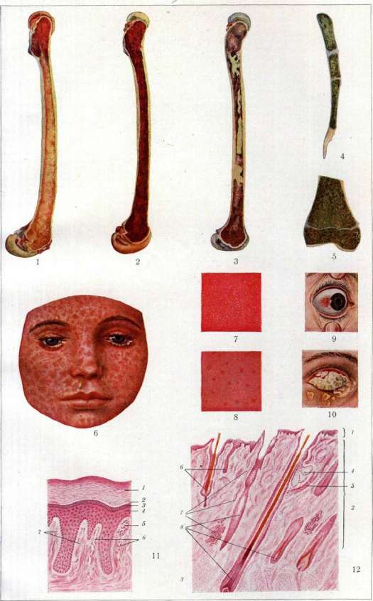
The cause of follicular catarrh and folliculosis in the vast majority of cases is a special constitutional condition of the organism, in which there is observed hyperplasia of adenoid tissue. The condition usually concerns school-age children having the so-called lymphatic-hypoplastic constitution, in whom along with manifestations of hyperplasia of lymphoid tissue (adenoids, enlargement of lymph glands, etc.) there is also development of follicles in the conjunctiva. A contributing factor to the development of follicular C. or folliculosis is often poor hygienic living conditions (prolonged stay in dusty, unventilated rooms, etc.), then intensified studies, especially in persons having an anomaly of refraction, etc. A whole series of ophthalmologists (the so-called unitarists) considers follicular C. identical to trachoma, viewing it as a mild abortive form of the trachomatous process. The clinical picture of follicular C. and folliculosis has in common the development of follicles in the area of the transitional folds, ♦24 Figure 1. Yellow bone marrow of the femur (normal); in the upper epiphysis and partly along the edges of the diaphysis-red bone marrow. Figure 2. Red bone marrow. Myeloid hyperplasia along the entire length of the femur. Figure 3. Bone marrow. Metastases of cancer (pinkish areas) and necroses (greenish areas). Figure 4 and 5. Green bone marrow in chloroleukemia (the boundary of the epiphysis is visible). Figure 6. Measles rash on the face (according to Pfaundler-Schlossmann). Figure 7 and 8. Symptom of Koplik-Filatov (figure 7-early period, figure 8-stage of disappearance). Figure 9. Phlyctenular conjunctivitis. Figure 10. Diphtheritic conjunctivitis. Figure 11. Structure of the epidermis (skin of a finger): 1-stratum corneum; 2-hyaline layer; 3-granular layer; 4-layer of prickle cells; 5-Meissner's corpuscle; 6-capillaries; 7-papillae (from the collection of the Clinic of Skin Diseases of the North Caucasian State University). Figure 12. General structure of the skin (hairy part of the head): 1-epidermis; 2-true skin; 3-subcutaneous fatty tissue; 4-sebaceous gland; 5-m. arrector pili; 6-falling out hairs; 7-sweat gland; 8-hairs cut in various planes. (See article Skin, Conjunctivitis, Measles, Bone marrow.)
See article Measles. Conjunctivitis. Measles, Bone marrow.
most often of the lower eyelids, sometimes also of the upper, and very rarely follicles also appear in the area of the conjunctiva of the cartilage, usually only in its corners. The follicles lie superficially, slightly elevated, the size of a pinhead, pale pink, semi-transparent; they are often arranged in rows in the transitional folds. In folliculosis, follicles appear among little changed or completely unchanged tissue of the conjunctiva; follicular catarrh, however, is accompanied by symptoms of catarrhal inflammation—the transitional folds of the conjunctiva are hyperemic, somewhat edematous, there is discharge, usually weakly expressed. The course of typical follicular C. and folliculosis is chronic; they can exist for years, and only follicular C. can be accompanied by subjective symptoms characteristic of C., but both processes pass without causing complications and without leaving traces. Differential diagnosis is difficult only between the initial stages of trachoma and follicular C. Treatment for folliculosis is unnecessary, for follicular C.—according to the usual rules for treating chronic C. It is important to eliminate factors contributing to the occurrence of C. Also belonging to follicular C. is a process peculiar in its etiology, the so-called bath C., or swimmer's C. (Badconjunctivitis of German authors). This C. was first described in 1899 by Fehr in Berlin, but has been studied in particular detail in the last 10-15 years. Bath C. belongs to infectious C., but to this day its causative agent is unknown. Many attribute etiological significance to intracellular inclusions, completely identical to the Prowazek-Halberstadter inclusions in trachoma. These inclusions in scrapings from the epithelial covering of the conjunctiva in bath C. are encountered with great constancy. Bath C. has repeatedly occurred in recent years in the form of epidemics in various cities of both Europe and America, and it was observed in persons who used public baths or swimming pools, often affecting entire groups of athlete-swimmers who were swimming in the same pool at the same time. Some share Lindner's view that the Prowazek intracellular inclusions, causing infection of the genital tract in certain cases, can pass from there through the pool water into the conjunctival sac and cause bath C. But infection can also be transmitted from persons suffering from bath C. through objects that come into contact with the pus of the conjunctiva. Patho-anatomically, in bath C., we are dealing with subepithelial diffuse infiltration by lymphocytes, among which follicle-like accumulations of cellular elements are observed. In late periods, there are also strong proliferations of the papillary bodies of the conjunctiva; in the regressive period, no scar changes are observed. The clinical course is characterized by the development after an incubation period of 1-2 weeks of an acute follicular inflammation of the conjunctiva, resembling the picture of acute trachoma. The process begins with swelling of the eyelids, the appearance of marked hyperemia of the conjunctiva, edema, especially of the transitional folds, where many follicles appear, which quickly spread to other parts of the conjunctiva, also affecting the area of the conjunctiva of the cartilages; purulent discharge appears, usually not abundant. In later stages, hypertrophy of the papillary bodies of the conjunctiva is also observed. In some epidemics, there is also a superficial lesion of the cornea in its upper segment in the form of small superficial infiltrates, which resolve favorably. The duration of bath C. is usually determined as 2-4 months, and it as a rule ends with complete recovery. The disease first affects one eye, but usually then spreads to the other. As a complication in this process, catarrhal lesion of the Eustachian tubes has been noted. Differential diagnosis is difficult only in relation to trachoma, but the outcome without any consequences sharply distinguishes this C. from trachoma. Prevention consists in disinfecting the water of public baths and swimming pools. Treatment—the prescription of various disinfectants in the form of drops and compresses. Conjunctivitis Pari-naud—a peculiar inflammation of the conjunctiva, first described by the French ophthalmologist Pari-naud in 1889. This disease is relatively rare and occurs at different ages. The etiology of this C. cannot yet be considered established, but by the majority its infectious origin is accepted. Pari-naud himself called C.—an infectious C., resulting from transmission from animals ('conjonctivite infectieuse d'origine animale'), considering the source of infection to be animals with which a person comes into contact. Confirmation of this assumption exists in many observations, but the nature of the infection is determined differently. Some tend to consider Pari-naud's conjunctivitis one of the forms of tuberculous conjunctivitis (infection with the tubercle bacillus of the bovine type), while the majority consider it as a special infection having nothing in common with tuberculosis, and thereby recognize the entire process as independent. In recent years, Bayer and Herrenschwand, on the basis of clinical and experimental data, attribute the etiological role to Bac. pseudotuberculosis rodentium, but there are not sufficient grounds to consider this microbe the sole cause of Pari-naud's conjunctivitis. Patho-anatomically, the process in typical cases is characterized by diffuse infiltration of the subepithelial tissue of the conjunctiva of the eyelids and transitional folds, predominantly by lymphocytes with great participation of plasma cells; in many cases the infiltration gives rise to follicle-like accumulations of cells. Authors who have observed a picture of tuberculous Pari-naud's conjunctivitis have described in such cases changes characteristic of the tuberculous process. Clinical picture of C. Pari-naud. After a short prodromal period, expressed by general malaise, moderate elevation of temperature, there almost as a rule develops on one eye inflammation of the conjunctiva with redness, swelling of the eyelids, and the conjunctiva of the cartilage and transitional folds becomes strongly hyperemic, edematous, and in it appear, especially in the area of the transitional folds, growths, follicle-like formations of various sizes, grayish or yellowish in color. Soon in the area of these formations, superficial defects in the epithelial covering—small ulcers—are often found. At the same time, on the side of the lesion, enlargement of the lymphatic glands is observed, first and foremost the preauricular, and then the parotis and glands of the neck. This one-sided lesion of the glands makes the process particularly characteristic. The enlarged glands subsequently sometimes undergo suppuration. Inflammation of the conjunctiva is usually accompanied by a small amount of seropurulent discharge. Complications from the cornea are extremely rare. The process usually runs without special subjective sensations, lasts for several weeks, sometimes months, and ends without leaving any consequences after it. Differential diagnosis can most often be made between trachoma and tuberculous conjunctivitis. The acute onset of the disease, disturbance of the general condition, lesion of the glands on the same side, and resolution of the process without scar changes in the conjunctiva exclude trachoma. As for distinguishing tuberculous conjunctivitis, this sometimes appears to be a difficult matter, since in a number of cases there is no doubt that tuberculous infection can also give the clinical picture of C. Pari-naud, but if one recognizes only those cases of C. Pari-naud in which tuberculous infection, syphilis, and sporotrichosis are excluded, as Morax proposes, then differential diagnosis of Pari-naud's conjunctivitis is sometimes possible only through bacteriological and patho-anatomical analysis of the case. The prognosis of C. Pari-naud is entirely favorable. Treatment—usual for conjunctivitis, one should only avoid caustic, strongly irritating agents; one can manage with astringent and disinfectant ones.
Related articles
Mentioned in
Cite this page
“Conjunctivitis.” Soviet Medical Encyclopedia. English translation of Bolshaya Meditsinskaya Entsiklopediya, 1st ed. (Moscow, 1928–1936), ed. N. A. Semashko. https://sovietmedicalencyclopedia.pages.dev/article/conjunctivitis/