Measles
Historical document, translated for reference. It reflects medical knowledge of the 1920s–30s and is not medical advice.
Summary
Measles is an acute infectious disease characterized by fever, characteristic rash on mucous membranes and skin, and inflammation of the respiratory tract and eye mucous membranes. The article discusses its history, attempts to identify the causative agent, and experimental findings.
Encyclopedia article (1928–1936)
MEASLES, (lat. morbilli, franç. rougeole, germ. Masern, engl. measles), a general acute infectious disease characterized by fever, a characteristic rash on the mucous membranes and skin (enanthema and exanthema), and inflammation of the mucous membranes of the respiratory tract and eyes. History. Based on available historical data, it should be considered that measles was known to humanity many centuries before the Christian era. In the 9th century of the Christian era, it was described under the name hash-bah by the Arab physician Rhazes. Physicians of the 14th-16th centuries described it under the names blac-ciae and morbilli. In the second half of the 17th century, Sydenham and Morton gave a detailed description of the clinical picture of measles, but, as before, this disease was very often confused with other exanthematous diseases, mainly with scarlet fever and smallpox. Only in the second half of the 18th century was measles identified as an independent nosological unit. Further in the history of measles, the year 1847 should be noted, when Panum published a report on a measles epidemic on the Faroe Islands, which gave much for understanding the clinic and epidemiology of this disease. The modern era is marked by attempts to find the causative agent of measles and the introduction of preventive vaccinations against measles (Weissbecker, 1896; Nicolle and Conseil, 1916; Degkwitz, 1920); the methodology of these vaccinations was developed in detail by Degkwitz. Etiology. The causative agent of measles is still unknown. The numerous works carried out in the 19th century and in the first decade of the 20th century basically come to two conclusions: according to some authors, the causative agent of measles belongs to the group of filterable viruses; another, more numerous group of researchers recognizes various bacteria as the etiological cause of measles. Unfortunately, control experiments did not confirm the findings made, and therefore until recently, the theory of the filterable virus was essentially poorly substantiated. The last decade has been marked by a number of new works, which, being based on modern achievements in microbiology, are of considerable interest. Caronia (1923) described a Gram-negative diplococcus as the causative agent of measles, which he managed to discover in the pharyngeal mucus, blood, and bone marrow of measles patients. A culture of the microorganism was obtained under anaerobic conditions on special 'catalytic' media (ascitic broth with a piece of guinea pig organ or with fresh blood). According to the author's description, this diplococcus in its development goes through a phase when it becomes ultramicroscopic and therefore can be filtered. Caronia believes that he has found specific antibodies in the blood of measles patients in relation to the microbe he described. The results of vaccinating healthy children for preventive purposes are particularly interesting. Thus, Caronia and Sindoni vaccinated 539 children who were in contact with measles patients under various conditions with a vaccine from their microbe; of the vaccinated, only 9 people later became ill. Meyer and Nobel, checking these observations, could not confirm them. In addition, Groer and Redlich, by vaccinating children in contact with measles patients with extracts from guinea pig organs, protected them from measles. Burgers and Bachmann showed that even unvaccated media from Caronia often give growth of a microorganism identical to the one described as the causative agent. Based on the experiments of Groer and Redlich, one could admit that the success of Caronia's vaccination depends on non-specific immunization; but recently the observations of Italian authors have received indirect confirmation in the works of Zlatogorov, Nasledysheva and Burova. The latter authors managed to isolate in 10 cases (out of 24) by blood culture and pharyngeal mucus from measles patients a Gram-variable diplococcus, strictly anaerobic. With this microbe, the authors successfully carried out neutralization experiments with serum from convalescents from measles, skin tests, and preventive vaccination with good results. Along with this, in a number of works starting from 1917, Tunnicliff describes a Gram-positive diplococcus with green pigment as the causative agent of measles. Unlike the microbe of Italian authors, this microorganism is anaerobic only in the first generations. It is isolated on blood agar. With this microbe, Tunnicliff not only carried out serological reactions and skin tests with positive results, but also, by immunizing goats, obtained a serum that gave in the hands of Houp and Hasul the same results as the vaccination according to Degkwitz. Although Long and Cornwell, using Tunnicliff's technique, came to negative results, Duval and Hibbard, as well as Belikov and Trushina, partly confirm Tunnicliff's observations. The latter authors from the blood and pharyngeal mucus of measles patients managed to isolate a Gram-positive diplococcus, almost indistinguishable from Tunnicliff's microorganism. Unfortunately, serological reactions, skin tests, as well as the use of vaccine and serum from immunized goats gave uncertain results. In addition, from inoculating rabbits with cultures, the same pathological changes in organs were obtained as from inoculating blood and pharyngeal mucus (filtered) directly from patients. - Close to the above-mentioned are the findings of Ferri and Fischer, Donges and Thomson, which deal with microorganisms of the streptococcus species. In addition, A. Fedorovich in 52 cases of measles was able to establish the presence of Tunnicliff's microbe in smears from conjunctival secretions, and this diplococcus was mostly located intracellularly. It should be noted that all the listed authors succeeded in obtaining positive findings only when taking material from the patient in the prodromal period or on the first-second day of the rash. - Based on the available data, especially taking into account that all the listed authors isolated microorganisms that do not differ significantly from each other, and that the isolation of microorganisms was possible only during a certain limited period of the disease, it should be recognized that this microorganism, although it may not be the causative agent of measles, has some close relation to it and therefore deserves thorough study. The theory of the filterable virus has recently been revived again in the works of Degkwitz. According to him, the virus can be cultivated on a special buffer medium for several generations. In the last generation, the original virus was in a dilution of 1:3,000,000, and yet in the experiment on humans, when a filtrate from a culture containing along with the virus also symbionts (cocci with green pigment, Tunnicliff's microorganism, pneumococcus) was inoculated, a positive result was obtained: 10-15 days after injection, a symptom complex developed in the vaccinated child resembling measles, but a typical measles rash was not observed. Inoculation of the filtrate of a culture of only symbionts did not give such a reaction. It was not possible to obtain a typical rash in monkeys either. Children vaccinated with the filtrate of the virus culture and symbionts did not react at all to subsequent inoculation of blood from a measles patient. Degkwitz tried to prepare an immune measles serum on sheep. Released for sale by the firm Hoechst, when used for vaccination of children for preventive purposes, it did not give positive results. In view of the fact that experiments on animals and the use of serum gave no evidence in favor of the specificity of Degkwitz's finding, the existence of this filterable virus of measles should be considered as yet unproven. - Despite the fact that the so-called causative agent of measles is still unknown, experiments on animals, as well as clinical and epidemiological observations, provide sufficient data for judging the properties of the measles virus. Experimental measles. The high infectivity of the blood and secretions of the mucous membranes of a measles patient has been repeatedly proven since 1758 by inoculating material directly from person to person. This method was even widely used by some physicians for the purpose of preventing natural infection and disease with measles. Thus, for example, Katona (1842) inoculated 1,122 children with blood from a measles patient mixed with secretions from skin blisters or tear fluid, and obtained in 1,115 of them a mild but typical disease. Of the experiments of transferring the virus to animals, besides old observations with uncertain and negative results, the recent works of Goldberger and Anderson, Hektoen and Eggers, Blake and Trask deserve serious attention, carried out on monkeys. The works of the listed authors gave a number of conclusions important from an epidemiological point of view; thus, it turned out that the blood is infectious shortly before the rash appears and during the first day of the rash; while on the respiratory mucosa the measles virus persists longer - infection of monkeys was still possible 48 hours after the appearance of the rash. The virus was also contained in defibrinated and filtered blood. The virus outside the body is very unstable: heating to 55° kills it within 15 minutes; it resists drying at room temperature only for a day.
In the experiments of the aforementioned authors, it was possible to transmit the virus from monkey to monkey over several passages. In this case, the animal that had contracted the disease from such a vaccination did not react to a second vaccination with infectious material: the scales of the desquamating epidermis proved to be non-infectious in experiments on humans (Maug; 1848) as well as in experiments on monkeys (Goldberger and Anderson).- Experiments on monkeys seriously support existing clinical and epidemiological observations on humans, and this is their main value. The rabbit, which until recently was considered an animal little suited for experiments with measles, thanks to the detailed works of Simon and Scott (Simon, Scott), was again used to study the measles virus. These authors, confirming observations on monkeys, experimentally proved that in the external environment in the natural secretion from the body - nasal mucus - the virus is also unstable. Experiments in the same direction by Zlatogorov, Burov, and Nasledyshcheva, as well as Belikov, Dzhvikov, and Trushina, showed that infected rabbits give the same changes in organs as # monkeys in the experiments of Black and Trask. Taking into account that Nevin and Bittmann (Nevin, Bittmann) were able to transplant the measles virus from monkey to rabbit and from the latter back to monkey, it should be thought that for experiments with measles not only monkeys but also rabbits are suitable. Epidemiology. Location of the measles virus in the body. The measles virus in the body is undoubtedly present in the blood of the patient and in the secretions of the mucous membranes (pharynx, respiratory tract, and conjunctiva). It is probably not contained in urine and feces. The scales of the desquamating epidermis are also non-infectious. In the external environment, the measles virus cannot persist for any significant length of time. The measles contagion is characterized by extreme volatility.-The source of infection, as with other diseases, in measles is mainly the sick person. Infection occurs through droplet infection, and in the first days of the incubation period and during the illness from the moment the rash disappears, the patient is not dangerous to those around him. Complications of the main disease, according to available clinical and partly bacteriological data, are caused by the introduction of microbes of secondary infection, due to which they have no significance for the spread of measles. For infection in the vast majority of cases, direct, albeit brief, contact is necessary (e.g., kissing, etc.); sometimes it is sufficient to be in the room of a patient without coming into contact with him (Dopter). The spread of infection through the air within the limits of a floor or several floors of the same building appears to be little proven. The often expressed opinion that measles infection can spread from floor to floor through ventilation channels should be considered insufficiently substantiated. In the question of the possibility of indirect transmission of infection through objects used by the patient (bed, linen, dishes, etc.), there are conflicting opinions. In general, such transmission of infection should be considered doubtful, since in measles the possibility of spread of infection by healthy persons or persons with measles in an abortive form (formes frustes) is not absolutely excluded. Some authors admit transmission of the infectious agent through healthy persons or those who have had the disease (bacillus carriers). However, corresponding data in favor of this view are not yet available. Susceptibility to measles is universal and total (in all ages). There is no innate immunity to this disease. Rare cases of absence of measles in the medical history of adults can be explained by the presence of active immunity acquired as a result of an abortive form of measles or unnoticed spontaneous immunization with the measles virus from patients or carriers. In former times, when measles was not yet so widespread among humanity as it is now, entire pandemics and severe epidemics of this disease were repeatedly described: e.g., in Germany in 1823-24 and 1826-28, in northern and central Europe in 1834-36, in North America in 1846-47, etc. The susceptibility to measles of all ages is particularly well seen in the classic example of the measles epidemic on the Faroe Islands in 1846, described by Panum. On these islands, remote from communication routes, the last epidemic of measles had been observed in 1781. When introduced to the islands from Copenhagen in 1846, measles spread extremely widely: out of 7,782 inhabitants, 6,000 contracted measles. None of those who came into contact with the sick escaped infection. Only those who had previously (in 1781) had measles were spared from infection. At the same time, people even of advanced age, but who had not had measles, suffered the disease in the same severity as small children. Similar examples are also the epidemics on the island of Martinique in 1841 and 1851.-Once contracted, the disease leaves behind immunity for life. Cases of repeated measles described in the literature appear doubtful, since the accuracy of the primary diagnosis is little reliable.
P. Belikov. Statistics and Geographical Distribution. 1. Mortality. Measles is the most common childhood infection. Despite its relatively low mortality, due to its strong spread, it claims many thousands of victims annually. Below in Table 1 are the absolute numbers of deaths from measles in some European countries for the last years before the imperialist war and for 1925-1927. Table 1. Countries 1911 1912 1913 1925 1926 1927 England and Wales . Belgium . Hungary . Germany Holland Spain . Italy . France . Switzerland Scotland ! 13,128: I 1,666! I 8,941 8,380 i 1,243 I 7,498 ,10,581 3,997 I 344 I 923 12,856 2,389 6,007 9,326 1,163 5,333 7,2^8 4,949 220 1,983 3,518: 247 i 3,926! 4,455) 5,942: 132J 3,622: 5S2 691 6601 In Table 2 are the mortality rates from measles per 100,000 population in some European countries for a period of almost 50 years. Comparison of these rates indicates that in most European countries there is a tendency to decrease mortality from measles. In the last decade it has been significantly lower than in all previous periods. Table 2. Mortality from measles in European countries (on average per year per 100,000 pop). Years 1871- 80 1881- 90 1891-900 1901- 10 1911- 13 1914- 18 1919- 23 1924- 28 н : as i a s н п к к : 1 «5 o a , on 38,0 69,5 44,0 - 31,9 60,81 41,4 50,4 25,72 18,2 33,3 30,9 34,1 21,0 29,9 25,5 33,3 29,0 14,6 19,7 27,0 29,2 - 11,4 16,4 19,6 12,8 10,4 6,8 11,0 10,4 10,3 8,8 5,5» 9,1 16,8* 12,3 14,6 15,5 8,3 5,0 4,6 2,9 35,1 38,4 47,0 33,8 29,7 34,6 24,0 19,2 1 1887-90, г 1392-1900, » 1924-27, * 1924- In Fig. 1 is shown the mortality from measles in England for a period of 78 years - from 1851 to 1927. The curve gives an idea of the decrease in mortality from measles in recent decades. In Table 2 are given the mortality rates from measles for the entire population. The same conclusion - about the decrease in mortality from measles - must «.. ».« Уз, > 28,1 ^2Я? ,8 3_____ Mortality from measles in ENGLAND for 1851----1927 in average per year per 100,000 pop 1111! Years 1851-60 66-70 T'в-вО 86-90 96-900 06-10 14-18 24-1927 | ф-65 71-75 81-85 91-95 96l-05 П-13 19-23 [ Fig. 1. but must be reached when calculating mortality from it for the child population. Per 100,000 children under 15 years of age died: in Bavaria in 1901-1910 74.1, in Württemberg-60.7; in 1921-1925 in Bavaria-48.5, in Württemberg-23.4. Mortality from measles in the same period is not the same in different countries. Below in Table 3 are given the mortality rates from measles in a number of countries for the latest (1923-1928) years. Table 3. Mortality from measles in different countries for 1923-28 (per 100,000 pop). Countries
1923, 1924, 1925, 1926, 1927, 1928: Austria... England... Belgium... Bulgaria... Hungary... Holland... Greece... Denmark... Spain... Italy... Lithuania... Norway... Northern Ireland... Czechoslovakia... Switzerland... Sweden... Scotland... Egypt... Mexico... USA..... 13.8, 12.0, 9.9, 4.7, 10.6, 2.3, 5.3, 18.7, 13.2, 0.3, 1.4, 16.0, 7.0, 3.7, 2.1, 22.8, 14.1, 56.0, 4.4, 10.8, 4.2, 12.4, 7.0, 5.6, 5.4, 3.6, 8.6, 20.3, 13.9, 2.9, 3.1, 17.0, 9.8, 1.4, 9.0, 32, 6, 1.5, 12.5, 8.8, 8.6, 6.3, 13.7, 11.0, 16.1, 7.5, 8.2, 1.4, 2.5, 21.9, 21.7, 1.3, 2.1, 18.0, 10.5, 2.4, 1.0, 10.5, 1.9, 42.8, 76.9, 2.3, 2.1, 8.9, 9.3, 8.0, 2.9, 12.0, 0.5, 4.2, 20.1, 14.8, 1.0, 0.3, 25.0, 11.7, 3.3, 19.0, 8.4, 63.4, 23.2, 8.2, 5.0, 9.2, 8.6, 1.9, 6.2, 13.5, 2.0, 7.9, 13.8, 7.6, 8.4, 5.0, 5.9, 8.4, 0.6, 21.2, 1.5, -2.0, 17.8, 1.7, 13.5, 11.5, 26.1, 4.1, 9.9, 5.8, 20.5, 45.3, 57.0. Of European countries, the highest mortality from measles is observed in Spain, Italy, Ireland, and Scotland; the lowest in Greece, Lithuania, Norway, Sweden, and Switzerland. The relatively low mortality from measles in Switzerland compared to other countries is also evident from Table 2. Among the non-European countries given in Table 3, mortality from measles reaches very high figures in Egypt and Mexico. Of all childhood infections, measles shows the most pronounced periodicity of epidemic waves. This is explained by the high susceptibility to measles. The contagious index for it, according to Gottstein, is 95%, i.e., out of 100 people susceptible to measles who come into contact with a measles patient, almost all one hundred people will fall ill after a certain period of time equal to the incubation period. Under modern conditions of close contact among children, measles in a short time covers all non-sick population, its curve rises quickly, at the same time the combustible material is quickly exhausted, and the epidemic wave goes into decline, only after a certain period of time, when a new non-sick generation accumulates, for the epidemic to flare up again. The smaller the populated territory and the less it comes into contact with other populated areas, the more clearly this periodicity of measles waves is expressed. In Table 3, where mortality from measles is given for a relatively small period (6 years), one can already note large fluctuations in mortality rates for individual years in most countries. This periodicity of measles waves is more clearly expressed when comparing mortality for individual years over a long period of time, and it is the more clearly expressed, the smaller the country. Figure 2 shows mortality curves from measles by year for the period 1906-1928 for Holland and Switzerland. The periodicity of measles waves is most clearly expressed in small Switzerland. This periodicity is even more clearly expressed when comparing mortality by year in individual cities, and here too, the smaller the city, the more clearly it is visible. In small towns and rural areas, the measles wave in certain years drops to zero, only to rise to a great height in the nearest years. In large cities, where it drops to such low figures. Growing new generations maintain measles at a certain level in them, but
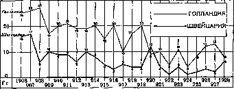
Figure 2. Mortality from measles in Holland and Switzerland for 1906-1928 (per 100,000 population). The social composition of the population is very diverse and where there is a constant influx of non-measles patients from outside, measles never
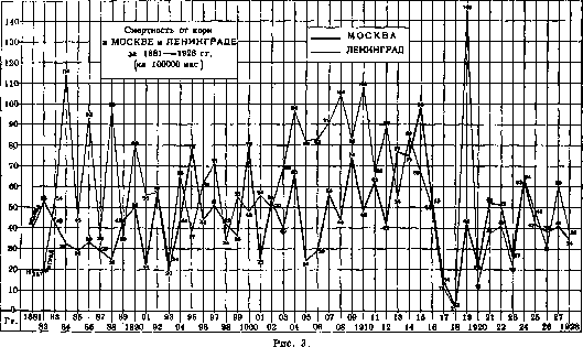
by year for 1881-1928, for Odessa from 1900 to 1928, for Königsberg from 1885 to 1900, and for Düsseldorf from 1913 to 1928. These curves clearly demonstrate the periodicity of measles waves; they are particularly clearly expressed on the last two curves, characteristic for small cities. This wave-like character of measles epidemics remains unchanged throughout the entire time available to our historical review. Kisskalt collected materials on mortality from measles in Königsberg from 1810 to 1900. For individual periods, the average mortality from measles per year per 100,000 population in Königsberg was: Table 5. Periods 1816-1850, 1851-1863, 1875-1900. Average mortality: 794, 127, 190. Years of maximum mortality: 1847, 1853. Here is noted: a certain tendency for a decrease in mortality from measles. Major epidemics lasted for a short time - from 1 year to 4-5 years (1828-32, 1860-63). However, Kisskalt is inclined to explain these long epidemics of the 19th century by inaccuracy of diagnosis: it is possible that doctors were dealing with smallpox or varioloid. Materials on mortality from measles in Moscow and Leningrad (Table 4) do not allow one to speak of a decrease in mortality from it in these cities. When calculating mortality from measles per 100,000 children under 14 years of age in the city of Moscow, the following indicators of this mortality (per year on average) are obtained: in 1892-1901 - 214, 1902-05 - 213, 1906-09 - 312, 1910-13 - 241, 1919-21 - 202, 1922-24 - 208, and in 1925-27 - 165. Table 6 gives mortality rates from measles for a long period of time in three large cities - Hamburg, London, and Paris. Table 6. Mortality from measles in Hamburg, London, Paris (per 100,000 pop). Years: 1851-1860..... 1, 23, -; 1861-1870..... 1, 36, -; 1871-1880..... 1, 31, -; 1881-1890..... 1, 33, -; 1891-1900..... 1, 21, -; 1906-1910..... 2; 1911-1913..... 2; 1925-1928..... Mortality from measles has noticeably decreased in Hamburg; in London it remained at approximately the same level for 60 years on average per year, and only in recent years is some decrease noted; in Paris it continues to remain high in the current century. This table gives an idea of the difference in mortality from measles in the same
Figure 4.
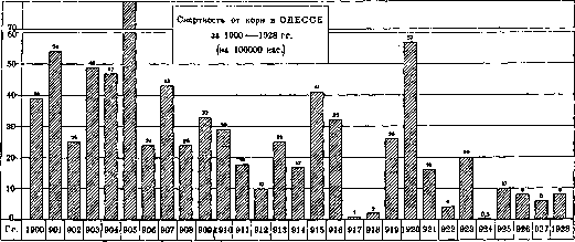
course of measles epidemics in small cities. This wave-like character of measles epidemics remains unchanged throughout
Figure 5.
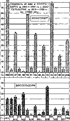
the entire time available to our historical review. Kisskalt collected materials on mortality from measles in Königsberg from 1810 to 1900. For individual periods, the average mortality from measles per year per 100,000 population in Königsberg was: Table 5. Periods: 1816-1850, 1851-1863, 1875-1900. Average mortality: 794, 127, 190. Years of maximum mortality: 1847, 1853. Here is noted: a certain tendency for a decrease in mortality from measles. Major epidemics lasted for a short time - from 1 year to 4-5 years (1828-32, 1860-63). However, Kisskalt is inclined to explain these long epidemics of the 19th century by inaccuracy of diagnosis: it is possible that doctors were dealing with smallpox or varioloid. Materials on mortality from measles in Moscow and Leningrad (Table 4) do not allow one to speak of a decrease in mortality from it in these cities. When calculating mortality from measles per 100,000 children under 14 years of age in the city of Moscow, the following indicators of this mortality (per year on average) are obtained: in 1892-1901 - 214, 1902-05 - 213, 1906-09 - 312, 1910-13 - 241, 1919-21 - 202, 1922-24 - 208, and in 1925-27 - 165. Table 6 gives mortality rates from measles for a long period of time in three large cities - Hamburg, London, and Paris. Table 6. Mortality from measles in Hamburg, London, Paris (per 100,000 pop). Years: 1851-1860..... 1, 23, -; 1861-1870..... 1, 36, -; 1871-1880..... 1, 31, -; 1881-1890..... 1, 33, -; 1891-1900..... 1, 21, -; 1906-1910..... 2; 1911-1913..... 2; 1925-1928..... Mortality from measles has noticeably decreased in Hamburg; in London it remained at approximately the same level for 60 years on average per year, and only in recent years is some decrease noted; in Paris it continues to remain high in the current century. This table gives an idea of the difference in mortality from measles in the same
MORTALITY FROM MEASLES IN LARGE CITIES OF EUROPE in 1926-28.
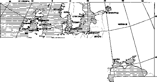
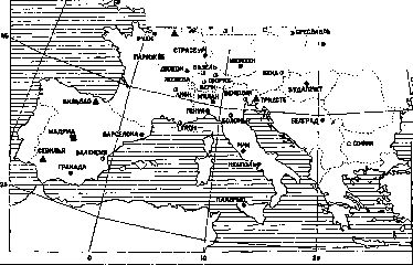
year on average per 100,000 pop. 0.7-2.0, 2.1-5.0, 5.1-10.0, 10.1-20.0, 20.1-43.1. Fig. 6. Mortality from measles (per 100,000 pop.) in some cities in recent years.
TABLE 7.
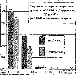
Fig.
Although somewhat less than in the second half of the 19th century, mortality from measles in Moscow and Leningrad continues to this day to be very high. Table 7 shows mortality rates in European cities. 1925 1926 1927 1928 Amsterdam Berlin Bern Budapest Warsaw Vienna Glasgow Geneva Leningrad London Madrid Manchester Moscow Odessa Oslo Paris Rome Stockholm Non-European Alexandria Cairo New York Mexico City Montreal Rio de Janeiro Auckland Sydney 11.0 4.6 3.7 11.6 25.6 2.4 39.5 0.0 48.0 7.8 9.6 17.5 41.5 9.5 0.4 23.1 31.9 27.0 0.2 81.3 86.9 2.2 66.8 8.8 56.1 0.6 17.3 2.0 1.8 4.4 3.8 0.3 41.4 0.0 31.0 20.7 36.6 21.3 38.3 9.0 2.7 20.8 13.3 0.0 6.9 14.1 15.1 1.9 2.4 3.9 6.7 1.0 4.5 13.5 2.0 4.6 6.7 11.8 7.1 29.3 2.4 60.0 3.9 3.9 21.8 41.0 6.0 6.7 20.0 18.7 0.9 0.9 0.4 13.0 0.6 82.3 6.0 14.6 4.0 0.4 5.4 2.1 2.7 8.4 3.7 1.5 35.7 0.0 38.0 29.8 31.9 16.7 33.6 8.0 1.6 20.0 1.8 4.7 3.7 9.6 112.4 5.8 18.6 9.7 25.4 0.5 8.8. In this table, covering a short period of time, one can see for most cities the characteristic fluctuations in mortality rates from year to year. These fluctuations are very weakly expressed in Leningrad and Moscow, which are distinguished by very high mortality rates for all years, as well as in Glasgow, Manchester, and Paris. Extremely sharp fluctuations are noted in some of the non-European cities listed in the table.
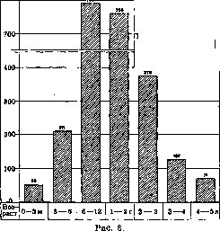
2. Mortality by age and sex. The main mass of deaths from M. falls on the age up to 4 years; in cities of the European part of the USSR in 1926, this age accounted for 96% of all deaths from M. (see Infectious diseases, Table 7, Fig. 3). Per 100,000 population of the corresponding age, died from measles in Moscow and Leningrad in 1926 (Table 8, Fig. 7): Table 8. Leningrad Age 0-1 year ... Moscow 405.4 1-4 years ... 281.7 14.3 10-14 years ... 0.7 433.2 179.1 15.4 0.0 0.2. In relation to the population of the corresponding group, the highest mortality from M. occurs in the age under 1 year. In Germany for 1925-27 years per 100,000 people of the corresponding age and sex died from measles: Table 9. Mortality from M. by age and sex in Germany in 1925-27. Male Female Age 1925 1926 1927 1925 1926 1927 0-1 year ... 128 105 1-4 years ... 45 45 15-30 years ... 0.1 0.02 0.1 0.1 0.1 0.1 31-60 years ... 0.04 0.04 0.03 0.1 0.04 0.03 61-70 years ... 0.1 0.1 0.1 0.05 0.05 0.05 71 years and older - 0.2 0.3 0.4 - 0.2. The most children die from M. at the age under 1 year. Individual cases of death are observed even in old age. Table 10 shows mortality rates by age and sex groups in England and Prussia (according to Prinzing) per 100,000 population of the corresponding age and sex. Table 10. Age 0-1 year 1-2 years 2-3 years 3-4 years 4-5 years 5-10 years 10-15 years England (1891-1900) Prussia (1892-1919) Male Female Male Female 292 297 155 94 47 1.7 1.8 1.7. The highest mortality occurs in the age from 1 year to 2 years. In children under 2 years, boys show somewhat higher mortality than girls. Table 11 (Fig. 8) shows mortality rates from M. in Paris from 1904 to 1913 by more detailed age groups. Table 11. Mortality by age in Paris in 1904-13 (average per year per 100,000 children of corresponding age). Age Died 0-3 months 3-6 months 6-12 months 1-2 years 2-3 years 3-4 years 52.6 210.6 789.8 757.5 376.1 126.7 Age Died 4-5 years 0-1 year 0-3 years 3-5 years 0-5 years 70.7 427.0 511.9 99.1 348.3. Mortality from measles is very low in children under 3 months; it is significantly higher in the age from 3 to 6 months, highest in the age from 6 months to 2 years, and with increasing age it decreases further.
3. Case fatality rate in M. Establishing the case fatality rate in M. appears to be a difficult task. Calculation of the case fatality rate in relation to registered patients does not give exact figures, since not all measles patients are registered. Hospital case fatality rate is higher than the actual one, since only the most severe measles patients are hospitalized. Therefore, indicators of case fatality rate calculated on the basis of hospital data need to be adjusted downward. By Binshtok, materials on mortality in hospitals of Leningrad for the period 1886-1926 were collected. The case fatality rate for measles during this period on average was 16%. By five-year periods it gave such fluctuations: Table 12. Case fatality rate in M. in hospitals of Leningrad. Years 1886-1890 1891-1895 1896-1900 1901-1905 Case fatality rate 2.7 5.3 15.7 20.8 Years 1906-1909 1910-1914 1915-1921 1922-1926 Case fatality rate 17.3 17.1 21.3 13.0 * No data for 1917 and 1918. The case fatality rate for measles shows large fluctuations depending on the age of the patients. Table 13 (Fig. 9) shows the corresponding indicators in hospitals of Petersburg for 1886-1909. Table 13. Case fatality rate in M. in hospitals of Petersburg by age groups. Age 0-5 years 6-10 years 11-15 years over 30 years. The highest case fatality rate was observed in the children's group under 5 years during these years.
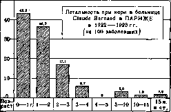
Table 14 shows the case fatality rate by age groups in hospitals of Paris. The highest mortality from M. is observed in the age under 1 year, and in individual hospitals up to 64% of all children of this age died. The case fatality rate is also very high in the age from 1 year to 2 years, reaching 50 percent of all patients. With increasing age, it decreases noticeably (Fig. 10).
4. Morbidity. In the USSR, from 400,000 to 500,000 cases of M. are registered annually, or from 30 to 40 cases per 10,000 population (see Infectious diseases). M. is registered at approximately the same level in other countries. Thus, in 1926 and 1927, per 10,000 population, measles patients were registered: in Denmark 37 and 53, in the United States of America - 60.1 and 37.6, in Canada - 47.3 and 29.6. In absolute numbers, the number of registered measles patients by individual countries is shown below in Table 15. Comparison of the figures of registered patients by individual years gives here also a clear picture of fluctuations in the height of measles waves. The smaller the country, the more clearly these fluctuations appear. Morbidity in
Figure 10.
Table 14. Died per 100 patients 0-1 year 1-2 years 2-3 years 3-4 years 4-5 years 0-2 years 2-5 years 1886-1889 1893-1898 1914-1923 1922-1923 1912 4.183 - 43.3 36.2 17.1 - 5.7 0 1 Over 5 years 38 14
1923.
1924.
Asian republics, Dagestan, etc.)
Figure 11.
47.6 Central-Chernozemn. 9.9 27.5 Lower-Volga. 20.0 45.4 Vyatko-Vetluzhskaya 7.5 38.8 Bash. republicka. 6.9
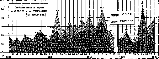
66.3 50.8 25.6 20.5 35.1 28.8 25.3 36.9 32.6 29.0 16.9 27.8 41.8 11.4 20.9 10.9 4.1 46.7 34.5 69.1 54.1 25.5 55.0 13.8 31.8 29.4 15.1 44.9 13.9 99.6 38.9 15.9 28.5 11.0 18.5 26.4 41.0 25.1 41.6 58.7 49.1 27.3 30.8 37.2 47.9 60.0 25.1 101.4 35.8 8.7 37.3 15.9 9.0 34.6 67.2, perhaps, can be explained by less complete registration of patients in them. 5. Morbidity by age and sex is illustrated in Table 19 (Fig. 12). Table 19. Morbidity by age and sex in the Moscow Governorate and in Moscow in 1926 (per 10,000 population of corresponding groups). Moscow Governorate Moscow Age 1906-10 | 1926 | 1926 m. f. | m. \ f. m. , f 0-1 yr. 1-4 yr. 5-9 yr. 10-14 yr. 15-19 yr. 20-29 yr. 180 203 99 37 4.6 1 17") 203 98 34 5.4 1.0 ,5 456 646 48 7 1 The most children contract M. at ages up to 5 years; significantly fewer from 5 to 9 years; after 15 years M. occurs in isolated cases. However M. is a "childhood" disease only insofar as almost everyone gets it in childhood, and subsequently it leaves a stable immunity. In those places where M. for some reason has not occurred for a long time, all those who have not had it get sick regardless of age (see above). During the world war M. gave rather significant morbidity in the troops. Thus, in the French army for the period from 1915 to 1918, contracted and died from M. and other "childhood" diseases: Table 20. Diphtheria; Diseases and mortality Measles Scarlet fever Number of cases . . . deaths . . . Mortality (per 100 cases)...... 76,081 1,470 1.93 35,010 662 26,065 650 More contracted and died from measles than from scarlet fever and diphtheria combined. In the army contracted 5 times more than among the entire population. This is explained by the large influx into the army of rural residents who had not had measles.
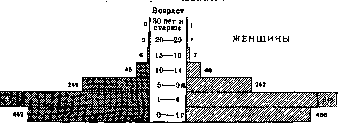
6. Morbidity and mortality from M. in cities and rural areas. M. as a rule is registered higher in cities than in rural areas. Table 21 shows morbidity rates for measles in cities and rural areas of the RSFSR for 1924-1927. For comparison, the same rates for pertussis are given. The highest mortality from measles and pertussis is observed in the poorest families. Bertillon calculated mortality per 10,000 inhabitants in different parts of Berlin (1886-95), Vienna (1891-97) and Paris (1886-95). For M. and pertussis these figures are given in Table 23. Table 21 Per 10,000 population Years Measles Cities 69.6 69.1 67.4 88.6*-78.1** . ** Other cities. Rural areas 16.4 37.3 24.1 25.3 Pertussis Cities | Rural areas 24.6 28.1 42.3 40.4*-16.6** ! 16.9 28.3 ; 30.6 1 29.2 * Administrative centers In % to the total Measles Cities 44.0 26.0 31.1 40.1 56.0 74.0 68.9 59.9 Rural areas Pertussis Cities 21.0 16.0 17.9 23.4 Rural areas 79.0 84.0 82.1 76.6 The difference in morbidity for both these infections in cities and rural areas is significant. Mortality from M. in cities is also higher than in rural areas. It was per 100,000 inhabitants in Württemberg (1899-1902): in cities with population over 10,000 inhab.-23.7, in the rest of the country-21.4; in Upper Bavaria (1899-1902): in cities-34, in rural areas-22; in England (1897-1901) per 100,000 children under 5 years in cities: 388 for boys and 358 for girls, in rural areas-143 and 130. In France (1906-13) died from measles per 100,000 inhabitants: in Paris-23.5, in cities with population from 30,000 to 1 million-16.4; in cities with population from 5,000 to 30,000 inhabitants-10.4, in communes with population below 5,000 inhabitants-6.6. Table 22 shows data on mortality from measles in cities and rural areas of North American United States (per 100,000 population). Table 22. Population centers | 1900 | 1905 | 1910 | 1915 | "~", "Figure 15", "Morbidity from measles by age and sex in Moscow in 1926 (per 10,000 people of corresponding groups). Cities....... 16.4 Rural areas j 9.8 9.1 3.4 13.4 : 7.1 11.6 3.4 This fact - higher morbidity and mortality from M. in cities than in rural areas-is noted everywhere with rare exceptions.
7. M. and social factors. Measles, like pertussis, belongs to those childhood infectious diseases in which the role of the social factor is most strongly expressed. Neefe compared mortality from childhood infections per 10,000 children under 15 years in different parts of Budapest for 1881-90 depending on the average annual income. Table 23. Mortality from M. and pertussis depending on well-being in Berlin, Vienna and Paris (per 10,000 inhab.). Berlin in 18 8 | M a ! p | o | o 3.2 2.9 3.9 2.6 3.6 1.5 2.1 1.1 1.9 0.7 0.9 Vienna Paris g n l ft and ft o o o p 11.2 1 1.4 6.7 2.8 7.4 1.3 5.6 2.3 8.6 1.5 3.7 1.2 6.4 1.1 2.4 0.8 2.5 0.5 2.0 o,b 0.3 0.1 0.9 0.3 Table 24. S Percent Died from M. per ! people with area; 100,000 children under 1 year Districts | chemistry |---------------------- | district | in | on | rooms | disease | houses | total Rich . . . Comfortable Poor . . . 35.7 45.6 55.9 47.6 : 58.6 115.5 I 57.0 133.8 | 98.2 106.2 172.5 232.0 with high mortality. In Table 25 is given mortality for measles in the city of Glasgow for 1893-1912 depending on the number of rooms in apartments. Table 25. Number of rooms in apartment ........ Mortality (per 100 cases) .... 12 3 4 and more 9.2 4.6 2.5 1.5 Higher mortality in poorer apartments is explained by lack of air, light and worse care for patients and, as a result of all this, more frequent complications with measles. 8. Seasonality of M. spread. In Fig. 13 is given monthly, 1928 35000-30000- \ / g , ch N / 1/ > > \ // / -x \ / / / / ch^ Months i XI from March to June, the minimum falls on September and October. In the RSFSR and Ukraine the minimum also falls on September, maximum morbidity is noted in the winter-spring period.
I. Dobreytser. Pathological Anatomy. Patho-anatomical changes in measles, if we do not count the skin rash, are concentrated primarily (often almost exclusively) in the respiratory organs, which are the most vulnerable to this infection. They arise already in the prodromal period in the form of the usual catarrh of the upper respiratory tract for this period, which in cases of moderate severity lasts until the rash fades or somewhat longer. In more severe cases, this catarrh can be accompanied by necrosis of the superficial layers of the mucous membrane, and such a necrotic character the process takes most often in the larynx (especially in the area of the vocal cords), but often also involves part or all of the trachea, and sometimes extends to the large bronchi. The mucous membrane at this time loses its luster, becomes cloudy, rough, and appears as if sprinkled with finely crumbled grayish decay. Where the necrosis extends beyond the limits of the epithelial cover and involves the connective tissue base of the mucous membrane, ulcers form with the same grayish uneven necrotic base. These ulcers are usually not deep and do not go beyond the mucous membrane, but can persist for quite a long time (2-3 weeks or more), without changing their necrotic character and showing very little tendency to clean and heal. Often on this basis, purulent perichondrites develop, leading to necrosis of the underlying cartilage and the formation of purulent cavities of various shapes and sizes in the wall of the larynx. The catarrh of the upper respiratory tract can easily spread downward along the mucous membrane of the bronchial tree, giving bronchitis, bronchiolitis, and catarrhal bronchopneumonia. However, in their pure form, these forms, apparently due to the comparative ease of their course, are far from often found as autopsy findings. Patho-anatomical examination mainly reaches more severe cases, where the process quickly takes on those special features that are rightly considered the most characteristic for the patho-anatomical picture of measles. The essence of these features lies in the great tendency of the inflammatory process to penetrate deep into the tissues, due to which the inflammatory changes are not limited to the mucous membrane of the bronchus, but also involve its elastic-muscular wall and peribronchial tissue. Thus, in the acute period, in addition to the appearance of catarrhal exudate in the lumen, one can always observe a strong dilation of the blood vessels, as well as some swelling and more or less marked (mainly round-cellular) inflammatory infiltration of both the bronchial wall itself, and especially the surrounding tissue. And since the penetration of the virus into the depths occurs most easily and quickly where the mucous membrane is most delicate and permeable, i.e., in the smallest branches of the airways, it is natural that in these latter the inflammatory changes in general, and in particular the formation of infiltrated cellular accumulations around the lumens, are expressed most strongly. The development of these infiltrates turns the bronchial wall together with the surrounding tissue into one solid wide cellular cuff and leads to the fact that on the surface of the section of such a lung, bronchioles become visible to the naked eye in the form of small grayish spots. Edema and infiltration usually do not limit themselves to the peribronchial tissue, but subsequently also spread to the adjacent alveolar septa, giving a more or less wide belt of interstitial pneumonia. To the latter, due to the appearance of effusion in the affected alveoli, exudative pneumonia is often added, which most often has the so-called desquamative or fibrinous, more rarely catarrhal character. In this way, around the affected bronchus, a new pneumonic cuff is formed, closely merging with the first and giving on the section of the lung already a larger grayish or yellowish-gray focus (so-called peribronchial pneumonia). With widespread involvement of the respiratory tract, such foci can merge with each other, leading to extensive, sometimes lobar and even total consolidations of the lung. The histological feature of these pneumonias is the frequent presence in the affected alveoli (mainly in small children up to 2-3 years old) of multinucleated giant cells, originating from the alveolar epithelium and sometimes very numerous (giant cell pneumonia). The process just described in the deep layers of the bronchial wall is very prone to a prolonged course, spreading along the peribronchial lymphatic pathways and gradually involving larger branches. Where changes exist longer, hyperemia gradually subsides, the infiltrate becomes drier, denser, first turning into granulation tissue, and then into fibrous connective tissue. Sections of such bronchi on the lung section have a paler, whitish-gray color and stand out even more, very much resembling tuberculous nodules, from which they differ only in that upon careful examination (especially with a magnifying glass) a central lumen can almost always be noticed in them; microscopically in such bronchi, metaplasia of the cylindrical epithelium into multi-layered flat is often observed. Interstitial changes in the adjacent alveolar septa lead to their thickening, and the exudate present in the bronchioles and alveoli often does not resolve, but deposits, over time undergoing organization and causing collapse and carnification of the corresponding areas of the lung. The course of measles bronchitis and peribronchitis, especially in its acute period, can at any moment be complicated by a necrotic process, similar to that observed in the larynx and trachea, and also localized mainly in the smallest bronchial branches. Here, the entire wall of the bronchiole is usually subjected to necrosis, and often also the peribronchial tissue with a larger or smaller number of adjacent alveoli, especially if the latter have pneumonic changes. Thus, in form these necrotic foci correspond to the outlines of the peribronchial cuffs and are well distinguished on lung sections due to their dry appearance and pale, sometimes almost white color, and in the latter case they often resemble tuberculous caseous foci (pneumonia morbillosa pseudo-caseosa). If the walls of bronchial branches, still passable for air, undergo necrosis, then in them (on the one hand due to the loss of elasticity by the walls, on the other - under the influence of cough shocks and associated frequent and sharp increases in pressure in the air-conducting system) bronchiectatic expansions easily develop, either diffuse or more limited depending on the spread of the necrotic process. Sometimes foci of necrosis appear in the pneumonic areas and independently of the bronchial system. - A characteristic feature of measles is the tendency of necrotic masses, wherever they appear, to undergo putrefactive decay, due to which in severe measles lesions, gangrene of the lungs often develops, more rarely gangrenous laryngitis. Similarly, purulent melting of tissue is possible, starting almost always from the wall of the bronchus and leading to the formation of sometimes very large abscesses. The outcomes of necrotic and purulent processes are especially unfavorable in terms of restoring the normal structure and function of the organ. In place of the necrotized bronchial wall, a more or less wide zone of granulation tissue always develops, and if this occurs over a significant length (which is most often the case), epithelialization of the necrotic surface turns out to be very difficult, and such a bronchus in most cases is doomed to obliteration (bronchitis et bronchiolitis obliterans). At first, the hollow granulation cylinder turns into a solid one, the corresponding part of the lung parenchyma collapses, the exudate in the alveoli, as well as the necrotized areas of lung tissue, undergoes organization, and in the end all traces of the former structure disappear without a trace in the resulting scar focus. - It should be noted that all the described changes in the respiratory organs in severe prolonged lesions mostly combine with each other in the most varied combinations and different sequences, creating extremely motley and bizarre patho-anatomical pictures. The entire process can drag on for months, even years, leading to the development of extensive scars located mainly along the course of the bronchovascular bundles and interlobular lung septa.
Measles lesions of the lungs are very often complicated by lesions of the pleura in the form of fibrinous, purulent, or productive obliterative pleurisy. - In general, diseases of the respiratory organs account for more than 70% of all cases reaching autopsy as the cause of death in measles. The digestive tract is affected in measles much less intensely and more rarely. Indeed, catarrhal anginas and pharyngitis are quite common occurrences, but they take on a necrotic character only in exceptional cases; occasionally, catarrhal, aphthous, or even ulcerative stomatitis are encountered. The reaction from the cervical lymph glands is also moderate. Consequently, secondary suppurative processes in the soft tissues of the neck (phlegmons, purulent lymphadenitis, retropharyngeal and lateral pharyngeal abscesses) are not often encountered here. Much more characteristic of measles is the lesion of the large intestine, found in approximately 20% of all measles autopsies. As a rule, these are catarrhal colitis with mucous (much less frequently mucopurulent or bloody) discharge. Their significance lies mainly in the fact that if they persist for a long time, they can cause severe exhaustion and intoxication and thus significantly contribute to a fatal outcome. The presence of a fibrinous or ulcerative process in the intestines should always arouse suspicion of an accompanying dysentery, and only after the latter has been ruled out can it be attributed to measles. - Otitis in measles, in contrast to scarlatinal otitis, almost never leads to lesions of adjacent bony parts (mastoid process, pyramid of the temporal bone) - a circumstance all the more deserving of attention, as their causative agent (as well as other purulent inflammations in measles) is very often streptococcus. - Among the later measles lesions, it is necessary to mention noma (see). It usually occurs in children, exhausted by prolonged pathological processes in the respiratory organs or intestines. - Changes in the skin in measles rash are not specific. This is a focal inflammatory process, expressed by hyperemia, edema, and cellular infiltration of the corresponding areas of the cutis propria, mainly its upper layers. The infiltrates are most strongly expressed around the vessels, less so around the excretory ducts of the sebaceous and sweat glands. In the epidermis, corresponding to the inflammatory foci, edema, leukocytic infiltration, and degenerative changes in cells are noted, often reaching necrosis (so-called focal necrosis of the epidermis). Subsequently, the altered epithelial cells undergo accelerated and improper keratinization (parakeratosis), as a result of which the degenerative-necrotic foci transform into horny ones, which are gradually pushed upward by the normally growing epithelial tissue from below and finally are shed in the form of fine branched scales. By this time, the inflammatory phenomena subside in the connective tissue layer of the skin. - Filatov-Koplik spots, appearing on the oral mucosa, owe their origin to the same changes that characterize the skin rash. The epithelium of the mucosa here also undergoes degeneration and partial necrosis with subsequent improper keratinization. And since both necrosis and keratinization deprive the epithelial layer of transparency, the underlying vascular network at the corresponding place ceases to be translucent, which gives the spots their characteristic whitish color.
M. Skvortsov. Clinical features of Measles. The pathogenesis of Measles is still insufficiently studied. The extraordinary cyclical nature of the process, the character of the skin manifestations, the reaction of the glandular apparatus, the condition of the blood, the state of the cardiovascular system—all this gives a number of authors the right to consider Measles a "specific reactive (serum-type) disease", similar to smallpox (Moro, Pirquet). The virus (microbe or toxin) has a unique selective property of affecting certain systems while having little effect on others. The most vulnerable systems are the digestive tract, respiratory tract, and glandular apparatus. The cardiovascular system is little affected. From the side of the nervous system, the animal part is affected with moderate intensity; peripheral lesions are a great rarity; from the side of the autonomic nervous system, a unique state of increased vagotonia is noted. This unique vagotropic influence of the virus can, to a certain extent, explain both the selectivity in the involvement of systems (respiratory and digestive tracts, mucous membranes) and the similarity of the picture of skin changes and blood with the manifestations of serum disease (state of vagotonia).- In complications, in addition to the effect of the virus (toxin) on the epithelium of the respiratory and digestive tracts (superficial necrosis) and peribronchial spaces (interstitial processes), a major role is played by infection of the body by other microbes—diplococcus, streptococcus, influenza bacillus, as well as the state of reduced resistance of the body to various microbes, particularly in relation to tbc (Pirquet's anergy), which is characterized by the disappearance of the tuberculin skin reaction. Course and symptoms. A typical case of moderate severity. The incubation period averages 8-10 days. Shorter and longer incubation periods are rare. Recently, due to the use of convalescent serum according to Degkwitz, cases of elongation of the incubation period (up to 3 weeks) have been described. The incubation period usually does not manifest clinically, although some authors observed in patients during it instability in weight and some fluctuations in temperature. The most characteristic are changes in the blood picture; they show that during this period, very active processes occur in the body, which serve as an indicator of the mutual reactions of the microbe and the human body.- Prodromal period (period of precursors). The first symptoms of this period are: elevated temperature and catarrhs of the mucous membranes. Temperature rises sometimes reach 39.5°. Fluctuations in temperature are quite significant. After remaining at high figures for 1-2, rarely 3 days, temperature shows a significant decrease, and sometimes even falls to normal, which usually precedes the onset of the rash. With the appearance of the rash, temperature rises again quite high (Figure 14). This unique wave is very typical for the measles process. However, here can be significant variations both in the intensity of temperature and in its duration. In a number of cases, especially in small children-asthenics and in adults, exhausted and weakened by other diseases, fever may be mild and atypical (Fig. 15).-Parallel with the rise in temperature, there is a picture of moderate general intoxication: lethargy, loss of appetite, headache, in children capriciousness, restlessness, poor sleep, irritability.- Catarrhs of the mucous membranes involve the conjunctiva, nose, larynx. The conjunctiva reddens, photophobia increases, tearing intensifies; eyelids swell; later a catarrhal-purulent discharge appears, so that in the mornings children's eyelids stick together and are glued by purulent secretion. From the nose, first slight, then abundant mucous, later mucopurulent discharge; sneezing. The face becomes somewhat puffy, and the entire appearance of the patient with red, purulent eyes, swollen eyelids, tear-salivation, nasal discharge becomes very characteristic. Laryngitis can vary greatly in severity (see below-complications). Very characteristic and of great diagnostic importance is the symptom described for the first time by N. Filatov in 1895, and later, in 1896, by the American physician Koplik (Filatov-Koplik symptom). This symptom consists of a unique bran-like desquamation of the epithelium of the mucous membrane of the gums and cheeks opposite the molars. Upon examination, the mucous membrane appears as if sprinkled with small splashes of lime, small bluish-white spots. If they are examined in the early stage, one can notice several small, pinhead-sized, white spots, surrounded by a narrow rim of redness. After several hours or a day, their number increases, they become coarser, the background merges [see separate table (Vol. XIII, p. 743-744), Figs. 7 and 8]. This is a unique desquamative catarrh of the cheek mucosa. This symptom appears on average 2 days before the rash (Filatov, Molchanov-Bosse, Lautz), but sometimes 4-5-7 days before the rash. The Filatov-Koplik symptom is extremely specific, does not occur in other diseases, and sometimes allows diagnosis to be made so early that by isolating the patient, one can prevent other children from getting sick. Therefore, this symptom has enormous practical importance. The frequency of the symptom is very significant. It may be absent in very small children, especially weak, premature, asthenic children, in combination of measles with other diseases (e.g. scarlet fever+measles). The presence of stomatitis can mask it. The Filatov-Koplik symptom can be confused with aphthous stomatitis and thrush (soor); but in the latter case, these spots are white-yellow in color and coarser, there is no red rim; in stomatitis, the spots are also coarser and quickly turn into an ulcer. The Filatov-Koplik symptom can last the entire prodromal period, one-two days of the rash, and then disappear. Often its duration is even shorter. Somewhat later than the Filatov-Koplik symptom, approximately a day before the rash, a spotted redness-enanthema-appears on the soft and hard palate in the pharynx, which also has great diagnostic importance. Sometimes in the prodromal period, vomiting and liquid stool are observed. In some cases, abdominal pains resembling appendicitis, the excretion of pus cells in the urine, jaundiced coloring of the skin, early otitis are observed. These phenomena are explained by the catarrhs of the mucous membranes of the corresponding organs. Period of rash. At the height of the process, which coincides with the first days of the rash, there are a number of symptoms typical of Measles, of which 3 are main: rash, angina, intoxication. - Measles rash. The onset of the rash coincides with the 12-14th day from the moment of infection. In rare cases, the rash appears later, on the 16-17th, even on the 20-21st day. The rash appears behind the ears, on the cheeks, on the forehead. At first, the rash is sparse, then within a few hours the rash becomes denser all over the face, sparing neither the chin nor the lips, which distinguishes it from scarlatiniform rash, which leaves the lips and chin free from rash ("scarlatiniform triangle"). Each papule begins with a small papule, especially clearly visible if the skin is examined in lateral light; in some cases, the papule is quite sharply expressed, clearly protruding above the level of the skin, which is noticeable if you run your finger over the skin. Later, the number of individual papules increases, each becomes larger, flatter, around them appears a zone of redness of irregular shape, the spots begin to merge. Although so-called confluent Measles occurs, especially on the back, there is never a solid redness; there are always free areas on the affected skin, which, along with the exudative nature of the rash and eye involvement, gives such a characteristic appearance to the face of a measles patient [see separate table (Vol. XIII, p. 743-744), Fig. 6]. The measles rash appears very abundantly on the hairy part of the head. By the end of the first-beginning of the second day, the rash densely covers the face and begins to appear on the trunk, where within 1-2 days it goes through the same process: an increase in the number of papules and the transformation of papules into merging, spotted-papular eruptions. The skin between the rashes, if the rash is not too dense, remains unaffected; however, often around the rashes the skin has a somewhat cyanotic appearance, and the spot itself is surrounded by a small unique anemic zone.
This is especially sharply noticeable on the limbs and gives pictures that very much resemble 'serum sickness'. On the 3rd day from the beginning of the rash, while remaining very abundant on the face and trunk and often giving on the back and chest entire fields of continuous exudative erythema, the rash begins to involve the limbs, spreading from top to bottom and repeating in each individual spot the same cycle of development. This gradual nature of the rash (from top to bottom: face, trunk, limbs) is very characteristic of measles, as is the cycle of rash development over 3-4 days. In this way, measles differs from rubella (measles) and serum disease, where there is no such gradualness and cyclicity. During the rash, itching is often observed. In some cases, a so-called 'prodromal' rash (Vor-exanthem of German authors) - scanty pale pink spotted eruptions on the face, sometimes on the trunk - appears a day or several hours before the rash. The measles rash can have many variations, being sometimes finer and sometimes coarser, sometimes merging into entire fields, being colored sometimes more palely and sometimes more brightly, and being very different in the intensity of exudation into the skin. Often on the skin, especially on the flexor surfaces of the limbs, small and pinpoint hemorrhages appear. They can sometimes be quite abundant, but if not accompanied by general severe phenomena, bleeding from mucous membranes, they have no particular diagnostic or prognostic significance. The Rumpel-Leede phenomenon is often positive. As rare variants, rashes that take on a pustular character or resemble pemphigus (Heubner) are observed.
There is a very distinctive tendency for the measles rash to appear on areas where there was already hyperemia of the skin, irritation, or any inflammatory skin processes (diaper rash, eczema). In exhausted individuals, very small children, atrophics, in severe states of malnutrition, with severe disorders of cardiac activity, the rash can be atypical, scanty, sometimes cyanotic from the very beginning. French authors describe so-called recurrent rashes (recrudescences) - eruptions in several stages with intervals of several days. The skin during measles is usually moist, measles patients are prone to sweating, the sebaceous glands work normally or excessively, which also distinguishes measles from scarlet fever, where the skin is dry, sweating and the work of sebaceous glands are reduced. As the rash fades, it pigments, leaving behind dark brown spots of various sizes and intensities at the site of the eruptions, which can be very valuable for diagnosis in the late stages of the process. The intensity of this process is very varied; the pigmentation lasts 1-1½ weeks, sometimes up to 2 weeks. Peeling is slight, is not observed in all cases, usually has a fine branched character, and appears more often on the face and trunk; on the palms and soles there is no peeling, which also distinguishes measles from scarlet fever.
Angina. Changes in the pharynx and throat appear already at the end of the prodromal period; a few hours or a day before the eruption, a faint spotted redness on the soft and hard palate (enanthema) can sometimes be noticed. Subsequently, the spots very quickly begin to merge, the tonsils become somewhat swollen, and by the time the rash is in full bloom at the height of the disease, there is a distinct general catarrhal angina. Usually no necrosis is observed, which sharply distinguishes measles from scarlet fever. Only in very rare cases in very weakened children and in atrophics can phenomena of superficial necrosis of the epithelium be observed, involving the pharynx, nasopharynx, sometimes descending into the esophagus and in some cases causing retropharyngeal and lateral abscesses. The tongue is moderately coated, moist, salivation is increased.
Intoxication. The phenomena of general poisoning are quite distinct; already in the prodromal period, changes in mood, restlessness, irritability can be noted in the patient. At the height of the disease, these phenomena are even more pronounced. Caprices and restlessness of the child, poor sleep, sometimes delirium - constant phenomena; in adults - complaints of headache and general malaise. However, very severe phenomena with unconscious state, severe delirium, convulsions and coma are observed extremely rarely, as are meningeal phenomena. The temperature in typical cases gives a very characteristic curve with two rises - at the height of the precursors and at the beginning of the rash. During the flowering of the rash, the temperature (remittent) makes rather wide swings, remains such throughout the rash period and begins to fall simultaneously with the fading of the rash (4-5th day from the beginning of the eruption), often critically. However, many variations in the nature of the temperature curve can be observed.
Other manifestations of the disease. Peripheral lymph glands usually do not participate; only in individual cases can a moderate general acute lymphadenitis be observed. Catarrhs of the mucous membranes (conjunctivitis, rhinitis, laryngo-tracheo-bronchitis), starting in the prodromal period, accompany the entire eruptive period and at the height of the disease can be very strongly expressed. However, significant variations are also observed here. At the beginning of the disease, nosebleeds are not uncommon. Cough, initially coarse and dry, later becomes moist. Usually it is quite significant, strongly irritates and bothers the patient. On percussion of the chest, a very characteristic picture of acute emphysema is found: the percussion sound is box-like, the heart is covered by the lungs, their borders are low. The cause of this emphysema is not yet sufficiently studied. On auscultation - a significant amount of dry rales, and during the height of the disease - often moist rales as well. The stool is usually more frequent, semi-liquid or liquid consistency, often with an admixture of mucus. This is especially noticeable in infants and small children. The liver is not enlarged, not painful. Jaundice is occasionally noted.
The cardiovascular system is little affected by the measles process. The dimensions of the heart are usually unchanged, the tones are clear, the pulse during the height of the disease is accelerated corresponding to the temperature. With systematic investigation of blood pressure, some lowering of pressure can be noted in the prodromal period and at the beginning of the eruption, then equalizing. In the period of convalescence, arrhythmias are often noted. This property of sparing the cardiovascular system sharply distinguishes measles from many infectious diseases, in which the cardiovascular system is affected quite distinctly (diphtheria, scarlet fever, typhus). And with these properties is explained the fact that cases of death from uncomplicated measles are almost never observed.
Changes in the blood are very characteristic (Comby, Hecker, Molchanov-Bosse, Lautz). Already in the incubation period (7 days before the rash), there begins a rise in neutrophils and a fall in the number of lymphocytes; soon (6 days before) hyperleukocytosis appears; in the prodromal period, leukopenia, eosinopenia, decrease in the number of neutrophils (4-3 days before) and a shift of the blood to the left according to Arneth-Schilling (4 days before). The table below shows how many days before the rash the changes appear. . ......
- \ 6
6 | Decrease in lymphocytes . j
б I .4
4,4 i 3
2,9
4 ! 4,3 Shift according to Arneth-Schilling
1,5
J
_
! Resistance of erythrocytes is unchanged (Weil), coagulability
The sedimentation rate of blood is slowed (Gornitskaya). The number of platelets decreases by the time of rash appearance and becomes normal by the fading of the rash (Chistovich, Shifer). The blood sugar picture is unchanged. During purulent complications, there is the usual leukocytic-neutrophilic increase, however, it is less sharp than in other infections (e.g., in scarlet fever). The amount of urine at the height of the process is moderately decreased. Sometimes so-called febrile albuminuria appears. Very often a positive diazo reaction is found. An increase in urobilin content, in contrast to scarlet fever, is usually not found. The excretion of NaCl is unchanged; the ability to concentrate and excrete water is not impaired; in the febrile period, some decrease in urea excretion. Symptoms from the central nervous system are moderately expressed. No changes are noted from the peripheral nervous system. Changes from the endocrine-vegetative apparatus are very characteristic. Already in the prodromal period, a number of symptoms can be observed that indicate a state of excitation of the parasympathetic department of the vegetative nervous system. These phenomena persist throughout the eruptive period and weaken with the disappearance of the rash. Functional examination of the vegetative nervous system shows that starting from the prodromal period and at the height of the process, there is a state of active vagotonia, which gradually weakens parallel to the disappearance of the rash. The adreno-sympathetic system is slightly affected. Reactions to pilocarpin are sharp, to adrenaline are weakly positive. These data bring Measles closer to serum disease, where a vagotonic syndrome is also seen—a type of disease opposite to that encountered in scarlet fever (Koltypan).-When examining the ear with a mirror at the end of the prodromal period and during rash appearance, slight hyperemia of the eardrum can be found.-In the prodromal period and at the beginning of the eruptive period, photophobia is even more pronounced in the presence of corneal or conjunctival disease; sometimes-phenomena of blepharospasm. Course of Measles. The duration of the incubation period is on average 8-10 days, of the prodromal period-2-4 days (and sometimes even 5-6 days), of the eruptive period-3-5 days. Pigmentation lasts for 1-11/2 weeks. Desquamation is very short. On the 2-3rd day of rash, all symptoms reach their greatest strength. With the fading of the rash (4-5th day), all symptoms begin to subside: temperature falls, catarrhs weaken, cough becomes soft, however, it can remain for quite a long time; general condition improves, appetite appears. However, in small children, lack of appetite persists for a long time, as well as a tendency to liquid stools and a state of tearfulness and nervous irritability. In the convalescent period—slightly expressed disturbances of cardiac rhythm. With the fall in temperature, urine and blood gradually return to normal. In small children, a noticeable loss of weight is observed, especially if the process was accompanied by diarrhea, and often a state of severe dystrophy develops as a manifestation of the harmful effect of the pathogen on the body tissues and partly from starvation. Forms of Measles. From this typical course of Measles, there can be significant deviations. The causes of this, as with respect to other diseases, remain insufficiently studied: here the virulence of the microbe, often associated with the character of a certain epidemic, sometimes apparently the influence of other microbial groups, then age, constitution, nutrition and previous disease can play a role. Measles can give cases of mild and severe course. To the first group are attributed the forms of so-called abortive Measles, where all manifestations can be expressed very mildly and the disease ends very quickly. In other cases, atypicality concerns only one period; e.g., the prodromal period can be expressed very mildly, catarrhs are insignificant, temperature is low. Sometimes atypicality concerns the eruptive period—the rash can be expressed very mildly and even be completely absent (measles without rash). Atypical cases are very common in children-atrophics and in very early infancy. These same forms can be observed in adults, exhausted by disease or hunger, in combinations with other infections. In other cases, Measles takes a more severe course and can quickly lead to death. A form adynamic, or toxic, is distinguished, where the phenomena of severe general poisoning and weakness of the cardiovascular system come to the fore; then the pulmonary form, with very rapid development of severe lesions of the respiratory tract. Very rare are the forms of hemorrhagic Measles with profuse bleeding from the nose, intestines, skin hemorrhages, usually ending in death. Death from uncomplicated Measles is an exceptional phenomenon. The cause of death is usually complications. Complications. Lesions of the upper respiratory tract (rhinitis, laryngo-tracheo-bronchitis)—constant symptoms of Measles. They can be called complications if they take a severe course, exacerbate or reappear after disappearance. Rhinitis only in rare cases takes a severe course or gives a necrotic form. In some cases, already in the prodromal period or in the eruptive period, the ordinary measles laryngitis takes a more severe course: cough becomes dry, barking, difficult noisy breathing appears, a picture develops resembling true diphtheritic croup, clinically differing from it only by the more rapid development of all symptoms, absence of deposits in the pharynx and negative data with respect to Corynebacterium diphtheriae. The process can reach asphyxiation and require surgical intervention (intubation, tracheotomy). However, these measures rarely give effect, since here we are dealing with a necrotic process in the larynx. Whether the cause of these necroses is the Measles pathogen itself or the toxin or whether other microbes play a role here remains insufficiently clarified. As a result of this process, ulcers form in the larynx, which cause reflex spasm (stenosis), and during intubation (as well as during tracheotomy) they give pressure sores, create impossibility of extubation and extraction. In both cases, pneumonia easily develops, and the case ends in death. Children are especially prone to this complication up to 3 years of age. It must be distinguished from true diphtheritic croup as a secondary, but very common disease in Measles, since Measles creates increased susceptibility to diphtherial infection. This true diphtheritic croup, developing later, usually in the period of pigmentation, gives a somewhat slower course. On examination of the mucous membranes, deposits in the pharynx or in the nose can often be noted, the culture gives growth of Corynebacterium Loffieri. Inflammation of the lungs—one of the most frequent complications. Mortality from Measles in children in the vast majority of cases depends on pneumonia. Pneumonia can appear very early, at the very beginning of the eruptive period. These cases are usually the most severe and most typical in their clinical picture and pathoanatomical changes. However, pneumonia can join Measles also in the period of pigmentation, after the fall in temperature. If pneumonia develops early, in the eruptive period, in the presence of high temperature, then its recognition can present considerable difficulties: pneumonia is indicated here by the joining dyspnea, increasing with movement, flaring of the nostrils, sometimes cyanosis of the lips, face and extremities. On percussion, the chest gives a boxy sound (tympanitis); due to acute emphysema, the heart is covered; on auscultation—many dry, and mainly moist, small, sonorous and subcrepitant rales, especially—concentrated in the posterior parts of the lungs. Breathing is vesicular. Later, focal phenomena can join in connection with the fusion of small foci into larger ones (shortening of sound, bronchial breathing). Parallel to this, the symptoms of cardiac weakness increase—pulse is frequent and weak. The liver enlarges; the patient can die within 2-4 days from the beginning of the disease. Measles pneumonias, however, can have a more prolonged, and sometimes even very prolonged course (several months) with prolonged fever of an irregular type, further complications (pleurisy, empyema), exhaustion and again lead to death or cause severe changes in the lung (carnification, abscess, cavity, gangrenous bronchitis, gangrene). Bacteriologically, various microbes are found, most often of Diplococcus Fränkel and Diplococcus streptococcus. In later periods of measles, pneumonias can proceed in ordinary forms—lobular, less frequently—lobar pneumonias. As complications of pneumonias, dry pleurisy, empyemas (Diplococcus streptococcus), lung abscesses, bronchiectases are observed. The digestive tract. Complications from the mucous membrane of the mouth are very common. Already at the beginning of the eruptive period, the specific desquamative catarrh can pass into a widespread stomatitis of the aphthous type, and sometimes ulcerative, which greatly hinders the feeding of small infants. In atrophics, sometimes a widespread superficial necrosis of the mouth and throat epithelium is found. Lips in children often crack and are covered with dirty-colored deposits-necroses (Staphylococcus!). Particularly dangerous is the complication known under the name of measles (see). Lesions of the gastro-intestinal tract in early age especially often take a severe course and can be the cause of death.
Dyspepsia in infants and liquid stool, in older children, and often in adults, is a very frequent phenomenon at the beginning of the process. These disorders sometimes can last for the entire illness. Often these diarrhea cases take on the character of colitis (mucus, tenesmus, sometimes blood); in their course they differ little from ordinary infectious colitis, increasing the patient's exhaustion. They last
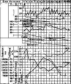
Figure 16. Case of measles complicated by otitis.
for a very indefinite time. Bacteriologically, these colitis in some cases are due to infection by the dysentery bacillus; more often the matter is obviously due to the influence of the measles contagion itself. Complications from the nervous system are rather rare. In recent years, there have been a number of reports about encephalitis after Measles (29 cases were collected by Bosse). The process more often develops during the period of fading rash, accompanied by new rises in temperature, cerebral symptoms. The spinal fluid shows no changes, is sterile. In a number of cases, complete recovery was observed, in others--mental retardation developed. Opinions about the genesis differ. It is possible that the process is connected with the effect of the measles poison (toxin) on a particularly vulnerable and prepared soil (Bosse). As very rare phenomena, peripheral paralyses have been observed. Otitis of otogenic origin purulent meningitis may be observed. Very rarely, nephritis are observed, more common, especially in small children, pyelitis.-- Organs of sense. A frequent complication is otitis. It can be serous and purulent. Otitis can appear already at the beginning of the period of rash, more often--by the time of temperature drop in the period of pigmentation.(fig. 16). Otitis is usually accompanied by a sharp rise in temperature, often pain and shooting in the ear and leukocytic-neutrophilic shifts in the blood. Unlike scarlatinal otitis, its course is more benign, although streptococci are often found in the pus. In general, about 60% of otitis in children does not lead to suppuration (Polteva); therefore, in case of serous acute measles otitis, one should not rush with paracentesis. Mastoiditis--an extremely rare complication. From the eye--blepharitis, but corneal and choroidal lesions are rare. The measles poison lowers the general resistance of the body; therefore, in measles convalescents, various skin lesions (abscesses, furunculosis, phlegmon) are often observed. The effect of the measles poison on chronic infection is very peculiar. Apparently any acute infectious disease, superimposed on a chronic one (tuberculosis, syphilis, malaria, rheumatism), can cause an exacerbation of the chronic process and give a flare-up. Measles has special properties of giving these exacerbations mainly in relation to tuberculosis (anergy). Flare-ups of the miliary process, appearance of local forms of tuberculosis in children after Measles have been described repeatedly. So apparently the matter stands also in relation to syphilis, malaria, gonorrhea (Pirke, Danilevich).-Mixed infections. Measles, like any other infectious disease, can give a concurrent course with another infection. This is more often observed in conditions of hospital treatment of children. Combinations of Measles and scarlet fever, Measles and diphtheria, Measles and chickenpox and a number of other rarer, sometimes even triple combinations are often encountered. Usually such combined cases proceed more severely, as if summing up their harmfulness. The course is especially unfavorable when Measles precedes another infection, which is particularly noticeable in relation to diphtheria, which usually proceeds more severely in measles patients. Diagnosis. For diagnosis, the incubation period, presence of prodromas (catarrhs), character of the rash (spotted-papular), peculiarity of the rash (three-day cycle), pigmentation, absence of necrosis in the pharynx are of importance. In the blood--leukopenia, slowing of coagulation, thrombocytopenia. Of particular importance in the prodromal period is the Filatov-Koplik symptom, not occurring in other infections. Differential diagnosis can present significant difficulties in relation to a number of diseases from the group of so-called acute infectious exanthems.- Measles rubella (rubeola morbillosa). Differences: longer incubation period (17-21 days), absence of prodromas and Filatov symptom; catarrhs are negligible; character of the rash is finely spotted, color more pink, rash appears immediately from the trunk; in the pharynx negligible hyperemia, pigmentation almost absent; typical swelling of the posterior cervical and occipital glands, in the blood plasma cells, no thrombocytopenia. The course is benign, usually without complications.- Serum disease can give significant diagnostic difficulties. Differences: in the history serum injection, time of appearance of serum rash--no later than 3 weeks from the moment of injection, rash appears from the site of injection, general swelling of glands; no catarrhs, Filatov-Koplik symptom and angina; rash is more exudative in nature.-Septic rashes. Presence of a source of sepsis (erysipelas, preceding disease, otitis); septic type temperature, chills, often high hyperleukocytosis of non-neutrophilic nature (but leukopenia can also be present), positive data from blood culture from a vein, frequent involvement of joints, septic pyemias, especially in small children, absence of catarrhs. Character of the rash is varied--the rash is sometimes spotted, sometimes spotted-papular, no cyclicity of rash. At the beginning, Measles can be confused with smallpox, when papules abundantly appear on the face, and catarrhs are not sharp. Differences: no signs of previously vaccinated smallpox, severe course, enlargement of the spleen, absence of catarrhs and Filatov-Koplik symptom, monocytic hyperleukocytosis, drop in temperature at the beginning of rash.-In rare cases, Measles can be confused with typhus. Prognosis and hospital mortality. Prognosis is determined by age, nutritional status, form of the disease, presence or absence of chronic infection. The younger the child, the more serious the prognosis, since mainly small children become victims of complications from the respiratory and digestive organs. Measles in a child under 3 years--always a dangerous disease. In older children and adolescents, Measles becomes dangerous when it affects an exhausted and weakened child from other infections. It is especially dangerous for tuberculosis patients. For adults, it can be dangerous during epidemic outbreaks affecting areas where Measles has not been present for a long time, and in subjects weakened by hunger, diseases or nutritional deprivation. Hospital mortality, which is more precisely studied among children--about 15-20%; in children under 2 years--significantly higher (40-60%). This high percentage is explained by the fact that the hospital receives almost exclusively severely ill children or with pulmonary and intestinal complications. Treatment. General hygiene, care and dietetic measures have great importance. The temperature of the room should be moderate (15-18°); it is necessary to monitor the cleanliness of the air; in summer in warm weather, the patient can be in the open air or with widely open windows; in cold weather, the room should be thoroughly and systematically ventilated, especially at night. Dimming the light should never be done; only in cases of severe photophobia, the patient's head is placed toward the light source, a temporary screen is set up, or an adult is given an eye umbrella. Bathing children is recommended daily or every other day, washing the face and hands every day. Great importance is attached to the care of the bed and mucous membranes, thorough rinsing of the mouth and pharynx; lips are smeared with boiled vegetable oil; eyes are washed with 2% boric acid, eyelids are smeared with sterile vaseline.- Nutrition of the patient is of great importance in childhood, especially in early age. For a breast-fed child, the most important measure is the preservation of breast feeding. In the early periods of Measles, food should be liquid and semi-liquid. For artificial and mixed feeding--mixtures with carbohydrates (2 parts milk + 1 part rice or oat broth with 5% sugar); for older children (after 6-7 months)--tea and coffee with milk, whole milk, kissels; in case of intestinal disorders--protein milk with 5-7% sugar, buttermilk with sugar and flour; kefir, concentrated broth, mixtures from x/a-2/з milk with dextrinized flour. For older children--porridges, kissels, broth, cottage cheese, simple sour milk; abundant drinking. After the temperature drops, one can quickly return to normal food, if there is no liquid stool and appetite appears. In case of colitis, it is important not to let the child starve. Food should be full and varied. Diet and treatment--as in ordinary acute colitis (see). Respiratory tract. Great importance is attached to clean, fresh air in the room. For laryngitis--hot baths (37-40° for 10 min.), in older children--inhalation of 2% soda solution; in cases of increasing stenosis--bromine preparations, thorough ventilation of the room, hot baths, creating a calm atmosphere around the child. Surgical intervention (intubation, tracheotomy)--only in cases of extreme necessity. In the treatment of pneumonia, the most important thing is to provide the child with the maximum amount of fresh air: constant ventilation of the room with widely open windows; frequent change of the patient's position is necessary, carrying small children in arms. In the latter, mechanical cleaning of the oral cavity and pharynx from mucus is very important. Hot water procedures [hot baths (40°)] have a good effect, in small children--mustard wraps, in older children and in cases of vascular disorders--mustard plasters. Compresses that restrict breathing (especially in small children and rachitic children) are not indicated. Internally--alkaline mineral waters (Borjomi), 2% soda solution with milk; in older children, in cases of dry irritating cough, narcotics (Codein, Pulv.) sometimes have to be used.
Treatment.-For otitis-5% carbolic glycerin; after paracentesis and with spontaneous perforation-ordinary therapy. Treatment of mastoiditis-according to general rules. Treatment of noma-see Noma. The child can be allowed out of bed if there are no complications, approximately one week after the onset of rash.-Specific treatment with serums has not yet become widespread. Prevention. Measures for prevention include three tasks: cessation of contact between the sick and the healthy and destruction of the infectious focus (isolation, disinfection), measures to enhance natural immunity, measures for specific prevention (active and passive).-In relation to the first task, early diagnosis and early isolation of the patient are of great importance. A case isolated from the children's collective in the early prodromal periods by the Filatov-Koplik symptom may not lead to further diseases; however, one isolated during the rash period usually leads to the disease in almost all children who have not had Measles. Discharge from the hospital without danger to healthy children, according to the rules established by the People's Commissariat of Health, can be made after the 7th day from the onset of rash.-Separation of those who have come into contact with measles is carried out for the incubation period-21 days. For the same period, admission of children to the collective where quarantine has been declared is stopped. Disinfection is not required; ventilation and ordinary cleaning of the room are sufficient.-Measures to enhance natural immunity include general hygienic-dietetic measures. It is necessary to widely propagate proper hygiene in early childhood, in particular the propagation of breastfeeding and constant presence of children in the open air. However, due to the insufficiency of all the mentioned measures, attempts to find ways to create artificial immunity (passive or active) in the child do not cease. This question cannot receive a proper solution until the causative agent is discovered and the nature of immunity is studied. Of the methods of active immunization, Hermann proposed introducing into the nose of healthy children mucus from the nasal cavity of measles patients; Dégkowitz used as a vaccine his culture from the pharynx of patients, Caronia-a vaccine of the microbe he had discovered. However, these methods have not become widespread and cannot be recommended. The greatest popularity has been gained by Dégkowitz's method (see Dégkowitz's vaccinations).
a. Koltysh.
Related articles
Mentioned in
Cite this page
“Measles.” Soviet Medical Encyclopedia. English translation of Bolshaya Meditsinskaya Entsiklopediya, 1st ed. (Moscow, 1928–1936), ed. N. A. Semashko. https://sovietmedicalencyclopedia.pages.dev/article/measles/