Smallpox (Variola)
Historical document, translated for reference. It reflects medical knowledge of the 1920s–30s and is not medical advice.
Summary
A historical overview of smallpox, detailing its ancient origins, global spread, and the impact of vaccination. It covers the clinical characteristics, the distinction between natural smallpox and related conditions, and the early 20th-century understanding of its etiology.
Encyclopedia article (1928–1936)
12 III. Statistics and geographical distribution. 39 VI. Animal smallpox. Smallpox, variola (from Latin varus—spot, nodule), is an acute infectious disease characterized by general intoxication, a specific type of temperature curve, and a specific rash on the skin and visible mucous membranes, which has a definite cyclic course. Human susceptibility to smallpox infection is extremely high. As a rule, having had smallpox once protects against infection for life, although in isolated, exceptionally rare cases, recurrent diseases are encountered. The disease in humans is given the name natural smallpox (variola vera) in contrast to animal smallpox (see below) and chickenpox (see)—a disease sui generis, having nothing in common with natural smallpox except for a purely external similarity in clinical manifestations. Some authors have proposed the name variola major for natural smallpox, in contrast to alastrim (see)—variola minor. Natural smallpox usually spreads epidemically and, as a rule, is accompanied by high mortality. I. History and epidemiology. Smallpox belongs to the oldest diseases of humanity. Long before the Christian era, it existed in China, which was apparently its cradle. Another ancient focus of smallpox is India, where the cult of the goddess of smallpox, Mariatale, has existed since time immemorial. Smallpox was not known to the classical world. Smallpox left the borders of its ancient foci only in the 4th century A.D. Having spread widely throughout the East, it appeared in Arabia in 572. In the 7th century, with the Arab invasion of Egypt, smallpox was repeatedly brought into this country by the armies of the conquerors. Soon, having engulfed the entire north of Africa, the disease penetrated into Spain and from there spread throughout Europe. The movement of masses of people during the Crusades greatly contributed to the dissemination of the smallpox contagion, especially in Germany and Austria. In 1138, according to the testimony of Aventinus, the entire army of Frederick Barbarossa perished from smallpox. By the time of the Crusades, special shelters for the treatment and isolation of smallpox patients appeared in Europe for the first time. Around the 15th century, smallpox built a firm nest for itself throughout Central Europe (Italy, Germany, France) and penetrated into Great Britain. Smallpox was brought to America by the Spaniards in 1517 and caused severe devastation among the natives who had not previously known this disease. In Mexico, over 3.5 million people died from it, about half of the entire population. In the 16th century, smallpox was brought by the English to North America; it penetrated into South America somewhat later. In general, in the 16th and 17th centuries, smallpox had already spread to almost all countries of the world known to Europeans, often manifesting itself in the form of formidable pandemics. Thus, the epidemic that began in 1614 engulfed all of Europe, Asia Minor, and Africa. Around this time, the disease also spread in Russia. It is reliably known that as early as 1610, smallpox was brought by the Russians to Siberia, where it caused such severe devastation among the native tribes that the majority of the population abandoned their homes and fled. But smallpox reached its highest development in the 18th century. Smallpox epidemics became such a common phenomenon in Europe that the majority of doctors of that time passed over them in silence. Germany, England, and especially France suffered greatly from smallpox, where the number of those who died, were maimed, and were disfigured by this disease constituted about 2/4 of the entire population. It is believed that since the epidemic spread of this disease, no less than 150 million people have died from it in Europe. Appearing for the first time in a populated area, smallpox struck residents regardless of gender and age. In endemic foci, however, it always bore the character of a childhood disease. With universal susceptibility to smallpox, those naturally immune to it were observed to be no more than 5%—the disease usually struck people in early childhood. Having had smallpox in childhood provided protection against this disease in adulthood. Only when a certain number of "smallpox-susceptible" children accumulated among the growing generation did a rise in smallpox cases among this group of the population follow. Usually, such outbreaks of smallpox occurred regularly periodically every 4-6 years. The idea that smallpox possesses contagious properties and that the disease is capable of being transmitted from one person to another originated as early as the 16th and 17th centuries (Avicenna and especially van Helmont), however, it received confirmation and universal recognition only in the 18th century thanks to the high authority of Boerhaave. Around this same time, attempts were made to fight smallpox through sanitary-preventive measures (isolation, disinfection), attempts that did not have success due to the strong contagiousness of the disease and the ubiquitous dissemination of the contagion. The introduction of inoculation (see) was hardly accompanied by greater effect. The ancient Hindus and Chinese already knew that a single bout of smallpox protects against a new disease from it. They also knew the fact that artificially inoculated smallpox is tolerated significantly easier than natural smallpox obtained through natural infection. The Arabs, Persians, and other peoples of the East knew about this, among whom artificial inoculations of weakened natural smallpox (variolation) borrowed from the Hindus and Chinese are still widely used today. In the 18th century, attention was turned to this in Europe as well, which was being devastated by smallpox epidemics. Despite the improvements introduced into variolation by European doctors, the latter proved to be far from an ideal remedy (mortality with it reached 2-3%, the inoculated sometimes became a focus for the spread of natural smallpox) and in the first half of the 19th century, it was everywhere supplanted by vaccination (see). The latter is the factor that had the most significant influence on the history of smallpox in the 19th century and later. The successful organization of vaccination, which by the beginning of the 19th century had received noticeable spread in all civilized countries of the world, was everywhere accompanied by a fall in the absolute incidence of smallpox and, in particular, a reduction in incidence among children (vaccination was carried out primarily among this group of the population). A classic example illustrating the incidence of smallpox at the turn of the 18th and 19th centuries and the beneficial role of vaccination can be the Swedish smallpox statistics (Table 1, cited from Gins). As can be seen from the table above, the reduction in smallpox mortality occurred mainly at the expense of the child population group (0-5 years), starting from the five-year period of 1801-05, after vaccination (see) was introduced in Sweden in 1800. The pandemic of 1870-74 (see below) raised interest in the scientific study of the vaccine and in all questions of the proper organization of vaccination. Starting from 1875, vaccination was legalized as a mandatory measure in Germany, France, and other countries. The idea that the quality of the vaccine is an essentially important prerequisite for the success of vaccination led to the establishment of model smallpox institutes in many countries (Austria, England). At the same time, the experience of the pandemic of 1870-1874 and later (especially in Switzerland, England) showed that such proven general hygienic measures against infectious diseases as mandatory notification, isolation, and disinfection are of little effect against smallpox and are not capable of preventing its spread. In the 20th century, large smallpox epidemics are already the lot of only culturally backward countries (British India, Central Africa). In most European states, smallpox has completely disappeared or cases of it are reduced to units (see below—statistics). A completely new epidemiological problem in recent years is the appearance of alastrim (see). Apparently, this is a low-virulence and reversible variant of natural smallpox, against which the same preventive measures are indicated as against natural smallpox (mandatory notification, vaccination, isolation, disinfection).
II. Etiology. Like other contagious diseases, smallpox is undoubtedly caused by a living pathogen. The close genetic and biological affinity between natural smallpox and cowpox (see below—Animal smallpox) allows us to speak of a common pathogen for both of these diseases, of the smallpox-vaccine pathogen. All efforts to obtain a culture of the smallpox virus on artificial nutrient media have proven fruitless. Reports by some authors that the smallpox virus is capable of multiplying in tissue explants need verification and confirmation. The question of the morphology of the smallpox pathogen has also not been finally clarified. The transparent content of young pocks, which abounds with the specific virus, is always devoid of banal flora (sterile). On this basis alone, it should be accepted that the various microbes (bacteria, Protozoa, etc.) isolated by individual authors from the content of pocks and recognized by them as allegedly specific pathogens of smallpox have no relation to the etiology of this disease. 65.7 10.2 11.5 The pyogenic staphylococcus and streptococcus sometimes encountered in pocks towards the end of their maturation are also devoid of etiological significance and are only the pathogens of secondary purulent processes. The content of pocks retains contagious properties after passing through a filter candle impermeable to very small representatives of the bacterial flora. However, such experiments do not always yield a constant result.
The concentration of the filtered material (usually scrapings and the liquid contents of pocks), the degree of its pulverization, the quality (porosity) of the candle, the speed of filtration, and many other factors have a decisive influence on the outcome of the experiment. Even in the case of success, the filtrate contains only a very meager amount of the virus, and special techniques are required for its detection. (Negri, for example, placed a small piece of cotton wool soaked in the filtrate onto the scarified cornea of a rabbit and sutured the eyelids closed over the cotton for 24 hours.) Table 1. Mortality from smallpox in Sweden from 1776 to 1825 (per 100,000 population). Age groups 1776-80, 1781-85, 1786-90, 1791-95, 1796-1800, 1801-05, 1806-10, 1811-15, 1816-20, 1821-25. Thus, the ability of the smallpox virus to pass through a candle can be considered proven, but filtration occurs with difficulty and is accompanied by a huge loss of virus. Therefore, some authors consider the smallpox virus to be conditionally filterable. Based on the filtration experiment, the size of the smallpox pathogen is determined to be approximately 0.2 µ. It lies within the limits of the resolving power of modern optical instruments, and therefore, like some other filterable bacteria (the pathogen of influenza, Malta fever, etc.), the smallpox virus can apparently be detected in the field of view of a microscope. There are many reasons to consider the peculiar bodies described in 1907 by Paschen in the liquid contents of human pocks to be the pathogen of smallpox. "Elementary," or "Paschen bodies" are extremely small, rounded, coccus-like formations [see separate table (Vol. IV, art. 353-354), fig. 6], dividing in the shape of a gymnastic dumbbell. In a fresh state (in a hanging drop), they appear as rounded, matte, weakly light-refracting bodies. In a dark field of view, they glow weakly. The bodies are motionless; the movement observed in the paraboloid condenser is undoubtedly Brownian molecular motion. The bodies yield to the action of staining substances common in microbiology with very great difficulty and only after very prolonged treatment. For their successful detection in dry preparations, the latter must be kept for some time in distilled water to remove serum and protein residues that hinder the clarity of the picture. Quite reliable staining results are obtained only by using mordants (e.g., Löffler's) followed by intensive treatment with Ziehl's carbol-fuchsin, carbol-methyl violet, etc. In preparations stained in this way, the "elementary bodies" appear as small, about 1/4 µ, rounded formations of a dark red or violet color. Usually, many forms of division are encountered, and a constriction or bridge connecting them is visible between the paired bodies. Paschen bodies are very well detected by staining according to Fontana-Tribondeau. The bodies take on a dark brown or black color, and the connecting constriction takes on a brown or slate color. Due to overstaining, the bodies seem somewhat coarser than with other staining methods. Initially, elementary bodies were discovered in the contents of vaccinal pocks in children, then in variolous pocks, in vaccinal pocks, and on the vaccinated cornea of rabbits, calves, and other animals. Especially in smears from children's lymph, from the contents of variolous pocks, they are found in huge quantities, and the microscopic picture resembles a pure culture of cocci. In internal organs (vaccinated brain, testis, etc.), elementary bodies cannot be detected by staining. The reason for this, according to Paschen, is as follows: the smallpox pathogen is found in 2 forms or stages—aerobic and anaerobic. The first possesses an affinity for superficially located tissues in which it can multiply (outer skin, mucous membranes, cornea); it stains and is easily detected under a microscope due to its larger size and the lipoid envelope enveloping it. The second form is found in internal organs, is devoid of an envelope, does not stain, and is invisible under a microscope. Paschen considers the bodies he discovered to be the pathogen of smallpox—an opinion joined by the majority of researchers. Others,
Gildemeister and Ungermann recognize Paschen bodies as specific to smallpox. Apparently, the question of the nature of these formations will be resolved only after it is possible to obtain them in a pure culture on artificial nutrient media. Another formation strictly specific to smallpox is the bodies described by Guarnieri (1892). Cellular inclusions in the epithelium of pocks were already found by Weigert in 1874 and Pfeiffer in 1887. Guarnieri confirmed these observations and, in addition, had the happy idea of using the rabbit cornea as a tissue with a simpler structure for studying smallpox bodies. Guarnieri bodies are best detected on sections from a scarified cornea infected with vaccine or smallpox virus. The inclusions are found in the protoplasm of the epithelium, predominantly in the layer of prickle cells. The shape of the inclusion is very diverse—circle, oval, sickle, pyramid, spindle, etc., the size ranges from a few tenths of a micron to the size of a cell nucleus [see separate table (Vol. IV, art. 353-354), fig. 5]. Inside the bodies, small, darkly stained inclusions in the form of grains, rods, or commas are sometimes visible, located in a structureless mass. According to Prowazek, this is a special stage of the pathogen, described by him under the name of "initial bodies," while in the opinion of other authors, these are the "elementary bodies" of Paschen. Guarnieri bodies most likely represent a colony of the smallpox microbe, enveloped by the products of the cell's reaction to the introduction of the parasite. Guarnieri was convinced that the formations he discovered were the parasite of smallpox, belonging to the Protozoa. This opinion did not receive confirmation from other researchers, but all authors are unanimous that Guarnieri bodies are strictly specific and pathognomonic for smallpox; they are not found in other diseases. At the same time, the idea arose that this feature of Guarnieri bodies could be used as the basis for a laboratory method to distinguish smallpox from smallpox-like diseases (varicella). In a practically convenient form, the method was developed by Paul in Vienna and has found some application in Austrian and German laboratories. The test material, usually the contents of pocks or crusts ground in physiological saline, is applied to the scarified cornea of a rabbit. In the presence of a live variolous virus, ridge-like or hemispherical transparent epithelial growths form along the incisions, which is usually confirmed no later than 36-48 hours after inoculation. The animal is killed, the eye is enucleated and immersed in a solution of mercuric chloride. If it is a case of smallpox, the aforementioned growths acquire a saturated white color, by which they differ sharply from the surrounding uninfected surface of the cornea (macroscopic Paul's test). In the absence of smallpox virus in the test material or in the presence of the varicella virus, etc., diseases, the above-described phenomena do not occur. In case of a doubtful or negative outcome of the experiment, the latter is supplemented by a histological examination of the cornea for the presence of Guarnieri bodies. According to Gins, in cases of clinically indisputable smallpox, the Paul test gives up to 80-90% positive outcomes. A negative result of the reaction has no diagnostic value. Inoculation of material from cases of varicella is always accompanied by a negative outcome.
M. Morozov. III. Statistics and geographical distribution. 1. Global distribution of smallpox. In the 17th and 18th centuries, smallpox was pandemic in nature. In Europe, about 5/6 of the entire population contracted smallpox, and up to 400,000 people died from it annually. Even in the second half of the 19th century, its epidemics caused terrible devastation. The last major epidemic in Europe broke out during the Franco-Prussian War (1870–71). Total losses from smallpox in France for 1869–71 are estimated at approximately 200,000 people. In Germany, over 160,000 people died from smallpox during 1871–72 (Prinzing). The epidemic was not limited to these countries but engulfed a significant part of Europe (Belgium, Switzerland, Austria, England, Holland, Denmark, Sweden, Finland, Russia) and penetrated into North America. Only gradually, as vaccination (see) spread, did smallpox cease to be a scourge of humanity. Table 2 presents figures for smallpox mortality in some European countries and Japan for the period from 1861 to recent years (average per year per 1 million population). In the table, one can note: 1) a sharp increase in smallpox in most countries in 1871–75; 2) a steady decline in subsequent years in those countries where systematic and proper vaccination was introduced (Fig. 1). [The diagram shows the figures for deaths from smallpox in Germany (for 1860–1870 in Prussia) for the period from 1860 to 1930. A sharp decline in mortality is noted after the introduction of compulsory vaccination (1874)]; 3) an increase in smallpox during the years of the imperialist war and the following years (e.g., in Germany, Italy, Finland). Table 3 presents the absolute figures for deaths from smallpox in some European countries for 1919–29. In the first years of this period, a significantly higher mortality from smallpox is noted than in subsequent years, which is the result of smallpox epidemics in these countries in connection with the imperialist war and the disruption of proper vaccination. The highest figures for deaths from smallpox in European countries up to recent years are noted in England, Spain, Portugal, and France. Table 4 presents the absolute figures for deaths from smallpox in some non-European countries for the period from 1919 to 1929. Exceptionally high figures are noted in British India, where, according to official data, from 40,000–50,000 to 100,000–136,000 deaths from smallpox are registered annually (from 166 to 552 per 1 million inhabitants).
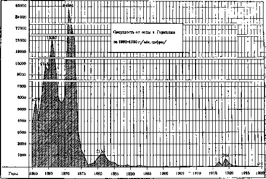
Figure 1.
In British India, smallpox is endemic and, until recent years, has not shown a tendency to decline. British India represents a type of country where smallpox is still left to its 'natural' course and where it produces regular rises and falls at certain intervals of years. Its rises are observed approximately every 5–6 years (Fig. 2). Table 5 presents the figures for deaths from smallpox in one of the provinces of India (in Bengal) for the period from 1883 to 1927. They are summarized by five-year periods. Table 5. Number of deaths from smallpox in Bengal from 1883 to 1927 (absolute figures). Years: 1883–87 — 9,896; 1888–92 — 22,759; 1893–97 — 23,509; 1898–1902 — 58,632; 1903–07 — 55,784; 1908–12 — 76,695; 1913–17 — 72,682; 1918–22 — 97,797; 1923–27 — 95,301. From these figures, it is evident that the number of victims of smallpox in India is growing with every five-year period. High mortality from smallpox is also registered in the Dutch and Portuguese Indies. In other colonial countries of Asia, smallpox epidemics also take on an extremely cruel character from time to time. In the Philippine Islands, for example, in 1919, 49,071 people died from smallpox, or 5.2 people per every 1,000 inhabitants. In Japan in 1928, the indicator
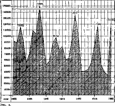
Mortality from smallpox in British India (abs. figures).
mortality from smallpox was 2, in Korea 4 per 1 million inhabitants. In Persia, from April to December 1929, 434 deaths from smallpox were registered. In Africa, smallpox is registered at high levels in Algeria, the Belgian Congo, Nigeria, Rhodesia, Sudan, and other colonies. In America, exceptionally high mortality rates from smallpox are registered in Mexico (in 1928—310, in 1929—358 per 1 million inhabitants). They are also high in Brazil. In the USA, the mortality rate from smallpox is low (1927—1 per million population), while the number of cases there is very high (see below). The prevalence of smallpox in those countries for which there is no exhaustive registration can be judged by the mortality from smallpox in large cities. Table 6 shows the mortality rates from smallpox in some cities in recent years (per 100,000 population).
Cities: Asia: Baghdad, Bombay, Delhi, Calcutta, Madras, Rangoon, Tehran. 25.6, 60.8, 36.4, 265.3, 5.6, 59.3, 8.4, 79.2, 42.2, 140.9, 49.9, 45.4, 103.8, 161.6, 18.4, 120.1, 147.0, 170.8, 34.6, 3.8, 13.2. Cities: Africa: Suez, Tunis. South America: Asunción, Bogotá, Guayaquil, Rio de Janeiro. Regarding smallpox in China, one can only judge by fragmentary materials. In Shanghai, in the International Settlement area, deaths from smallpox among Europeans were registered at 61.1 in 1928 and 39.5 in 1929; among the Chinese, 16.8 in 1928 and 19.8 in 1929 per 100,000 population.
2. Morbidity. Table 7 presents figures for smallpox cases in various countries for the period from 1919 to 1929. Table 7 gives an idea of the rise in smallpox associated with the imperialist war. Its epidemic in 1919 and the following years was observed, besides the USSR, in Bulgaria, Germany, Italy, Latvia, Lithuania, Poland, Romania, Finland, Czechoslovakia, Yugoslavia, as well as in most non-European countries (see also Tables 3 and 4). In the last decade, a major outbreak of smallpox was also observed in Switzerland, where it lasted from 1921 to 1925, and in Greece.
In the vast majority of countries, smallpox is gradually declining. The exceptions are the USA, where no downward trend in smallpox has been noted until recent years, and England, where the number of cases is growing every year. In Holland, where for a number of years smallpox was observed only in isolated cases, a major rise was noted in 1929 in connection with the disruption of proper vaccination. Austria, Bulgaria, Denmark, Norway, Finland, and Sweden have completely eradicated smallpox (since 1924, not a single case has been observed or only isolated cases have been noted), as have Australia and New Zealand among non-European countries.
Table 8 gives an idea of the intensity of the spread of smallpox in individual countries, showing the morbidity rates of smallpox for those countries where it has had a more or less strong spread in recent years. (It should be noted that the registration of smallpox in colonial countries, e.g., in India, is far from complete.) This table gives an idea of the maximum morbidity rate observed after the World War in different countries. In the USSR, it reached 30.0, in Romania 13.0, in Italy and the USA 10.0, in Korea 7.0, in Egypt 6.0, etc. In Germany, the maximum morbidity rate did not exceed 0.8 per 10,000 population. High morbidity rates in recent years have been observed in England, Canada, and the USA. Morbidity in them in recent years, in particular, is significantly higher than in the USSR.
It is characteristic to compare the trend of morbidity in recent years in the USSR, England, and the USA: while in the former the rate is gradually decreasing, in England and the United States of America, on the contrary, it is increasing from year to year (Fig. 5). According to materials published by the Health Section of the League of Nations, in 1929 and 1930, the total number of cases and deaths from smallpox registered was: [Table data omitted].
These data are inaccurate. For Europe, there is no information on the number of cases in Spain, or on the number of deaths in Italy, Lithuania, Finland, France, and the European part of the USSR. For Asia, there is no information on either cases or deaths for Afghanistan, Mongolia, China, and Persia. For Africa, information on cases in many countries is incomplete, and information on deaths is absent for most colonies. For America, there is no information for Argentina, Venezuela, Colombia, Peru, El Salvador, and small South American republics; for Chile, there is no information on the number of cases. For Australia, there were no cases in 1929–1930.
The following figures give an idea of the changes in the number of smallpox cases over the last 10 years: in 1920, in 30 countries of the world (excluding British India), for which information could be collected, 317,970 cases of smallpox were registered; in 1930, in the same countries, 64,356 cases of smallpox were registered; 131,676 people died from smallpox in 1920 in 33 countries (including British India), and in 1930, about 65,000 people died in the same 33 countries.
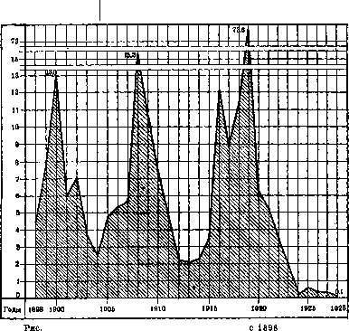
Smallpox in the USSR. Before the World War, there was no compulsory vaccination in Russia. Its improper implementation, the coverage of a relatively small part of the country's population, and the almost complete absence of revaccination were the cause of the extremely high morbidity of smallpox. Every year in Russia, about 100,000 cases of natural smallpox were registered (about 7 cases for every 10,000 population). At certain intervals—every 5–7 years—significant outbreaks were observed. After the revolution, the morbidity began to decline and dropped in the USSR in 1927 to 1.0, in 1928 to 0.7, and in 1929 to 0.4 per 10,000 population. Morbidity by five-year periods (on average per year) from 1891 to 1929 is shown in Table 9 (information for 1917 and 1918 is extremely incomplete and has been omitted). Figure 6 shows the morbidity in the USSR (Russia) by year from 1890 to 1929, calculated for each year per 10,000 population.
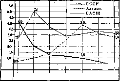
Table 9. Morbidity of smallpox in the USSR (Russia) from 1891 to 1929 (average per year). Years: 1891–95, 1896–1900, 1901–05, 1906–10, 1911–13, 1914–16, 1919–23, 1924–28, 1929. Absolute figures: 108,610, 115,851, 95,713, 128,779, 90,979, 102,438, 105,617, 34,680, 6,406. Per 10,000 population: 9.1, 8.9, 6.8, 8.4, 5.6, 8.1, 14.2, 1.2, 0.4. Fig. 5. Smallpox morbidity in the USSR, England, and the USA for 1923–29 (per 10,000 inhabitants).
rises in smallpox, and it took on the character of an epidemic. The number of cases increased to 150,000 or more, or up to 10-12 per 10,000 population. For the entire country, such epidemics lasted 3-4 years. This was the case, for example, in 1890-93, 1897-1900, and 1908-10 (Fig. 6). During the World War and the Civil War, vaccination was sharply disrupted, as a result of which an unprecedented rise in smallpox was observed. In 1919, according to incomplete data, the morbidity rate reached 30 per 10,000 population; in the following 2 years, it was 18 and 12 per 10,000 population. In 1919, the first decree on compulsory vaccination was issued, and in 1924, the second decree (see Vaccination). As a result of the systematic implementation of vaccination, smallpox began to steadily [decline]. Republics: BSSR, Azerbaijan, Armenia, Georgia, Turkmenistan, Uzbekistan, Tajikistan.
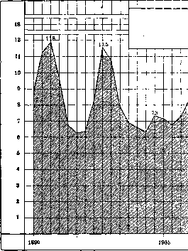
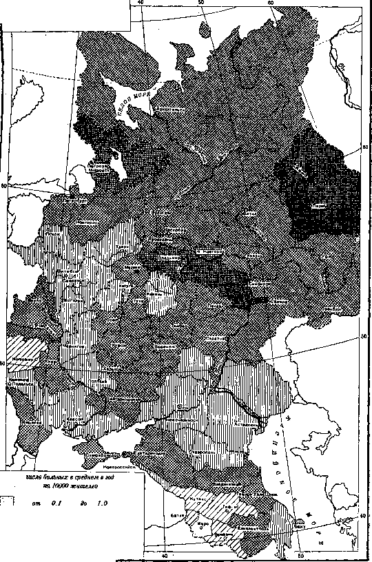
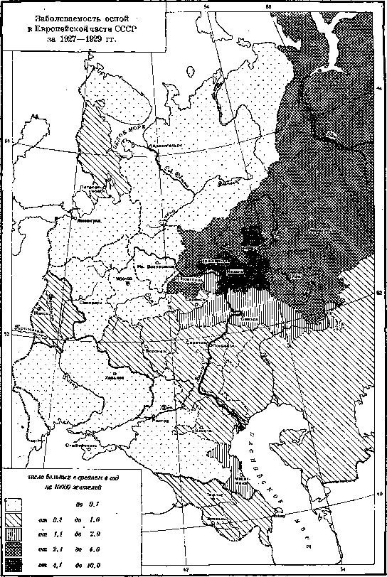
Figure 5. Smallpox morbidity in the USSR, England, and the USA for 1923-29 (per 10,000 inhabitants). 1891-95: 108,610 (9.1); 1914-16: 102,438 (8.1); 1896-1900: 115,851 (8.9); 1919-23: 105,617 (14.2); 1901-05: 95,713 (6.8); 1924-28: 34,680 (1.2); 1906-10: 128,779 (8.4); 1929: 6,406 (0.4); 1911-13: 90,979 (5.6). The decline of smallpox does not proceed at the same speed in all republics of the USSR. For individual republics, the morbidity figures for recent years are as follows (Table 10): Table 10. Republics, Absolute figures, Per 10,000 population. Smallpox continues to be registered most highly in Uzbekistan and Tajikistan; its morbidity is lowest in Ukraine. In the RSFSR, smallpox continues to be registered most highly in certain national regions where medical organization and vaccination are still organized most weakly. Table 11 shows the morbidity of smallpox by individual regions of the RSFSR (per 10,000 population). Figure 6. Smallpox morbidity in the USSR (Russia) from 1890 to 1929 (per 10,000 inhabitants). The comparative morbidity of smallpox by individual regions of the European part of the USSR (Russia) for 1911-13 and 1927-29 is provided. Of the individual regions of the RSFSR, smallpox is registered most highly in the Tatar and Bashkir Autonomous Republics, in the Votyak and Mari Autonomous Regions, and in the Chuvash Autonomous Republic. In recent years, smallpox in the USSR has been registered almost exclusively in rural areas. Table 12 gives an idea of the decline of smallpox in cities, where the rates of deaths from smallpox in the cities of Leningrad and Moscow for the period from 1881 to 1929 are given. Table 12. Mortality from smallpox in Leningrad and Moscow from 1881 to 1929 (average per year per 100,000 inhabitants). Before the World War, smallpox was observed annually in both of these cities. Epidemics recurred with a certain regularity. A sharp rise is noted during the World War, and especially during the Civil War. This epidemic reached its maximum in 1919: in Moscow, 1,747 people died of smallpox that year; in Leningrad, 2,085 people. Starting from 1924, smallpox, as a result of properly conducted vaccination, has practically disappeared in both cities. The isolated cases that are observed in them in recent years are almost exclusively among visitors. A complete absence of cases in recent years is also observed in a number of other cities (Figs. 7 and 8). In particular, not a single case of death from smallpox was registered in 1926-1928: in the RSFSR—in Arkhangelsk, Vologda, Ivanovo-Voznesensk, Zinovyevsk, Kiev, Kremenchug, Nikolayev, Odessa, Poltava, Kharkov, and Kherson; in Belorussia—in Bobruisk, Vitebsk, Gomel, Minsk, and Mogilev; in Transcaucasia—in Tiflis. Figure 7. Mortality from smallpox in Leningrad and Moscow for 1883-1929 (per 1 million population). Figure 8. Mortality from smallpox in Kiev and Odessa from 1900 to 1928 (absolute figures). Smallpox morbidity in the Russian and Red Army (per 10,000 personnel).

Figure 9.
Kaluga, Kozlov, Kostroma, Kursk, Orel, Pyatigorsk, Sevastopol, and Stavropol; in Ukraine—in Berdichev, Vinnitsa, Zaporozhye, etc. Smallpox decreased sharply in the army: in 1911, for every 10,000 personnel, 1.3 people contracted smallpox; in 1912—0.7 people; in 1929–1930 in the Red Army—0.03 people (Fig. 9). When studying the curves of smallpox morbidity or mortality from it over a long period of years, one can note a sharp change in their character after the introduction of mandatory smallpox vaccination in the USSR: before the World War, both in Russia as a whole and in individual provinces and cities, periodic rises and falls in smallpox morbidity were observed, which is characteristic of those countries where smallpox vaccination is carried out unsystematically and covers only a small part of the population. Starting from 1924, smallpox everywhere shows a sharp and persistent decline, and the periodicity of its rises disappears (Fig. 4, 7, 8, 10). 3. Morbidity and mortality from smallpox by age. Smallpox is registered mainly in early childhood, but in epidemic years it gives a relatively high [Table: Smallpox morbidity in the city of Moscow by age groups, Age, Abs. figures, Per 100 sick of all ages, Per 10,000 people of corresponding age 1917 1919 1920 1917 1919 1920 1917 1920 Under 1 year... 7.1 7.5 7.7 } 24.3 18.1 1-4 years 18.3 18.9 19.2 5-9 » 6.4 7.5 14.9 4.3 7.4 10-14 » 4.4 3.9 3.9 3.0 1.7 15-19 » 14.8 7.9 5.5 6.7 2.1 20-29 » 19.9 17.4 16.1 5.6 2.8 30-39 » 16.2 19.1 15.9 6.1 3.7 40-49 » 10.0 11.4 10.8 6.1 3.4 50-59 » 3.1 3.9 2.2 2.9 1.1 60 years and older 0.5 0.8 1.1 Unknown age 0.3 1.4 2.6 - - Total 3 254 100.0 100.0 100.0 6.2 4.1]
SMALLPOX (VARIOLA)
[Table 14. Mortality from smallpox in the city of Leningrad by age groups. Absolute figures, Per 100 deaths of all ages, Per 100,000 people of corresponding age. 1910, 1919, 1920, 1910, 1919, 1920, 1910, 1920. Under 6 months: 76, 97, 25, 15.3, 4.6, 7.0, 291.4, 411.4. 6-12 months: 65, 192, 17, 13.0, 9.2, 4.8. 1-4 years: 133, 635, 122, 26.7, 30.5, 34.2, 99.1, 374.7. 5-9 years: 40, 197, 37, 8.0, 9.4, 10.4, 28.2, 60.1. 10-14 years: 17, 43, 7, 3.4, 2.1, 1.9, 12.0, 9.7. 15-19 years: 71, 9, 4.6, 3.4, 2.5, 11.1, 13.1. 20-29 years: 192, 36, 15.0, 9.2, 10.1, 15.1, 27.3. 30-39 years: 51, 219, 29, 10.2, 10.5, 8.1, 14.9, 23.4. 40-49 years: 16, 233, 30, 3.2, 11.2, 8.9, 7.9, 30.3. 50-69 years: 2, 97, 17, 0.4, 4.7, 4.8, 1.9, 24.9. 60 years and older: 1, 73, 20, 0.2, 3.5, 5.6, 1.3, 54.2. Unknown: 36, 6, 1.7, 1.7. Total: 2,085, 27.0, 48.0.] morbidity and in older age groups. The corresponding figures, compiled from materials of the cities of Moscow and Leningrad, are given in Tables 13 and 14. In all cities of the European part of the USSR with a population of over 50,000 people in 1927, per 100,000 population of the corresponding age, deaths from smallpox were: under 1 year—43.0; from 1 year to 4 years—17.7; from 5 years and older—0.5. Where smallpox vaccination is carried out correctly, smallpox has ceased to be a "childhood" disease; individual cases are observed among adults who have not been revaccinated for a long time. 4. Lethality. Smallpox in the same years proceeds with unequal severity in different countries. In one and the same country, different lethality is observed for it in different years. If we summarize the number of all registered cases of smallpox diseases and deaths from smallpox for the period from 1921 to 1928, then for individual countries the following lethality indicators are obtained (Table 15). Table 5. A great influence on the level of the lethality indicator in one country or another is exerted by
10. Smallpox morbidity in the Moscow province (per 10,000 population).
Up to 1928. Fluctuations in lethality by individual years in %. British India: 610,335 cases, 208,823 deaths, 34.2% lethality, fluctuations from 23% to 53%. Korea: 18,778 cases, 5,531 deaths, 29.4% lethality, fluctuations from 19% to 31%. Italy: 6,472 cases, 22.9% lethality, fluctuations from 3% to 29%. Japan: 6,571 cases, 21.9% lethality, fluctuations from 14% to 24%. Egypt: 5,313 cases, 21.0% lethality, fluctuations from 14% to 29%. Romania: 3,750 cases, 19.4% lethality, fluctuations from 7% to 25%. Germany: 16.5% lethality, fluctuations from 0.1% to 59%. 104,161 cases, 1.1% lethality, fluctuations from 0.5% to 2%. England: 50,312 cases, 0.3% lethality, fluctuations from 0.2% to 2.8%. Switzerland: 5,493 cases, 0.3% lethality, fluctuations from 0.1% to 1.8%. (* 1926-28; ** 1921-27; *** 1925-27). The completeness of the registration of diseases and causes of death varies. Nevertheless, it is indisputable that in some countries in recent years smallpox has been proceeding with a very low lethality (England, Switzerland, USA). It should be noted that in the same countries before the World War, the lethality from smallpox was significantly higher: in England in 1911-13 it ranged from 7.4% to 8.7%, in Switzerland in 1909-12 from 7.1% to 14.3%. In the USA, along with very mild forms of smallpox, outbreaks with very high lethality are observed. Thus, in 1924-25 in Minneapolis, an outbreak of smallpox was observed, in which 1,430 cases and 365 deaths were registered; the lethality was therefore 25.5%. In the hospitals of the city of Leningrad (Petersburg) from 1886 to 1926, 23,442 smallpox patients were treated, of whom 5,740 died, or 24.5%; for individual five-year periods of this time, the lethality fluctuated from 21.9% to 31.0%; for the years 1922-26, it was 23.8% (Binshtok). The lethality from natural smallpox is not the same in different age groups. According to data from Leningrad hospitals, for the period from 1886 to 1909, the lethality from natural smallpox for different ages was: Table 16. Age: Under 5 years, 54.3% lethality; 6-10 years, 26.2% lethality; 11-15 years, 13.3% lethality; 16-20 years, 12.6% lethality; 21-30 years, 13.9% lethality; Older than 30 years, 18.3% lethality. The highest lethality is observed everywhere in the younger age groups. Lethality is not the same for different forms of smallpox. It is highest in its hemorrhagic form (see below - clinical aspects of smallpox). 5. Seasonality of smallpox morbidity. The monthly distribution of smallpox cases is not the same in different countries. In the USSR and other European countries, as well as in Canada and the USA, the maximum number of cases occurs in the first quarter, and the minimum in the third. In British India, the maximum number of cases is registered in the second quarter, and the minimum in the fourth. In Mexico, the maximum number of deaths from smallpox is registered in the third quarter, in Northern Rhodesia (Africa) in the fourth. Table 17 shows the figures for the distribution of smallpox cases by quarters of the year for some countries in different parts of the world. Table 17. Distribution of smallpox cases by quarters of the year in different countries (absolute figures). The indicated seasonal distribution of smallpox cases is explained by living conditions and increased contact among the population in certain months of the year. Figure 11 shows curves of the monthly distribution of smallpox cases in some countries (I. Dobreitser). IV. Pathological anatomy. Upon external examination of smallpox corpses, the first thing that catches the eye is the skin rash. In its typical and completed form, the smallpox rash has a pustular character, i.e., it represents foci of purulent inflammation with the exit of exudate into the thickness of the epidermis, with the separation and partial melting of the latter. Pustules are always preceded by a papular period, when there is no separation of the epidermis by exudate, and the whole matter is limited to acute inflammatory phenomena in the papillary and subpapillary layers of the skin. Microscopically, various changes are observed in the skin depending on the degree of maturation of the exanthema. In the initial periods, when the rash still has a papular character, inflammatory phenomena are found in the papillary and subpapillary layers of the skin, as well as swelling of the epidermis, with its cells taking on a spherical shape. Soon after this, the protoplasm in the epithelial cells takes on a reticular appearance, and later liquefies completely (reticulierende und ballonierende Degeneration; Unna); the complete liquefaction of such cells and the simultaneous transudation of serous exudate into the thickness of the epithelium leads to the development of a vesicle visible to the naked eye, initially with transparent contents; in such places, from the side of the epidermis itself, one notes discomplexation, i.e., its splitting, the formation of fissures or chambers in it. In these chambers, the liquid is under some pressure, as evidenced by the strong stretching of the preserved epithelial cells that delimit the chambers and run vertically from the surface of the pustule inward. It is precisely this tension and some inelasticity of these epithelial attachments that explain the slight depression of the vesicle from the surface in its central parts. In the neighboring parts of the epidermis, the epithelial cells undergo various degenerative changes, such as: vacuolization, pyknosis of nuclei, coagulation of protoplasm, and the formation of homogeneous and lumpy masses of detritus, etc. Subsequently, the transparent (serous) exudate that has arisen in the epidermis, and sometimes under it, is rapidly filled with leukocytes emigrating from the underlying tissues of the skin itself, and the vesicle, which was hitherto clear, becomes turbid, seropurulent, and finally purulent (approximately on the 9th-10th day of the disease). This suppurative period lasts for several days; the skin around it swells strongly, the face, for example, becomes unrecognizable. By the end of the second week, the formation of crusts begins, after the falling off of which pigmented, slightly depressed, rounded scars remain, which forever give the skin of these subjects, especially their faces, a characteristic appearance. It should not, however, be thought that smallpox pustules necessarily leave visible scars. Since the destructive process is concentrated mainly in the thickness of the epidermis and does not touch the skin itself (usually even the deepest cells of the germinative layer of the epidermis are preserved), all the prerequisites for healing without a trace are present; if, however, disfigurement by scars is often indeed observed (e.g., in the area of the nasal wings), this is explained by the depth of the suppuration itself, when there is a complete exposure of the papillae with partial melting of the papillae themselves. In these cases, the crust falling off the pustule exposes a defect of the skin itself, and this defect is covered with a new yellowish, already diphtheritic coating with subsequent formation of a scab and scar. These same phenomena of deep scarring can also be observed in varioloid and chickenpox (especially with repeated scratching), in which the just-described scarring is usually not observed. Eruptions are also observed on the mucous membranes [see separate table (Vol. XXII, pp. 439-440), Fig. 1] (mouth, nose, larynx, pharynx, esophagus, vagina, colon), but here the development of typical pustules usually does not occur, because due to the absence of a dense surface layer of epithelium, the contents of the vesicles quickly break through to the surface, and ulcers are formed. Sometimes ulcerations and necroses of the mucous membranes in smallpox are very extensive, which, however, may also be associated with a secondary infection coming from the mucous membrane itself. In rarer cases, an admixture of erythrocytes and even the prevalence of the latter are noted among the elements of the exudate, the so-called hemorrhagic or black smallpox, which is distinguished by the great malignancy of its course.
One should distinguish so-called purpura variolosa from hemorrhagic Smallpox; it proceeds as a severe, always fatal sepsis with vivid manifestations of hemorrhagic diathesis; the changes in the skin here have the character of hemorrhages with necrosis of the epidermis, but without the development of typical pustules (death in 1-2 days). Such cases can present great difficulties for differential diagnosis (in relation to hemorrhagic forms of sepsis), especially since the finding of streptococcus in them is not a rarity. Based on these findings, the opinion is even expressed that hemorrhagic forms of Smallpox are generally a product of mixed infection, which is clearly incorrect, since any infection can be hemorrhagic. Regarding the spleen, a significant enlargement is noted (3-4 times or more) due to congestion and hyperplasia of the pulp cells (with phenomena of myelosis), and regarding the parenchymal organs (kidneys, liver, myocardium)—various degrees of degenerative changes. Standing apart are the changes in the testicles, in which one often finds pictures of acute necrotic orchitis with sharp edema and disintegration of the tubule epithelium, sometimes over very large areas. Areas of serous-fibrinous infiltration with necrotization of formed elements are also found in the bone marrow (osteomyelitis variolosa). The question of so-called post-vaccinal encephalitis (encephalomyelitis postvaccinalis) deserves special attention (SEE Encephalitis).
I. Davydovsky. U. Clinic. The clinical picture of Smallpox presents an extremely great variety both in its course and in the manifestation of its individual symptoms depending on the severity of the infection and the organism's reaction to the introduced infectious agent. In some cases, we see very mild forms with an abortive manifestation of the symptoms characterizing the disease, in others, exceptionally severe ones, when death occurs before the manifestation of the typical signs of the disease. Between these boundaries, there exists a series of transitional forms. All cases of Smallpox can be reduced to three characteristic types, differing quite sharply from one another.
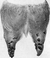
Each of these forms, in turn, can give deviations toward greater or lesser severity of the course. These forms are varioloid (variolois), natural Smallpox (variola vera), and hemorrhagic Smallpox (variola haemorrhagica). All forms of Smallpox are caused by one and the same virus, and their severity depends on the strength of the poison and the susceptibility of the organism. Infection from a mild form (varioloid) can produce a hemorrhagic one and vice versa. The strict regularity of the course of Smallpox allows one to distinguish the following stages of the disease: 1) latent (incubation), 2) initial (prodromal), 3) eruption, 4) irritation, 5) suppuration, 6) drying of crusts and scarring.
To describe the basic clinical picture, one can take a typical case of natural Smallpox. The incubation period lasts on average about 2 weeks. Characteristic for Smallpox in its initial period are considered pains in the sacrum and lumbar region. Sometimes they are so strong that the patient complains only of them, sometimes their intensity is not great and they are discovered only upon questioning. Some patients note constant pain, others discover it only upon movement. The pain is localized in the sacrum and the lower part of the lumbar region, sometimes spreading to the entire back and extremities. The causes of these pains lie, apparently, in hyperemia of the spinal cord and in the pressure exerted by blood-engorged vessels on the roots of the spinal nerves. These pains usually disappear with the appearance of the eruption. The temperature in the very first days rises rapidly to high limits and holds at constant figures (39-41°) for 4-5 days. The pulse quickens significantly, reaching up to 120 beats per minute and usually remaining satisfactory in fullness; cardiac activity shows no signs of weakening. The appetite disappears, the tongue becomes dry and coated. Catarrh of the fauces and tonsils is noted. Sometimes, especially in children, nausea and vomiting are observed. In the majority of cases, there is constipation. The spleen and liver are often enlarged. The amount of urine is decreased, its specific gravity is increased. The amount of urea, uric acid, and urates is increased; protein is often encountered. In women, the untimely appearance of menstruation is not uncommon. In the throat and nose, there is a sensation of dryness, sometimes nosebleeds. Breathing is quickened. Regarding the nervous system, besides the above-mentioned painful sensations, a phenomenon of confusion of consciousness, reaching a state of delirium, is sometimes noted.

On the skin, the eruption of a so-called prodromal rash is often noted; this rash appears most often on the 2nd-3rd day of the disease, lasts 1-2 days, and disappears by the time of the eruption of the actual smallpox rash, sometimes lingering into the first days of the eruption of the latter. Some authors (Trousseau, Filatov) consider the eruption of a prodromal rash a favorable omen, noting that its intensity is inversely proportional to the abundance of smallpox pustules; others (Auche) do not attach any prognostic significance to it. The frequency of the appearance of a prodromal rash is not the same in different epidemics: in some it is not encountered at all, in others rarely (2.4%), and in others relatively frequently (28.4%, Flerov). The character of this rash is quite varied. It can be in the form of a diffuse erythema or separate measles-like spots, predominantly on the chest and extremities, preferring their extensor sides; it can resemble a scarlet fever rash, having the appearance of a fine-spotted, sometimes petechial character. The localization of this form is observed predominantly in the so-called Simon triangle, i.e., it occupies the lower part of the abdomen and the inner surfaces of the thighs; in other cases, the rash spreads to the chest and lateral surfaces. Rashes of the character of urticaria or continuous (erysipelas-like) redness have been observed. A hemorrhagic prodromal rash usually takes place in so-called purpura variolosa. On the 4th-5th day from the beginning of the disease, the eruption of the actual smallpox rash occurs. The course of this period is strictly cyclic for this disease, which sharply distinguishes it from other diseases. The eruption usually begins on the forehead, spreading to the face, neck, trunk, upper and lower extremities. The rash appears in the form of red spots the size of a little more than a millet seed, rising above the skin, having a dark red shade. On the next day, protruding even more above the surface of the skin, it takes the form of papules surrounded by hyperemic skin, differing from measles by a denser consistency and a darker color. On the third day, a vesicle filled with light yellow serous fluid forms on the papule. This vesicle, in the further course, increasing in volume, can reach the size...
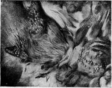
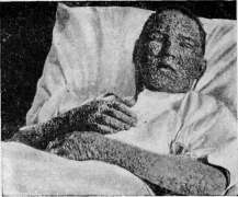
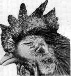
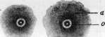
Figure 1. Degenerative changes of the epithelium during vaccination: a-reticular; b-ballooning colliquation. Figure 2. Goat pox on the udder (according to Zwick). Figure 3. Chicken pox (according to Zwick). Figure 4. Abbreviated scheme of the course of the vaccine process during vaccination and revaccination (according to Pirquet): 1-reaction during vaccination; 2-early reaction; 3-accelerated reaction; 4-control, a-areola, o-pock. Figure 5. Sheep pox; papules on the udder, on the anterior surface of the tail (according to Zwick). Figure 6. Confluent smallpox in the stage of suppuration (according to Jochmann).
size of a pea. Some of them remain round in shape, others form a characteristic depression, the so-called smallpox umbilication. On the 4th-5th day, the contents begin to cloud, turning into pus, and the vesicle transforms into a pustule, having the color of mother-of-pearl or a pearl. The development of the rash proceeds in a definite sequence, in accordance with its eruption: first macules on the face, and then on the entire body with a gradual transition into pustules and the formation of crusts, thanks to which one can observe simultaneously on the face pustules, and on the lower extremities papules, and sometimes macules. The skin around the smallpox eruptions is hyperemic, then becomes edematous, and the pustule sits on a sharply hyperemic base surrounded by a bright red rim (areola). The quantity of the rash varies from a few spots to a confluent rash covering the entire body; sometimes individual smallpox lesions merge, forming purulent blisters of greater or lesser size, in individual severe cases giving rise to entire purulent fields the size of a palm or larger. On the palms and soles, covered with a thick layer of cornified epidermis, the pustules form more deeply and during development sometimes cause painful sensations due to pressure on them from the horny layer. Simultaneously with the skin eruption, and more often preceding it, an eruption is observed on the mucous membranes. Hyperemia appears on the mucous membranes with separately protruding red spots, passing through the same stages of their development. The mucous and corneal membranes of the eye, the mucous membranes of the nose, mouth, fauces, pharynx, rectum, and vagina are affected. These eruptions cause corresponding disorders of the affected organs. Suppuration on the mucous membranes is accompanied by edema, which can cause a number of severe phenomena in patients. On the 9th-11th day of the disease, the drying of the smallpox lesions begins, proceeding from the face in the same order as the eruption occurred. Part of the pustules bursts, part is mechanically ruptured by the patients themselves, some dry up without opening. The pus that has flowed out from the opened papules dries, forming a crust of the color of pus, which subsequently takes on a dark brown color. On the palms and soles, upon drying, black-brown spots form under the layer of cornified epidermis. After drying, the crusts fall off, forming superficial scars in cases of shallow suppuration, and deeper ones in cases of deep suppuration. With damage and scratching of the pustules, even deeper scars are formed. On the soles and palms, the falling off of the crusts proceeds slowly due to the slow sloughing of the layer of cornified epidermis. The scars remaining after the sloughing of the crusts initially have a reddish-brown color, with the red color gradually disappearing and turning into brown, and the person recovered from smallpox is covered with such pigmented scars and spots. This pigmentation disappears after a few months, and the scars become white and, wrinkling, disfigure the skin, making it pockmarked. The duration of the drying period is from 19 to 25 days of the disease, and the falling off of the crusts from 25 to 35 and more (Flerov). With the appearance of the smallpox rash (on the 5th-6th day), the temperature usually drops to normal; in severe cases, it only gives a decrease, staying around 38°. In the period of suppuration, the temperature rises again and stays at high figures the whole time with significant remissions until the stage of drying, when it returns to normal, if there are no processes complicating the disease (Fig. 12). The general condition of the patient after the appearance of the smallpox rash and the condition of all organs improve significantly. In mild cases, patients feel almost recovered. The improvement in condition lasts until
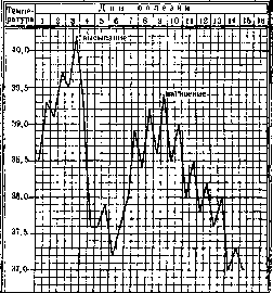
Figure 12. Temperature curve in smallpox.
the onset of suppuration, when the disappeared or weakened painful phenomena reappear. Heart activity weakens, the pulse quickens, becomes softer and irregular. The heart dilates, the heart sounds become muffled, and phenomena of myocardial degeneration develop. The appetite that had appeared disappears again. The tongue is dry and covered with coatings, as is the mucous membrane of the mouth. Swallowing and chewing are difficult, as a result of which salivation appears; thirst increases, urination decreases, and the urine possesses the properties of febrile urine. Constipation is observed more often, diarrhea less often. Pathological phenomena from the nervous system increase: headache, insomnia, delirium, and sometimes a comatose state. In the drying period, the painful phenomena begin to decrease, and in cases proceeding without complications, by the time the scabs fall off, the patient feels satisfactory. Depending on the severity of the course of the disease and the character of the rash, the described form can be divided into discrete smallpox (variola discreta), when the smallpox pustules sit separately from one another, and confluent smallpox (variola confluens), when the pustules merge with one another (see separate table, figure 6). With the second form of smallpox, a severe course is usually observed from the very beginning of the disease. The phenomena of the prodromal period are sharply expressed. With the appearance of the smallpox rash, the temperature, while decreasing, does not fall to normal. Due to the abundant eruption on the mucous membrane of the mouth, significant salivation is observed (Trousseau). The face and extremities show sharp edema. The process of eruption itself proceeds more slowly. In the drying period, part of the pus, flowing out from the bursting pustules, dries up, forming scabs, and part undergoes decomposition, emitting a sharp stench. When the process spreads over the entire body, the severely ill patient presents a 'terrifying appearance.' The patient is in a soporous or delirious state, the face is swollen, the eyes are closed and glued together by pus, and a foul-smelling liquid is discharged from the open mouth. The same discharge is observed from the blocked nose, which is impassable for breathing. The face, torso, and extremities are covered in places with foul-smelling scabs, and in other places with the same liquid pus. In many pustules, hemorrhages are noted (variola haemorrhagica). Cardiac activity decreases sharply, accompanied by phenomena of toxic myocarditis. Breathing is difficult due to the eruption in the nose and upper respiratory tract. The lesion of the nervous system takes the form of sharp depression or excitation, reaching the point of violent delirium. In relatively rare cases of recovery, the process of cleansing from scabs and scarring of deeply penetrating pustules proceeds slowly, and the temperature, usually falling lytically from the beginning of the drying period, slowly declines, bearing the character of hectic fever. Hemorrhagic smallpox (variola vera haemorrhagica). Hemorrhages accompanying the smallpox rash can be observed in any period of its development. All these cases belong to very severe forms of the disease, and the earlier the hemorrhage appears, the less hope there is for a favorable outcome. The form with hemorrhages in the prodromal period (purpura variolosa) leads rapidly (within 3-4 days) to a fatal outcome. These cases are encountered relatively rarely. Authors who found various microbes (most often streptococci) in the blood of those who died from purpura variolosa consider this form a mixed disease; others, who found the blood to be sterile, recognize these cases as a hypertoxic form of smallpox. Young, strong people are more often affected, and often those who have been vaccinated, whose organism has not only lost immunity but has also acquired an extremely high susceptibility to smallpox. After a shortened incubation period of 6-8 days, a stormy prodromal period ensues, accompanied by symptoms characteristic of the disease of natural smallpox. By the end of the first or on the second day, a scarlatiniform rash most often appears on the extremities, usually spreading over the entire body. On the 2nd-3rd day of the disease, hemorrhages into the skin and bleeding from the nose, lungs, intestines, bladder, and female genital organs occur. Sometimes one can note the eruption of a smallpox rash on the mucous membrane of the throat; such an eruption is usually not observed on the skin, because the patient perishes before the onset of this period due to phenomena of general intoxication and increasing decline of cardiac activity. In the hemorrhagic form of smallpox in the papular period (variola vera haemorrhagica papulosa), hemorrhage occurs into the freshly erupted smallpox rash. Variola vera haemorrhagica vesiculosa is characterized by hemorrhages into the contents of the vesicles. The most frequent form of hemorrhagic smallpox—variola vera haemorrhagica pustulosa—is accompanied by hemorrhage in the suppurative period. Depending on the severity of the case, the hemorrhage covers a larger or smaller part of the pustules, which take on a dark violet color and form dark brown scabs upon drying. In all these forms, as in purpura variolosa, phenomena of hemorrhagic diathesis are observed, and all kinds of bleeding can occur. The dark brown color of the affected skin in hemorrhagic smallpox has long given this form the name 'black smallpox,' which causes special horror of it on the part of the population. The mild (abortive) form of smallpox—varioloid (variolois)—is a disease of vaccinated individuals in whom immunity is still preserved to a certain degree, but is so weakened that it does not give the organism full protection against smallpox. This form is very rarely observed in the unvaccinated. In countries where vaccination is carried out, varioloid is the usual form of smallpox disease. The incubation and prodromal periods have the same course as in smallpox. On the 4th-5th day of the disease, a rash appears in a limited quantity (sometimes a few smallpox pustules); the temperature falls to normal, giving an increase only during subsequent complications, which are observed extremely rarely (Fig. 13). The rash sometimes turns into vesicles; extremely rarely, the contents of individual vesicles become cloudy. In the majority of cases, the smallpox pustules undergo regression, or the rapidly forming scabs fall off without leaving any scars. On the mucous membranes, catarrhal phenomena are usually noted; sometimes individual smallpox pustules are observed on the mucous membrane of the throat. This form provides, along with others, a lasting immunity, and for those around, it is just as contagious as the other forms, and in persons predisposed to smallpox, it can cause the most severe forms. The mild form also includes smallpox without a rash (variola sine exanthemate), which can be established either on the basis of anamnestic data (contact with a smallpox patient) or as a result of infection from this form of typical smallpox. Regarding changes in the blood, leukocytosis is noted, especially noticeable in the suppurative period, due to lymphocytes; the number of polynuclears is below normal. In the pustular period, Türk's irritation cells and basophils appear. The number of eosinophils is slightly increased above normal. According to Plavinsky's observations, fluctuations of leukocytes give two waves: the first in the initial period; it decreases, without reaching normal, by the period of eruption, and the second in the suppurative period; in this same period, Türk's irritation cells and normoblasts are encountered. Nägeli and others observed leukopenia in the initial stages, and leukocytosis subsequently, and believed that the smallpox poison causes leukopenia, and leukocytosis is the result of a secondary infection. Schilling considers hyperleukocytosis and neutropenia with a shift to the left and atypical mononucleosis to be characteristic of smallpox. Complications. Smallpox, as a toxic process, and subsequently a septic-pyemic one, can give all kinds of complications, which in the second half of the disease bear mainly a purulent character due to secondary infection by pyogenic microbes. The most frequent complications are lesions of the skin and subcutaneous tissue: abscesses, furuncles, phlegmons. The latter sometimes occupy extensive areas and penetrate deep into the subcutaneous layer. The formation of skin gangrene is observed. All more or less severe cases are accompanied by a lesion of the heart muscle with all the subsequent phenomena characteristic of acute myocarditis. Endo- and pericarditis are observed less often. Lymphadenitis is frequently encountered; abscesses in the liver and spleen are relatively rare. The respiratory organs give relatively frequent complications: edema of the larynx, simulating diphtheritic croup, retropharyngeal abscesses, bronchitis in almost all cases, and often catarrhal pneumonia. From the side of the digestive organs, abscesses of the tonsils, abscesses and phlegmons of the tongue, inflammation of the parotid glands, abscesses of the esophagus, purulent ulcers in the intestines, and peritonitis are observed. The genitourinary organs give lesions of the kidneys, starting with albuminuria and ending with severe nephrosis-nephritis, pyelitis, renal infarcts, and abscesses of the kidneys. In men, orchitis is encountered; in women, premature appearance of menstruation; and in pregnant women, premature birth. From the side of the sensory organs, a very severe lesion of the eyes is observed. A more or less sharply expressed conjunctivitis is a common companion of smallpox.
The development of the smallpox rash on the mucous membrane, causing a more or less deep lesion of the tissue, can also cause corresponding deformity of the eye. An eruption on the cornea can, upon suppuration, cause its perforation and lead to panophthalmitis, but even without such perforation, keratitis can entail iritis and chorioiditis with subsequent panophthalmitis. Inflammation of the middle ear is frequently observed, sometimes with involvement of the mastoid process. The nervous system is also involved in the process, giving rise to all kinds of sensory and motor sphere disorders in the form of neuritis, anesthesia, convulsions, and paralysis. Lesions of the brain and its membranes (myelitis, meningitis, encephalitis) are encountered. The psychic sphere also suffers. Besides the above-mentioned states of depression or excitation, sequential psychoses are observed, more often in the stage of drying of the rash or in the period of convalescence. Such acute psychoses usually pass without a trace. (Psychoses in smallpox—see Infectious psychoses.) Zenker's degeneration of muscles, purulent processes in them, and joint lesions are observed. Other infections sometimes join smallpox. Erysipelas and diphtheria are encountered more often than others. Other infectious diseases are observed less frequently. Any secondary infection, complicating the course of smallpox, thereby worsens the prognosis. Diagnosis of smallpox. In clearly expressed forms of smallpox, diagnosing the disease usually presents no difficulties, but in atypical cases and in varioloid, significant difficulties may arise. Diagnosis is especially difficult in the prodromal period and is not always easy in varioloid. To assist in the diagnosis of smallpox, as with other infections, the most thorough collection of the anamnesis of the given disease must come, with special attention paid to the presence of acute infections in the environment surrounding the patient, to the possibility of his contact with smallpox patients, or his stay in areas affected by this infection. The onset of the disease in a number of acute infections has much in common, and only the path of exclusion or the possibility of establishing contact of the sick person with a smallpox patient can lead to thoughts of smallpox infection. All complaints of patients and some signs characteristic of smallpox (pain in the sacrum and lower back) are not an exclusive symptom of this disease, being encountered in others as well. The only reliable sign must be considered the appearance of a prodromal rash, especially in the presence of a smallpox rash on the mucous membrane of the throat, usually preceding the skin eruption. In view of the variety of the character of the prodromal rash, it must be differentiated from the form to which it most closely approaches: measles, scarlet fever, erythema, toxic and medicinal eruptions. It is very difficult to distinguish purpura variolosa, which is a severe (hypertoxic) form of smallpox intoxication, from other septic skin hemorrhages. Diagnosis is aided by the presence of the main features of the smallpox disease, anamnestic data, and sometimes the presence of a specific smallpox lesion of the throat mucosa. Scarlet fever of a hemorrhagic character, in contrast to purpura variolosa, is excluded by the absence of angina and the "raspberry" tongue typical for scarlet fever. In the period of the eruption of the smallpox rash, confusion with other eruptive diseases is possible, although in this period, diagnosis becomes significantly easier. It is necessary to analyze all painful symptoms and anamnestic data especially carefully. Special attention should be paid to the time of eruption, localization, and character of the rash, comparing all these data with clinical phenomena, especially with temperature: The smallpox rash erupts on the 4th-5th day of fever, and with the appearance of the rash, a significant decrease in temperature is observed, sometimes to normal. The rash usually appears first on the mucous membrane of the throat, and then on the face, gradually moving to other areas of the skin. The rash undergoes a strictly cyclic evolution, characteristic specifically for this disease, and in each period of its development has a strictly defined appearance. Most often, one has to differentiate natural smallpox from chickenpox. Natural smallpox erupts on the 4th-5th day of the disease; in chickenpox, the rash appears almost simultaneously with the onset of the disease and spreads very quickly over the whole body, often affecting it more than the face; frequently in the following days, a repeated eruption is observed. The very appearance of the rash in these diseases has its own peculiarities. In chickenpox, the rash has the appearance of a transparent blister sitting on a red ring, and is similar to a blister from a burn by splashes of boiling water, whereas the smallpox vesicle is surrounded by a ring of infiltrated skin. Sometimes in severe forms of chickenpox, some blisters suppurate, and the formed pustules may have an umbilicated depression, which makes them very similar to smallpox ones, but the comparative speed (3-4 days) of the formation of these pustules and the polymorphism of the rash, when one can see simultaneously freshly erupted blisters, and suppurated ones, and ruptured ones covered with crusts, gives full opportunity for making a correct diagnosis. Temperature in chickenpox does not decrease upon the appearance of the eruption, as is the case with natural smallpox. It seems more difficult to make a differential diagnosis retrospectively in the presence of only crusts, especially in mild cases of smallpox and severe cases of chickenpox. In such cases, one has to take into account anamnestic and epidemiological data. Sometimes natural smallpox is confused at the beginning of the eruption with measles. The distinction serves as more sharply expressed phenomena of catarrh of the mucous membranes and the presence of Filatov-Koplik spots in measles patients and the eruption of a characteristic rash on the mucous membrane in smallpox. Temperature in measles eruption not only does not decrease, as is the case in smallpox, but on the contrary, often rises even higher. Smallpox can be confused with a syphilitic eruption. The main distinction of the syphilitic rash is its polymorphism and the property of syphilitic papules to often give peeling on their surface, which never happens in smallpox. Usually, a syphilitic rash is not accompanied by an increase in temperature, but individual cases accompanied by a febrile state can be misleading. In acutely proceeding acne syphilitica, there is no lesion of the throat mucosa; furthermore, the character of the temperature curve typical for smallpox draws attention; anamnestic data and enlargement of lymph nodes resolve the question, and the finding of syphilis spirochetes confirms the diagnosis. Various dermatites, bearing the character of papular, vesicular, and pustular, can give cause for confusion with smallpox, but the absence of typical fever, initial phenomena, and localization serve to distinguish them. Dermatites are for the most part limited to one part of the body; rare forms affecting the whole body do not possess the regularity in the distribution of the rash that is characteristic of smallpox and can serve as a sign for its recognition. Acne pustulosa occupies mainly the face and torso, and interspersed with pustules are nodules, in the center of which one can find a comedo. The so-called acne varioliformis, related to molluscum skin lesions, similar to a smallpox blister with an umbilicated depression, can be mistaken for smallpox. The absence of temperature and lesions of the throat mucosa, as well as the protrusion from these blisters when squeezed with nails of a sebaceous plug, followed by a glandular lobular formation, exclude smallpox disease. Medicinal rashes are for the most part not accompanied by fever, and if such does occur, it does not bear the form characteristic of smallpox, disappears upon cessation of the poisoning, and has different anamnestic data. Septic rashes, which can take the most diverse forms, usually give the most indefinite picture both in the form of the rash and its localization, and in the character of the temperature curve, which distinguishes them from smallpox. In doubtful cases of smallpox, the following methods can serve as auxiliary diagnostic methods: 1. Detection of Guarnieri bodies in the examined material or inoculation of the material onto the cornea of a rabbit with subsequent examination (see above). The disadvantage of this inoculation method is the troublesome microscopic examination with all the procedures for histological processing of the cornea, preparation of sections, etc. Besides this, a negative result does not exclude smallpox disease, since in 20% of cases the bodies are not detected. 2. Paul's method (see above). 3. Paschen suggests subjecting the contents of papules or vesicles to examination. In smears after appropriate processing and staining, elementary bodies can be detected. 4. Tieche, noting that in people possessing immunity to smallpox and vaccine, 4-7 hours after inoculation of the contents of smallpox pustules, an allergic reaction appears at the site of inoculation (redness and swelling), which is not obtained with other material, proposed using this phenomenon as a diagnostic method. Force and Beckwith proved that this phenomenon can be obtained in guinea pigs sensitized by subcutaneous inoculation of the vaccine (Morozov). The most suitable material for examination is young smallpox pustules. The smallpox pustule is opened with a sterile needle or scalpel and its contents are dried (without heating) in the form of a thick drop on a microscope slide, covered with another slide, and sent to the laboratory. Smallpox crusts are peeled off with a sterile needle and enclosed between two microscope slides.
All these methods are applicable from the first days of the eruption until the period of crust detachment, although the most distinct results are obtained before the pustular period. The prognosis in smallpox disease depends on whether the patient has undergone preventive vaccination or not, and the time elapsed since the last vaccination, in other words, depending on the state of the organism's susceptibility to the smallpox poison. Vaccinated individuals have a better prognosis. By age composition, children have a worse prognosis than adults (see statistics above). The shorter the time that has passed since a successful vaccination, the more favorable the prognosis. In unvaccinated individuals, the prognosis is very serious. Very often, severe forms develop in them. More severe forms also yield a more serious prognosis; thus, purpura variolosa gives an absolutely unfavorable one—mortality up to 100%. The same can be said about hemorrhagic forms in the papular stage; hemorrhagic smallpox, manifesting as such in the vesicular period, gives a somewhat better prognosis. Variola vera haemorrhagica pustulosa and the confluent form have a lethality of 50%. A relatively better prognosis is given by the discrete form of smallpox, although the percentage of lethality is still high. There are not enough grounds to establish a prognosis based on the phenomena of the prodromal period, since sharply pronounced symptoms can precede not only severe but sometimes also mild forms of smallpox. Mairinger notes that a measles-like rash is observed in milder cases, and a scarlet-fever-like one in more severe cases, especially when it spreads beyond the boundaries of Simon's triangle. The eruption period provides somewhat more grounds for prognosis. A decrease in temperature to normal, a sparse and rapid eruption speak for a mild form of the disease; incomplete fall in temperature, slow and abundant eruption give reason to assume a more severe form of the disease. Subsequent periods increase the validity of the prognosis; the suppuration period finally establishes the severity of the disease picture and makes it possible to finally clarify the prognosis. Of course, when clarifying the prognosis, not only the form of the disease must be taken into account, but it is necessary to consider the state of all organs, especially the cardiovascular system. The appearance of complications or the addition of other infections makes the prognosis more unfavorable. In varioloid, the prognosis is quite favorable, and if cases of death were noted, they depended not on the given disease, but on other underlying diseases from which the patient suffered. Complications in varioloid are observed very rarely. In all cases of prognosis, one should keep in mind and inform about the disfigurements and defects that may remain in those who have recovered from smallpox. Death can occur at different times of the disease: in hemorrhagic forms very quickly—on the 4th-5th day of the disease, in confluent forms—on the 10th-13th day. The onset of death is caused by toxic poisoning of the vascular and nervous system and toxic myocarditis. Later death occurs from complications due to damage to vital organs or subsequent sepsis and pyemia. Treatment. There is still no specific remedy for the treatment of smallpox. Attempts at vaccine therapy both in the initial period of smallpox and in the eruption period, despite favorable reviews by individual authors, must be considered unsuccessful. Experiments with treatment using serum from vaccinated calves and serum from calves that had recovered from smallpox (Auche) also did not yield success. Observations on the influence on the course of smallpox of serum from horses vaccinated with scrapings of calf pustules, ground in physiological saline solution, started in 1914 by Vlasyevsky at the Sokolnicheskaya (now Krasno-Sovetskaya) hospital in Moscow, were not finished due to wartime circumstances (Flerov). Treatment with red light (Finsen), which eliminates chemically active, skin-irritating rays, caused contradictory reviews, and in the end, it must also be considered ineffective. In the most severe form of smallpox—purpura variolosa—no therapeutic measures have any effect; the same can be said about cases of variola vera haemorrhagica papulosa. Thus, the whole matter of treating smallpox boils down to alleviating the patient's severe condition, with proper care, a correct regimen, and hygienic environment playing an exceptionally large role. The patient requires rest, bed rest in a well-ventilated room, an appropriate diet, monitoring of heart and bowel activity, and relief of various painful symptoms. In the initial period, the usual treatment for acutely febrile patients is carried out; ice on the head, sponging with water with vinegar or vodka; small doses of narcotic agents that act sedatively on headaches and lumbar pains are applicable. In case of severe nervous phenomena—warm baths, narcotic agents. In this period, cardiac activity usually does not yet require therapeutic intervention. Mild forms of smallpox, apart from care and hygienic maintenance, usually do not require treatment; in more severe ones, measures of general and local treatment are applied. The general treatment is the same as for other severe infections. Antipyretics have no significance; the best remedy is warm baths or cool sponging. Depending on indications, it is appropriate to use corresponding cardiac agents: caffeine, camphor, strychnine, digalen, adrenaline, etc. In order to raise blood pressure, periodic infusions of physiological solution, sodium chloride, or 10% glucose solution in small quantities (200-300 g), sometimes with the addition of adrenaline (10-15 drops of a 1.0:1000.0 solution). Regarding the respiratory organs, care for the nasal cavity is important, and great attention should be paid to the state of the pharynx and larynx. The nose must be cleaned, washed with disinfectant solutions, or applied in the form of nose drops; in case of strong swelling of its mucosa—infusion of a solution of cocaine and adrenaline. The pharynx must be rinsed or washed with disinfectant solutions. In the pustular period, edema can develop in the larynx, leading to stenosis and symptoms of croup with all its consequences. In view of the possibility of diphtheritic infection, in each such case, it is necessary to examine the laryngeal discharge for Loeffler's bacilli. In case of diphtheritic infection, the use of specific serum is necessary, and in its absence—the usual treatment for laryngeal edema: inhalations, expectorants, distracting external agents in the form of mustard plasters to the feet or mustard foot baths. In threatening cases—intubation, tracheotomy. Great attention must be paid to the care of the oral cavity: cleaning the tongue, teeth, oral mucosa in the form of wiping with an appropriate disinfectant solution, rinsing, washing. In case of dryness and cracking of the lips and tongue, lubrication with glycerin or almond oil is recommended. Bowel activity is regulated by enemas or mild laxatives. Bladder activity requires observation and regulation in the sense of taking appropriate measures in case of retention phenomena. Severe phenomena from the nervous system are alleviated by the use of sponging, warm baths, and various medicamenta nervina et narcotica (bromine, chloral hydrate, codeine, morphine, pantopon, sulfonal, medinal, veronal, adalin, luminal, etc.). Eye care requires exceptional attention, as eye damage in smallpox is one of the extremely severe complications that can lead to blindness. As a prophylactic measure
It is necessary to wash the eyes from the very beginning of the disease with weak solutions of disinfectants used in ophthalmic practice. According to data from the Red Soviet (formerly Sokolnicheskaya) hospital in Moscow, good results are obtained by instilling a 1.0:1,000.0 solution of methylene blue 3–4 times a day as eye drops. The use of these drops significantly reduced complications involving the eyes. Complications in other organs caused by the smallpox process are subject to appropriate treatment. Skin care during the illness and the treatment of skin complications are of great importance. As mentioned above, there is no agent capable of arresting or shortening the process of development and course of the smallpox rash and reducing scar formation. In the papular and vesicular period, local treatment is not required; in the pustular period, lukewarm baths or wet wraps alleviate the patient's severe condition and reduce (especially baths) the disgusting odor from him. After a bath, thorough drying of the skin surface without rubbing is required. Skin care during the suppuration period should be conducted according to the surgical method in the form of dressings—sterile dry or wet ones using Burow's solution or boric acid solutions, which can prevent the development of phlegmons. Some results in terms of alleviating the general condition are obtained from the use of mercury plaster or gray mercury ointment and rubbing with alcohol solutions, and it is necessary to monitor the lesions of the oral cavity particularly carefully, rinsing or wiping it with a solution of calcium chloride. Lubrication with boric vaseline and dry warm compresses alleviate the feeling of pain and tension. To relieve itching and the sensation of tension in the skin, it is recommended to apply glycerin, which also acts as a bactericide, or a solution of Kalii hypermanganici, and dusting with Zinc. oxydat. Disinfecting powders form thick crusts together with pus, under which pus accumulates, which often leads to the formation of phlegmon. To reduce pain in the limbs, warming compresses of disinfectant solutions are successfully used, sometimes cold lotions of the same. It should be strictly observed that patients do not scratch their rash, which contributes to the formation of abscesses and phlegmons, and upon healing leads to deeper, disfiguring scars. For children, and sometimes adults, it is necessary to apply dressings to the hands to avoid scratching. In mild forms, no special diet is required; it must be commensurate with the ability to digest and the action of the intestines. The more severe the case, the stricter the diet, and in very severe cases, nutrition should consist of liquid and semi-liquid, easily and well-digestible food. As the temperature falls and pathological symptoms disappear, it is necessary to gradually increase nutrition. Forms of diseases similar to smallpox: 1) Smallpox in Negroes usually does not differ in its course from that in Europeans. In Cameroon, Plehn observed two epidemics that stood out for their unusual picture: in the absence of prodromal and pustular
fever, the rash had the character of confluent smallpox with a very low mortality rate. 2) Sanaga smallpox. In 1900, in Cameroon, Plehn observed an epidemic disease accompanied by a prodromal period very similar to that of smallpox; after 3–8 days of a febrile state, a pustular rash erupted. A similar disease was observed in 1911 in Africa by Vorwerk. 3) Samoa smallpox, described by Prowazek, is similar in course and picture to mild forms of smallpox. 4) Alastrim (white smallpox), see Alastrim.
M. Kireyev. Prevention. The main measure in the fight against smallpox is properly organized vaccination (vaccination and revaccination). Its mandatory nature is established by the laws of all union republics (see Vaccination). Already now in the USSR, major successes have been achieved in the fight against smallpox (see above - statistics). For the near future, the task is set for its complete liquidation in the USSR. By a decree of the Council of People's Commissars of the RSFSR dated August 19, 1930, the People's Commissariat of Health and the State Planning Committee of the RSFSR are proposed, when revising the five-year health plan, to provide for measures ensuring "the complete and universal eradication of smallpox no later than the end of 1932." The Councils of People's Commissars of the autonomous republics, regional and provincial executive committees, as well as the People's Commissariat of Health of the RSFSR, are proposed by the indicated decree to "ensure measures for the fight against smallpox: 1) with the necessary credits from the local budget for the payment of vaccination personnel and their travel expenses, 2) with the necessary personnel by organizing short-term courses and using the industrial practice of medical faculty students, students of medical technical schools, as well as by all-out involvement of the Russian Red Cross in this work." According to the International Sanitary Convention signed in Paris on June 21, 1926, each government is obliged to immediately announce to other governments and simultaneously to the International Office of Public Hygiene the existence of a smallpox epidemic, indicating the locality where it appeared, the time of its appearance, the number of confirmed cases of the disease and deaths, and the measures taken. The Regulation on the Sanitary Protection of the Borders of the USSR provides for the mandatory notification of local sanitary institutions by medical personnel of every case of smallpox or death from it. A smallpox patient is immediately isolated (hospitalized). At the same time, smallpox vaccination is performed for all medical personnel and all patients of the medical institution where the patient is isolated. Vaccination is also performed for everyone who has come into contact with the patient. If smallpox has taken on the character of an epidemic outbreak (several cases of smallpox), then vaccination is performed for the entire population of the given area (universal vaccination). A smallpox patient is considered safe for others after the complete falling off of the smallpox crusts, but not earlier than 40 days from the onset of the disease. All secretions of the patient: sputum, mucus, urine, feces, as well as all items contaminated by them, are subject to thorough disinfection at the patient's bedside (concurrent disinfection). The room where the patient was located, as well as all items that were in this room, are subjected to terminal disinfection.
room. It is mandatory to completely exterminate flies in the room where the patient is located. In the area where smallpox has appeared, old clothing and rags taken out of this area are subjected to disinfection. Regarding goods and luggage arriving from abroad by land or sea, the International Sanitary Convention of 1926 provides, in the case of smallpox, for the disinfection of recently used undergarments, rags, clothing (consumer goods), recently used bedding, as well as rags not transported as wholesale goods. Regarding ships that either during the voyage or at the moment of arrival had a case of smallpox on board, the indicated convention provides for: 1) medical examination; 2) immediate disembarkation and isolation of patients; 3) vaccination of persons who, in the opinion of the sanitary authority, are not sufficiently protected by recent vaccination or a previous case of smallpox, and subsequent medical observation of them (up to 14 days); 4) disinfection of items (see above) and 5) disinfection of those parts of the ship in which smallpox patients lived and which, in the opinion of the sanitary authority, are considered infected.
I. Dobreytser. VI. Animal Smallpox. Cases of smallpox are encountered in cows, horses, sheep, chickens, and certain other domestic and wild animals (monkeys, pigeons). The fundamental question concerns the etiology of these diseases and the relationship between human and animal smallpox. Recent research has resolved this in the sense that a close genetic affinity exists between the smallpox of mammals, birds, and humans. At the same time, thanks to these studies, much clarity has been brought to the theoretical foundations of Jenner's doctrine (see Vaccination), practical methods of smallpox vaccination have been improved, the foundations of specific prophylaxis for animal smallpox have been developed, etc. The etiology of animal smallpox is poorly studied. The causative agents of sheep, horse, and chicken smallpox are better known than others. These are filterable viruses. Morphologically and tinctorially (by staining), they are indistinguishable from one another, as well as from Paschen bodies, which can serve as a prototype for them (see above, etiology of smallpox). These are very small, rounded, non-motile formations, 1/4 micron in size, occurring in enormous quantities in the contents of fresh eruptions in smallpox. Like Paschen bodies, they are stained only with the help of mordants (Loeffler's method of staining flagella, Fontana-Tribondeau silver staining) and are Gram-negative. In preparations, along with individual specimens, they are often found in the form of clusters or heaps, sometimes in the stage of division (diplococcus form). Cultures of these viruses on artificial nutrient media have not been obtained. The pathological-anatomical and histological picture of animal smallpox is diverse depending on the stage of the process, the properties of the pathogen, the individual and species characteristics of the sick animal, etc., and boils down mainly to focal inflammatory and proliferative phenomena in the epidermis and in the connective tissue base of the skin (and mucous membranes) with the formation of papules and vesicles. The changes in the epidermis common to smallpox (and vaccine) in humans (reticulating degeneration and Unna's ballooning colliquation) [see separate table (cols. 39-40), fig. 1] are encountered in a pronounced form only in sheep and partly in cows. Characteristic of animal smallpox is the involvement of the entire connective tissue base of the skin in the pathological process, from the subcutaneous tissue to the papillary layer. The capillaries and lymphatic vessels are dilated and strongly engorged; the base of the skin and the epidermis are permeated with white blood cells, among which multinuclear leukocytes, eosinophils, and lymphocytes predominate. Around the circumference of the pock, energetic proliferation of the epithelium occurs, which, together with the edematous infiltration of peripheral cells, is the cause of the umbilicated depression in the center (Delle). To resolve the fundamental question indicated above, particularly valuable conclusions were obtained by inoculating smallpox infection, specific to a given zoological species, onto another, whereby a number of conditions necessary for the success of this experiment were clarified. The virus must possess high virulence and be introduced in sufficient quantity. It is useful, and sometimes even necessary, to introduce an intermediate host (usually a rabbit). The first generation of smallpox on a new host is always accompanied by a weak, poorly specific reaction, and only after 2-3 new passages does a fully typical eruption develop. The failures and contradictory results of corresponding experiments noted in the literature are apparently explained by the non-observance of the above-mentioned conditions. The relationship between natural smallpox and cowpox has been studied with the greatest completeness. It has been proven that the variola pathogen, when transferred to a cow, turns into cowpox virus. The experiment is facilitated by the introduction of a rabbit as an intermediate animal. Usually, only on the 3rd or 4th passage does an eruption develop that is fully characteristic of rabbit smallpox (lapine), which is easily transmitted to a cow. In light of these data, the old doctrine of the existence of 'native' cowpox turns out to be untenable: the latter must be regarded as variolo-vaccina, and its pathogen as a variant of the natural smallpox virus that has lost its volatility and ability to cause general disease in humans, but without the loss of antigenic and protective properties. According to old observations, under natural conditions, this variant arises almost exclusively during the milking of cows—through the introduction of smallpox infection onto the skin of the udder and teats by the hands of milkers who have had natural smallpox (in a herd, as a rule, only dairy cows fall ill; bulls and heifers remain healthy). Besides the cow and rabbit, the human smallpox virus is transmitted to the monkey, horse, pig, sheep, and probably also a number of other mammals. Conversely, humans are apparently susceptible to all species of animal smallpox without exception, with the infection always being accompanied only by a benign local process. Under natural conditions, the danger of human infection with animal smallpox virus is quite limited. At the present time, when, under the influence of vaccination and other prophylactic measures, a significant decline and even complete extinction of smallpox epizootics is observed almost everywhere, only cases of the so-called Melker-knoten—'milker's nodules'—have retained practical significance. Equally rare are cases of animal infection with human smallpox. The close biological affinity between human and animal smallpox is proven with particular persuasiveness by the fact that, experimentally, strains with well-defined properties of cowpox have been obtained from all species of mammalian smallpox; in the very recent past, it has been possible to achieve the same success in relation to the virus of bird smallpox—the variety that has deviated most from the main type (variola hominis)—and through successful experiments (Blaxall, Sobernheim) with the transmission of alastrim (see) to a cow (via a rabbit), a bridge has been built between this benign variant of human smallpox and other species of animal and human smallpox. The variability characteristic of all microbes is unusually sharply expressed in the smallpox virus. Changes easily occur under the influence of the biological properties of the host. Thus, in humans, sheep, and monkeys, smallpox proceeds as a severe general infection with an abundant eruption over the entire surface of the outer integuments; the same occurs in goats and pigs, but the phenomena are less pronounced, and in horses and especially in cows, the disease, as a rule, manifests itself only as a benign eruption of a purely local character, and the general condition is almost unaffected. Cowpox—see Vaccine. Sheep pox (variola ovina) [see separate table (cols. 39-40), fig. 5], in terms of the severity of the disease, the abundance of eruptions on the skin and mucous membranes, and its epizootic spread, has a great similarity to human smallpox. After a 4-8 day incubation period, the disease begins suddenly with chills and fever up to 41-42°. On the 3rd or 4th day of the disease, a roseolar rash appears on the skin in the form of rounded spots, in the place of which nodules soon form, and then typical pearly-colored pocks with an umbilicated depression in the center. On the 8th or 9th day, their contents become cloudy, and they turn into pustules. The eruption is accompanied by severe catarrhal phenomena of the conjunctiva, the oral mucosa, and the upper respiratory tract. After 3 days, the process of regression of general and local phenomena begins. The pocks dry up and fall off after 6-8 days, leaving deep scars in their place. Similar to humans, mild forms of smallpox (ovina sine exanthemate) and very severe, malignant ones (ovina haemorrhagica, s. nigra, and ovina gangraenosa) are observed in sheep. Complications are most frequent in the lungs, the gastrointestinal tract, the genital organs (abortion), the nervous system, and the sensory organs. With the addition of secondary infection, pyemic processes arise. The average duration of the disease is 3-4 weeks. Mortality is up to 10-20%, and in severe epizootics, it reaches 50%. Infection occurs through the air (by droplet infection), and also apparently through objects contaminated with the smallpox virus. The sheep pox virus is very stable. When stored in a dark, cool place, it retains its virulence for 2 years. In 50% glycerin, the virus dies at 25° within 12 days. Diluted acids, iodine solution, and putrefaction quickly kill the virus. In dark stables, the virus retains its infectious properties for 6 months. Under natural conditions, the sheep pox virus is transmitted only to the goat and apparently to humans (a certain parallelism has been noted in the development of human and sheep smallpox by year). Experimentally, it is possible to transmit the sheep pox virus to the horse, donkey, cow, rabbit, pig, and chicken. Goat pox (variola caprina) [see separate table (cols. 39-40), fig. 2] clinically has a great similarity to sheep pox; in other cases, it takes the character of a local disease with localization on the udder. The incubation period is 6-7 days. The course is in the majority of cases favorable. Mortality usually does not exceed 5-15%. The duration of the disease is 2-3 weeks. The infection is transmitted by contact. According to most authors, caprina owes its origin to sheep and cow pox (Arnold). Transmission to humans is possible. Pig pox (variola suilla) affects young animals primarily. The appearance of pocks is preceded by fever (up to 41.5°), loss of appetite, discharge from the eyes and nose, and redness and swelling of the visible mucous membranes. The rash appears in the form of spots, then a nodule forms in the center of the spot, and after 2-3 days, a fully developed pock with an umbilicated depression appears. In addition to the skin, the rash can be localized on the mucosa of the mouth and lips, the trachea, and the bronchi.
Variola confluens and v. haemorrhagica are observed. Swinepox originates from human smallpox, cowpox, and perhaps also from goats. Horsepox (variola equina) is encountered very rarely. Clinically, it manifests as a disease of the mucous membranes (stomatitis equi pustulosa contagiosa) and as a skin disease. In the first case, after a 4-8 day incubation period, a spotted rash appears on the oral mucosa, which becomes swollen. Then, nodules form at the site of the spots, and slightly later, vesicles with clear or purulent contents. The pocks sometimes burst, turning into ulcers that heal quickly without scarring. The submandibular lymph nodes swell. The general condition of the animal suffers little. A slight increase in temperature occurs only at the beginning of the disease, during the eruption period. In the cutaneous form of smallpox, nodules, pustules, and ulcers are observed on various parts of the outer integument, especially often on the flexor surface of the fetlock joints. The appearance of the rash is preceded by redness and swelling of the affected skin surface. Mature pocks burst, the purulent secretion covering them dries up, forming scabs, upon the falling off of which recovery occurs. The disease, as a rule, proceeds favorably and ends within 10-14 days, rarely dragging on for up to 3-4 weeks. Horsepox has been known for a long time. Jenner knew about it, who apparently confused it with 'grease' (papulo-vesicular eczema on the flexor surface of the fetlock joints in horses) and, furthermore, mistakenly considered it the sole primary source of cowpox (the latter, as indicated above, always owes its origin to human smallpox). It is possible, of course, that the horse, as an animal susceptible to the vaccine, is sometimes an intermediate link in the spread of this infection among cows and humans. Cases of direct infection of horses with human smallpox have also been described. Fowlpox and avian diphtheria (epithelioma contagiosum et diphtheria avium), previously considered two different diseases, are now recognized as identical in etiology. 1. In epithelioma contagiosum, dense grayish-white nodules the size of a pea form on the comb, wattles, and beard [see separate table (col. 39-40), fig. 3]; later they acquire a grayish-red color and a pearly luster. Clusters of nodules resemble warts or mulberry-like growths. Their content is a fatty, mushy mass of yellow color. Usually, the edges of the eyelids, the conjunctiva, and the cornea are severely affected by the pathological process, which can lead to panophthalmitis. From the corner of the beak, the rash sometimes spreads to the oral mucosa. In other cases, it is also observed on the feathered areas of the skin of the neck and head. In its pure form, epithelioma contagiosum runs a benign course. Birds lose weight, but their general condition suffers little. Within 3-4 weeks, the disease ends in recovery. 2. Diphtheria avium is characterized by acute croupous-diphtheritic inflammation of the mucous membranes of the head. Initially, small, sharply defined round or oval spots of yellowish-white color are discovered. Merging with each other, they form a pseudomembranous coating of various shades of white, yellow, and brown on the mucosa. Simultaneously, disorders of breathing and swallowing arise. Upon removal of the coating, a red, easily bleeding ulcer remains. The disease is often complicated by catarrh of the nasal mucosa, croupous pneumonia, keratitis, and panophthalmitis with subsequent atrophy of the eyeball. An increase in temperature is usually observed towards the end of the disease. Death occurs due to exhaustion. The incubation period is 6-8 days, the duration of the disease is from 2 to 3 weeks, sometimes 1-3 months and longer. The lethality reaches 50-70%. Simultaneous cases of epithelioma contagiosum and diphtheria avium are observed. The pathogen is distinguished by great stability: in dry scabs, it remains virulent for up to 1.5 years, and it is preserved in glycerin for months. The spread of the contagion occurs through drinking water, feed, and poultry house premises contaminated by nasal secretions and particles of smallpox scabs. Apparently, intermediate carriers also play a certain role. Besides chickens, the disease is common among geese, ducks, turkeys, guinea fowl, pigeons, and other wild and domestic birds. According to old literature, smallpox epidemics among humans sometimes owed their origin to diseased chickens. In the Middle Ages, the belief in the identity of human and bird smallpox was widespread (Arnold). Immunity in animal smallpox is little studied. Given the close biological affinity between human and animal smallpox, the antigenic and protective properties of the viruses of these diseases differ little from each other. Thus, in the presence of cross-immunity between natural smallpox and the vaccine, the latter protects sheep, goats, and birds against the smallpox infection specific to these species. However, there are deviations from this rule, caused by the sharply expressed biological variability of the smallpox virus. Negligible individual characteristics of the host—even within the limits of one and the same zoological species—are sometimes the cause of the emergence of new races of the smallpox virus (especially in the organism of birds), and these biological changes are often accompanied by a weakening or even loss of immunizing properties. This undoubtedly explains, in part, the contradictory results in cross-immunization with pigeon and chicken smallpox viruses. It is also known that the vaccine protects against natural smallpox in 100% of cases, while natural smallpox protects against the vaccine in only 60%. In the organism of animals with smallpox, shortly after the onset of the disease, virucidal, complement-fixing substances, agglutinins, and precipitins (predominantly in the form of thermoprecipitins) are discovered. Thus, the essence of immunity in animal smallpox is the same as in human smallpox—it is a general, predominantly humoral immunity, in which organs (especially the bone marrow) and blood participate. The duration of immunity ranges from several weeks to 9 months (rabbit), reaching up to 10 months in birds and up to 1 year in horses. The fight against animal smallpox is conducted almost exclusively through specific prophylaxis (active immunization with live attenuated or sensitized viruses, simultaneous vaccinations, i.e., the simultaneous introduction of a protective or therapeutic serum and a live virus, etc.). Passive immunization for prophylactic purposes through the introduction of therapeutic sera is used relatively rarely and is considered not very reliable. M. Morozov.
Related articles
Cite this page
“Smallpox (Variola).” Soviet Medical Encyclopedia. English translation of Bolshaya Meditsinskaya Entsiklopediya, 1st ed. (Moscow, 1928–1936), ed. N. A. Semashko. https://sovietmedicalencyclopedia.pages.dev/article/smallpox/