Encephalitis
Historical document, translated for reference. It reflects medical knowledge of the 1920s–30s and is not medical advice.
Summary
This article from the 1928–1936 Soviet Great Medical Encyclopedia defines encephalitis as an anatomical-clinical symptom complex caused by various etiological factors, including infection, intoxication, and trauma. It distinguishes between primary and secondary forms of the disease, noting that the division is often conventional, and discusses the role of the blood-brain barrier in the development of toxic encephalitis.
Encyclopedia article (1928–1936)
Encephalitis, inflammation of the brain, is an anatomical-clinical symptom complex caused by various etiological factors: infection, intoxication, and trauma. The significance of each of the aforementioned factors is assessed differently, and the role of intoxication and trauma as causal moments in the pathogenesis of the encephalitic process is disputed by the majority of authors. The first stage in the etiological differentiation of encephalitis is the distinction between an encephalitic disease and an encephalitic reaction. An encephalitic disease is an etiological form with a definite anatomical substrate, clinical picture, and course. An encephalitic reaction, morphologically identical under different etiological factors, arises primarily or secondarily; in the latter case, it occurs on the basis of some underlying process in the nervous system (tumors, vascular disorders, etc.). Thus, an encephalitic reaction is only part of the underlying anatomical-clinical symptom complex and has no independent significance. In cases where the encephalitic disease dominates the main part of the anatomical-clinical picture, it is considered a primary encephalitis, such as, for example, epidemic encephalitis, polioencephalitis, etc. In another series of cases, encephalitis complicates an underlying disease—foci of local infection, endocarditis, suppuration, etc.—as a result of which secondary localizations of infection arise in the central nervous system—secondary metastatic encephalitis. Very close to these latter are encephalitic complications in general infections. There are no fundamental differences between primary and secondary encephalitis; this division is to a significant degree conventional, since secondary encephalitis, such as colibacillary, influenzal, etc., can proceed morphologically and clinically like primary encephalitis, in which all indicators of the underlying disease are reduced and phenomena from the nervous system with the character of an encephalitic disease come to the fore.
In morphological terms, the differences that exist between primary and secondary encephalitis are determined by the character of development and the dynamics of the pathogenetic process, and not by the essence of the latter. Primary infectious encephalitides are in the vast majority of cases caused by neurotropic filterable viruses—epidemic encephalitis, poliomyelitis, rabies, typhus encephalitis, etc. Primary encephalitides possess the properties of contagiousness and epidemic spread in the presence of appropriate conditions in the external environment, susceptibility of the macroorganism, and properties of the microorganisms. Secondary encephalitides are caused by diverse microorganisms; they complicate general infectious diseases or local infectious processes in the lungs, intestines, heart valves, and other internal organs during the generalization of the process or by a metastatic route—mycotic emboli. Thus, secondary encephalitides differ from primary ones by the presence in the organism of a primary localized focus or generalized infection, the absence of contagiousness, the absence of epidemic spread of the infection, and partly by the anatomical-clinical picture, which in the majority of cases has a focal character, with microorganisms very often being discovered in the foci. According to the character of the pathomorphological process, non-purulent and purulent infectious encephalitides are distinguished, with primary encephalitides belonging exclusively to the first, non-purulent group, while secondary encephalitides can be both purulent and non-purulent. According to the dynamics of the pathogenetic process, primary encephalitides are divided into acute and chronic; secondary encephalitides are most often acute.

The problem of primary toxic encephalitis has not yet been solved until recently. The majority of authors consider toxic encephalitis to be secondary, reactive, occurring on the basis of vascular disorders caused by the action of toxins on vascular walls, as a result of which necrosis of the nervous tissue develops with subsequent reactive processes. Some authors, including Spielmeyer, allow for the possibility of the development of primary toxic encephalitis in a limited number of cases and under special conditions. The occurrence of toxic encephalitis is determined by the functional state of the blood-brain barrier, which facilitates or, conversely, hinders the penetration of toxins into the nervous tissue. In this regard, the methods or pathways by which toxins penetrate the nervous system are of great importance. In cases where toxins penetrate bypassing the blood-brain barrier—endoneurally or directly into the cerebrospinal fluid or into the nervous tissue—toxic encephalitis constantly develops. The introduction of the same toxins paraneurally, i.e., through the mouth, intravenously, or under the skin, does not cause encephalitis. Any mechanical damage to the central nervous system primarily affects the innervation of the vessels, causing spasm of the latter with phenomena of prestasis and stasis, as a result of which ischemic necrosis of the nervous tissue and a reparative encephalitic reaction develop. Furthermore, in the case of trauma, the permeability of the vascular-endothelial barrier changes, as a result of which the accompanying infection penetrates into the nervous tissue and causes an encephalitic reaction.
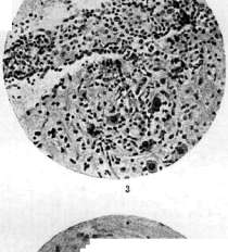
Figure 1. Hemorrhagic encephalitis: perivascular infiltrate of histioid mesenchymal-glial cells; epithelioid, plasma cells, plasmablasts. Figure 2. Hemorrhagic encephalitis: perivascular hemorrhages; degeneratively altered vascular walls. Figure 3. Hemorrhagic encephalitis: vascular thrombosis (white thrombus). Figure 4. Epidemic encephalitis (Economo type): vascular-inflammatory reaction, glial proliferation. Figure 5. Encephalitis (Economo type): perivascular infiltrate of lymphoid character. Figure 6. Lethargic encephalitis: nodular glial reaction. Thus, intoxication and trauma, although they can cause primary encephalitis under special conditions, nevertheless, their role is mainly predisposing and facilitating the penetration of infection into the nervous tissue. Factors contributing to the occurrence of infectious encephalitis are all those factors which, by changing the reactive state of the organism, influence the permeability of the blood-brain barrier (infection, intoxication, physical and mental trauma, diseases of the vascular system, nutritional disorders, overwork). From a pathomorphological standpoint, acute non-suppurative encephalitis is expressed by an active vascular-inflammatory process with alterative and proliferative changes in the ecto-mesodermal elements of the nervous tissue. The cells of the vascular infiltrate consist mainly of free histioid mesenchymal (including epithelioid cells) and glial cells with an admixture of hematogenous extravasate elements (see separate table, Fig. 1). In chronic forms of encephalitis (neurosyphilis, tuberculosis), proliferative processes in the glial tissue predominate (glio-granulomas, active and replacement gliosis). The process in encephalitis is localized mainly in the gray matter of the cortex, subcortical ganglia, and brainstem nuclei (epidemic encephalitis, polioencephalitis, rabies, typhus encephalitis, herpetic encephalitis, Borna disease in horses). Another group consists of encephalitis with predominant localization in the white matter of the brain—focal encephalitis (Strümpell-Leichtenstern type), encephalomyelitis, allergic encephalitis—post-vaccinal, measles, etc. The encephalitic reaction has a diffuse character in some cases and a focal character in others. The encephalitic process can selectively affect individual parts of the central nervous system—the cortex and subcortical parts (Strümpell-Leichtenstern type encephalitis), the midbrain, and the region of the third ventricle (Economo type encephalitis). The route of virus spread in infectious encephalitis is twofold: hematogenous and lymphogenous. Primary encephalitis is a general disease of the entire organism, with the process predominantly localized in the central nervous system; therefore, at the beginning of the disease, the infection of the organism is always generalized and hematogenous, while the lesion of the nervous system is carried out mainly by the lymphogenous route. With hematogenous spread of the virus, infection of the central nervous system occurs only on the condition of a breach of the blood-brain barrier; with lymphogenous spread, the lesion of the nervous system occurs bypassing the latter, along the perineural spaces of peripheral nerves, roots, and along the subarachnoid spaces of the spinal cord. The causative agents of primary infectious encephalitis penetrate the body through mucous membranes (nasopharynx, intestine, urogenital organs) or damaged skin (rabies). Acute primary encephalitis is observed in the form of epidemics (epidemic encephalitis, polioencephalitis) or in the form of sporadic cases. The main pathomorphological type of acute infectious encephalitis is focal infiltrative encephalitis, which is caused by a local partial breach of the blood-brain barrier during hematogenous spread of the virus and is expressed by a normergic reaction of the nervous tissue. In some cases, due to sensitization of the nervous tissue, which occurs during the initial hematogenous generalization of the infectious process, the normergic reaction of the nervous tissue passes into a hyperergic one with a predominance in the picture of encephalitis of a hemorrhagic component and extensive necrosis with simultaneous suppression of exudative processes. In diffuse forms of primary infiltrative encephalitis (epidemic encephalitis of the Economo type, herpetic encephalitis, polioencephalitis, rabies, etc.), there is a widespread breach of the permeability of the blood-brain barrier with a hyperergic reaction of the nervous tissue, expressed mainly by exudative and, to a lesser extent, proliferative changes. Selective involvement in these forms of individual parts of the nervous system is determined by the affinity of the virus, the nature of the vascular supply of the given area, and the functional state of the barrier and neurogenic mechanisms. Upon desensitization, the hyperergic reaction of the nervous tissue passes into a hypoergic one, which is characteristic of allergic encephalitis (post-vaccinal, measles, etc.). In such cases, the vascular-inflammatory reaction is completely absent or very weakly expressed, giving way to active proliferation of glia and focal perivascular and widespread degeneration of the nervous tissue. A natural classification of infectious encephalitis should be based on the etiological principle, although the latter is not always possible to carry out, since not all causative agents of various forms of encephalitis are sufficiently known. The classification scheme of encephalitis given below groups individual etiological forms of encephalitis—primary viral encephalitis; allergic encephalitis is distinguished into a special group, based on the nature of the nervous tissue reaction; and encephalitis during general infections of the organism and metastatic encephalitis are placed in a separate group. I. Primary viral encephalitis: 1) Epidemic encephalitis: a) lethargic encephalitis (Economo type), b) focal encephalitis (Strümpell-Leichtenstern type). 2) Encephalitic form of poliomyelitis. 3) Herpetic encephalitis. 4) Typhus encephalitis. 5) Rabies. 6) Epidemic encephalomyelitis. 7) Japanese encephalitis. 8) Psittacosis. II. Allergic encephalitis: 1) Post-vaccinal encephalitis. 2) Measles encephalitis. 3) Encephalitis after chickenpox. 4) Encephalitis after mumps. 5) Encephalitis in chronic sepsis. III. Secondary encephalitis in general infections and metastatic encephalitis. I. Individual forms of primary encephalitis—1) Acute focal hemorrhagic encephalitis (Strümpell-Leichtenstern type) is observed epidemically and sporadically. The presence of lethargic forms ('nona') during the epidemic of hemorrhagic encephalitis in 1890, combined and transitional forms, speaks for the closeness or identity of the causative agents of hemorrhagic and lethargic encephalitis and allows them to be combined into one form of epidemic encephalitis. The difference in tissue reaction in both cases depends on the reactive state of the macroorganism and changes in the properties of the virus. Sporadic cases of hemorrhagic encephalitis can be caused, in addition to a filterable virus, by various banal bacteria penetrating the central nervous system during general infections or local foci. In the pathomorphological picture of the hemorrhagic form, alterative changes predominate—hemorrhages, softening (see separate table, Fig. 2). Symptomatology. The onset of the disease is apoplectiform; sometimes there are prodromal symptoms: general cerebral phenomena, high temperature, cerebral paralysis, epileptiform seizures, violent movements, static and dynamic ataxia, aphasic and gnostic-apractic disorders. Diagnosis. Sinus thrombosis and sinus phlebitis differ from focal encephalitis by the absence of focal deficits, the presence of a source of thrombosis or sinus phlebitis (local foci), edema of the face and scalp, and congestive phenomena in the fundus of the eye. Encephalitis differs from brain abscess by the absence of a source of abscess, polymorphism, and a constantly increasing picture of the disease, and the preservation of consciousness in abscesses. Encephalitis differs from brain tumor by the absence of general cerebral phenomena, changes in the fundus of the eye, the course of the disease, and the multiplicity of symptoms. Encephalitis differs from cerebral hemorrhage, embolism, and thrombosis by the polymorphism of the picture, the multiplicity of foci, the febrile onset and progressive course of the disease, and the absence of etiological moments causing vascular lesions (old age, arteriosclerosis, syphilis, heart valve lesions). The comatose form of encephalitis is differentiated from uremic and diabetic coma. Encephalitis differs from brain syphilis by the absence of positive serological reactions in the blood and cerebrospinal fluid. Treatment of primary forms—see treatment of epidemic encephalitis; for secondary forms—treatment of the underlying disease, lumbar punctures, intravenous administration of urotropin. Wernicke's superior hemorrhagic polioencephalitis (very often developing on the basis of alcoholism) and inferior polioencephalitis represent a special localization of hemorrhagic encephalitis (see Polioencephalitis). 2) Acute epidemic encephalitis (Economo type)—see below Epidemic encephalitis. 3) Acute and subacute encephalomyelitis. Symptomatology. The onset of the disease is acute, amidst full health. Temperature is normal or subfebrile. Paresis and paralysis of the cranial nerves and limbs of a central and radiculo-neuritic character, sometimes ataxia in the limbs. Tonic and pain reflexes in the lower limbs. Tendon reflexes are increased; sometimes there are reflex-spastic phenomena and pathological reflexes; in rare cases, sphincter disorders.
Sensory disorders of the radicular or spinal type are observed, as well as local tenderness of the nerve trunks. There is mild pleocytosis in the cerebrospinal fluid, and in some cases, protein-cell dissociation; colloidal reactions are often positive. Pathoanatomically, in the brain and spinal cord, mainly in the white matter, there are scattered foci of vascular inflammation with a predominance of glial proliferation, relative preservation of the axis cylinders, and perivascular foci of demyelination of nerve fibers. Encephalomyelitis is caused by various pathogens from the group of neurotropic filterable viruses. The course of the disease is acute or subacute with remissions, more rarely gradual; in severe cases, it is a steadily progressive course. It must be differentiated from cerebrospinal syphilis, polyradiculitis, and acute multiple sclerosis. Encephalomyelitis differs from acute multiple sclerosis by the atypia of the multiple sclerosis symptom complex, the presence of pain and tonic reflexes, sometimes sensory and sphincter disorders, and the presence of epidemic outbreaks of encephalomyelitis, which are not encountered in acute multiple sclerosis. For treatment, see below: Epidemic encephalitis. 4) Herpetic encephalitis. In animals, the fever blister virus easily causes the picture of acute encephalitis in experiments; in humans, individual cases of herpetic encephalitis have been described. Ravaut, in 21 out of 26 patients with genital herpes, found changes in the cerebrospinal fluid in the form of pleocytosis. All described cases of herpetic encephalitis had a favorable course and outcome; no amyostatic stage was observed after them. Apparently, herpetic diseases, including those of the nervous system, are possible without eruptions, which complicates and sometimes makes etiological diagnosis of this disease impossible. 5) Typhus encephalitis. Cerebral phenomena develop during the febrile period of the disease, less often after the temperature drops, and as an exception, during the recovery period. The disease begins with a rapid rise in temperature, reaching high figures, often with chills, asthenia, pain in the limbs, headaches, dizziness, insomnia, enlargement of the spleen, increased pulse rate, and a drop in blood pressure. Simultaneously with the appearance of the rash, the following develop: a) general cerebral phenomena, b) focal symptoms, c) meningeal symptoms. Then, increasing clouding of consciousness appears with psychomotor agitation, coma, delirium, hallucinations, aggressiveness, cataleptoid states, tremors, choreic movements, tonic convulsions and contractures in the limbs, paralysis (hemiplegia), epileptiform convulsions (partial), bulbar phenomena, and disorders of respiration and hearing. Meningeal phenomena develop, and inflammatory changes in the fluid (lymphocytosis), the Weil-Felix reaction, and the hemolysin reaction in the cerebrospinal fluid are noted. Pathoanatomical changes: serous meningitis (lymphocytes, plasma cells, macrophages, mast cells). In the nervous tissue, there are foci of gliogranulomatosis, destructive thrombocapillaritis with a perivascular reaction of macroglia, microglia, and mesenchymal elements. Along the course of mainly venous vessels, single- and multi-nucleated cuffs form from lymphoid and plasma cells, with degeneration of nerve cells and nuclei. Inflammatory changes in typhus are localized mainly in the gray matter of the cortex and basal ganglia; in the spinal cord, foci of gliogranulomatosis are found mainly in the white matter. The greatest number of foci is found in the medulla oblongata in the region of the X, XI, and XII nuclei and the olives, in the vicinity of the Sylvian aqueduct and the molecular layer of the cerebellum. The formation of nodules is caused by primary damage to the vessels; parasites (rickettsiae) are found in the endothelium of the latter. Gliogranulomatosis and perivascular cuffs develop at the end of the first week, after which their regression is observed. 6) Trypanosomal encephalitis (tropical sleeping sickness) (see Sleeping sickness). The course is with remissions; the total duration of the febrile period is from several weeks to several years, and remissions last for months. Symptoms of the disease in the febrile period include enlargement of the lymph nodes, edema, erythema, increased pulse rate, and trypanosomes in the blood. Changes in the cerebrospinal fluid include pleocytosis; clinically, there are transient facial nerve paralysis, tremors, disorders of statics and gait, depression or psychomotor agitation, and cachexia. The second stage of the disease is characterized by drowsiness, gradually passing into deep sleep, and paralysis of the limbs and sphincters. Pathoanatomical changes include encephalitis and diffuse leptomeningitis. 7) Japanese encephalitis. An epidemic took place in Japan in the summer months of 1924. It began with a high temperature, a soporous or comatose state, delirium, agitation, convulsions in the limbs, and meningeal phenomena. Drowsiness and paralysis of the eye muscles are absent. In the blood, there is leukocytosis; in the cerebrospinal fluid, there is lymphocytosis. Histopathologically, there is round-cell infiltration in the cortex and focal infiltrates in the brainstem. The disease is caused by a specific virus, which is found in the blood, bile, and salivary glands of patients and penetrates the central nervous system via the hematogenous route. Mortality is 54%. 8) Psittacosis (parrot encephalitis) - see Psittacosis. II. Allergic encephalitides. 1) Vaccinal encephalitis (post-vaccinal) (Luck, 1924; Bastiaanse, 1925; Turnbull and MacIntosh, 1925), most often occurs in those being vaccinated for the first time at the age of 3 to 13 years; encephalitic phenomena develop 10-13 days after vaccination. The incubation period is not the same in different countries; the spread of vaccinal encephalitis in different countries is not uniform: in some, a cumulation of cases is observed, in others, sporadic cases are encountered, or there is no vaccinal encephalitis at all. The largest number of cases was observed in England (by 1930, 190 cases). Clinical picture of disseminated encephalomyelitis: rise in temperature, epileptiform seizures, pyramidal paresis of the limbs, sometimes drowsiness, oculomotor paralysis, pupillary disorders, nystagmus. Pathoanatomical picture: formation of perivascular foci of microglial proliferation and foci of demyelination of nerve fibers with partial destruction of axis cylinders. The prognosis for vaccinal encephalitis is serious; the mortality rate varies in different countries from 58% in England, 43% in America and Holland, to 25% in Germany (in 1928-29). Damage to the central nervous system during smallpox vaccination is caused by the penetration of the smallpox virus into the central nervous system through the blood-brain barrier. Initial hematogenous generalization of the vaccine after inoculation sensitizes the nervous tissue, which, upon subsequent disruption of the barrier, reacts allergically. Activation of a latent virus in vaccinal encephalitis has not yet been proven bacteriologically; morphologically, no form of viral encephalitis is known that would be expressed by the above-described picture of glial proliferation and focal demyelination. Acute disseminated primary encephalomyelitides, with which some authors identify vaccinal encephalitis, show mainly vascular-inflammatory changes, while glial proliferation and focal demyelination are secondary phenomena appearing only in cases of acute multiple sclerosis. Treatment: blood serum of vaccinated individuals. 2) Measles encephalitis. The most frequent clinical phenomena of measles encephalitis are cerebral paralyses, which usually develop at the end of the measles rash or a few days after the temperature drops. Hemiplegias are established in an apoplectiform manner, in full volume, and are often preceded by tonic convulsions, frequently in the limb that subsequently becomes paralyzed. In addition, various tremors in the head and limbs, choreoathetosis, and acute general ataxia are observed. General convulsions are rare and occur only in infants. Pathoanatomical changes in measles encephalitis correspond to the changes in vaccinal encephalitis. Measles encephalitis arises under the same conditions as vaccinal, i.e., upon sensitization of the nervous system by initial hematogenous dissemination of the measles virus with subsequent weakening of the blood-brain barrier and penetration of the virus into the nervous tissue via both hematogenous and lymphogenous routes. In rare cases, encephalitis is found in chickenpox (varicella), epidemic parotitis, scarlet fever, and serum sickness. The pathogenesis of encephalitis in these cases corresponds to the pathogenesis of vaccinal and measles encephalitis. Treatment: blood serum of those recovering from or who have had measles. 3) Encephalomyelitides during anti-rabies vaccinations. During or, more often, a few days after anti-rabies vaccinations, in a portion of cases (0.28% of cases according to Remlinger), phenomena from the central and peripheral nervous system develop. The clinical picture of these complications corresponds to polioencephalomyelitis, mono- and polyradiculoneuritis—rise in temperature, general cerebral phenomena, delirium, coma, paralysis of the cranial nerves, bulbar phenomena, paraplegic paralysis of the lower limbs, tenderness of the nerve trunks, and trophic disorders.
In the most acute cases lasting from 2 to 8 days, the pathological-anatomical changes correspond to acute meningoencephalomyelitis; in more slowly progressing cases, a picture of hyperergic inflammation is observed with extensive necrosis of nerve tissue, perivascular demyelination of nerve fibers, and significant proliferation of microglia, especially around venous vessels, with a comparatively weakly expressed vascular-inflammatory reaction. The fixed virus, which is contained in the antirabic vaccine, upon becoming generalized, sensitizes the nerve tissue, which, upon the weakening of the permeability of the blood-brain barrier, penetrates subarachnoidally and causes an allergic reaction. The inflammatory reaction in the peripheral nerves speaks for an increased permeability of the peripheral barrier as well. The prognosis for complications from the nervous system during antirabic vaccinations is very serious, with the lethal outcome occurring not only from the process itself but also from the complications that join it (bedsores, sepsis). III. Encephalitis in general infectious diseases (secondary E.). 1) Grippal E.—see above—hemorrhagic encephalitis. 2) E. in anthrax. Upon the generalization of anthrax in the body, a metastatic hemorrhagic meningoencephalitis arises. Already macroscopically, the predominant localization of the process on the convex surface of the cerebral hemispheres and the impregnation of the meninges and the adjacent brain substance with blood and edematous fluid is striking. Microscopically, a serous-hemorrhagic meningitis is discovered with the fraying of the tissue of the pia mater. In the nerve tissue, there is enormous dilation of the perivascular spaces by exudate, in which a mass of Gram-positive anthrax bacilli is discovered. The blood vessels are sharply gaping; some of them are thrombosed. Cellular proliferation and emigration of leukocytes in the vessel walls are usually weakly expressed. With primary localization of the infection in the conjunctiva, a picture of ophthalmitis is observed with thrombophlebitis of the veins of the eye and the circular sinus of the base of the brain, resulting in hemorrhagic anthrax meningoencephalitis. Anthrax meningoencephalitis sometimes proceeds fulminantly in the form of a stroke and ends immediately in death. Clinically—a picture of acute meningoencephalitis. Treatment: administration of anti-anthrax serum. 3) Malarial encephalitis. The symptomatology of lesions of the central nervous system in malaria is polymorphic—general cerebral phenomena, coma, delirium, focal symptoms: hemiplegia, aphasia, motor phenomena of irritation, trismus, athetosis, disorders of vision and hearing, bulbar phenomena, myelitic symptoms. The above-listed symptoms are in some cases observed periodically only during an acute febrile attack; in other cases, they remain as residual. Nervous symptoms develop simultaneously with the febrile attack; immediately after it, they can also develop instead of a febrile attack (larvated forms). Nervous complications are observed at the very first febrile attack, as often happens in the comatose form, or are discovered after several febrile attacks, sometimes after one of the later relapses. Consciousness during nervous complications may not be altered, or nervous phenomena develop during a coma. Macroscopically, a smoky coloration of the entire brain is discovered, especially of the gray matter. In the vessels, stasis is discovered, and in the swollen pale erythrocytes, plasmodia and blood pigment are contained in large numbers. Such stasis with an abundant quantity of erythrocytes filled with malaria plasmodia is characteristic of malarial coma, usually in tropical malaria. Stasis is sometimes accompanied by small hemorrhages and perivascular necrosis. The endothelium of the vessels is altered: proliferation, desquamation, and fatty degeneration. In the vessels, there are cellular and hyaline thrombi. Vascular-inflammatory changes are encountered only in severe cases. In the ganglion cells of the cortex, there are diffuse tigrolytic changes and swelling. Perivascular disintegration of nerve fibers occurs with predominant damage to the myelin sheaths and preservation of the axis cylinders. Glia shows regressive and progressive changes, primarily in the area of the vessels. In the gray matter, large progressive forms of glia are observed, which often unite into stellate formations; the number of satellite cells accompanying vessels in the white matter is especially increased. The formation of "malarial granulomas" (Dürck) is observed: in their center is an altered vessel, around which a necrotic zone forms; the myelin sheaths are mostly destroyed, while the axis cylinders are preserved. The focus is surrounded on the outside by a proliferated syncytial glial mass or radially arranged glial cells. Outside the glial wall, a belt of erythrocytes is located. The predominant localization of these nodules is the subcortical white matter, the corpus callosum, the molecular layer of the cerebellum, and the white matter of the spinal cord. Dürck's granulomas represent one of the later phases of the development of ring-shaped perivascular hemorrhages and necrosis, to which the proliferation of glial elements is added. Granulomas are not specific; they are encountered in various diseases accompanied by small hemorrhages and necrosis. In the pia mater, inflammatory changes are often encountered in the form of diffuse lymphocytic, rarely purulent, infiltrates with subsequent sclerosis and clouding of the pia mater. The diagnosis in doubtful cases is made on the basis of blood examination—detection of parasites or changes in the white blood cell formula (leukopenia, eosinophilia, and monocytosis). Treatment—of the underlying disease (malaria). 4) Septic (purulent) encephalitis. The path.-anat. picture of purulent E. consists of a combination of multiple miliary abscesses on the basis of mycotic emboli of the cerebral vessels and metastatic foci of hemorrhagic E. In some cases, purulent meningoencephalitis develops upon the spread of the process from purulently inflamed pia mater. Sometimes it is difficult and even impossible to distinguish fresh multiple abscesses, e.g., in endocarditis, from multiple foci of E. The distinction of purulent E. from focal and infiltrative E. is as follows: a) abundant infiltration of the vascular walls, with polynuclears predominating; b) abundant content of fibrin in the perivascular exudate; c) significant proliferation of glial tissue around the foci and the formation of characteristic nodules from microglia; d) presence in the nerve tissue of polynuclear leukocytes that have emigrated from the vessels, diffusely infiltrating the nerve tissue; e) embolic mycotic plugs in the vessels with hemolysis of the extravasated blood, necrosis of the tissue surrounding the vessels, glial reaction around such vessels; infiltration of the brain tissue with polynuclears with subsequent melting of the latter (formation of miliary abscesses). The causative agents of septic E. are various pyogenic bacteria. The same bacteria that cause an abscess can also cause a non-purulent E., which can exist alongside or simultaneously with an abscess. The clinical picture is polymorphic—in some cases of focal, in others (mainly chronic) of infiltrative E. 5) Syphilitic encephalitis. Chronic syphilitic meningoencephalitis is pathologically expressed in two forms: 1) non-specific diffuse cortical meningoencephalitis, which is the main form of diffuse syphilis of the nervous system. In the nerve tissue, a vascular-inflammatory reaction is observed, as well as degenerative phenomena in nerve cells and fibers, and proliferation of microglia and plasmatic glia. 2) Gummous encephalitis is characterized by focal localization, the formation of miliary and larger-sized gummas in the vessels and substance of the brain by way of the fusion of individual gummous foci and the development of gummous tissue. The clinical picture in diffuse syphilitic meningoencephalitis is close to progressive paralysis, differing from the latter by the preservation of pupillary reactions, the character of psychic changes, the composition of the fluid, and the reversibility of the process. On the basis of syphilis, acute and subacute meningoencephalitis are also observed. Histopathological pictures: diffuse lymphoid infiltration of the pia mater, inflammatory changes—less often miliary gummas and focal softening—in the substance of the brain. Clinically, acute diffuse syphilitic meningoencephalitis is very difficult to differentiate from infectious meningoencephalitis; nevertheless, the accent of the pathological process on the involvement of the meninges, often a gradual onset with prodromal phenomena, a significant increase in cellular elements in the fluid, mainly lymphocytes, serological changes in the fluid, and very weakly expressed meningeal symptoms, often with the absence of an increase in temperature, speak for syphilitic meningoencephalitis. 6) Tubercular encephalitis. Usually, the inflammatory process in tuberculosis spreads from the pia mater to the substance of the brain, although inflammatory foci in the brain are also observed, developing outside of a direct dependence on the inflammatory process in the meninges.
The histopathological picture of acute tuberculous meningoencephalitis is nonspecific, and only in the process of development do tubercles, caseous degeneration of inflammatory foci, and characteristic changes in the vessels form in the shape of intimal proliferation of the vessels (endovasculitis), obliteration of the lumen of small veins and arteries with subsequent hemorrhages and ischemic softenings. Clinically, the differentiation of tuberculous encephalitis, accompanied by focal deficit phenomena, is extremely difficult and must be carried out mainly with solitary tubercles and brain tumors. The absence of phenomena of increased intracranial pressure and inflammatory changes in the cerebrospinal fluid speak in favor of tuberculous encephalitis. Virchow described encephalitis of intrauterine origin in newborns, characterized by hemorrhages mainly in the white matter and in the subcortical ganglia. Recently, objections have appeared in the literature against considering these changes to be of encephalitic origin, since they are encountered in the majority of deceased newborns. Epidemic encephalitis. Etiology. The causative agent of epidemic encephalitis has not yet been discovered up to the present time. Levaditi, Hirschfeld, and Strauss obtained encephalitis experimentally by injecting an emulsion of washings from the nasopharynx of encephalitis patients intracerebrally into animals; they also obtained a culture of the virus in Noguchi medium—spherical, immobile formations about 0.25µ in diameter (staining according to Loeffler and Giemsa). McIntosh and Turnbull transmitted epidemic encephalitis to monkeys by injecting them subdurally with an emulsion of the brain of encephalitis patients. The virus of epidemic encephalitis is close to, but not identical with, the virus of herpetic encephalitis and influenza. The question of the relationship between the causative agents of pandemic influenza and epidemic encephalitis remains open. Although from the clinical and pathological-anatomical sides both forms are different, nevertheless, changes in the vascular system during influenza are a prerequisite for the disease of encephalitis, or influenza may provoke encephalitis by activating the virus of the latter. The place of the influenza causative agent as an activator can also be occupied by other pathogenic agents. A limited number of people contract epidemic encephalitis depending on congenital immunity to encephalitis and the peculiarities of the structure of the vascular and lymphatic systems. The greatest number of cases falls on the 3rd-4th decade of life. Sex and age have no significance in the etiology of the disease. Exogenous moments, by weakening the organism, reduce resistance to infection with epidemic encephalitis. Lethargic encephalitis, described in 1917 by Economo, is not a new disease. In 1712, Camerarius described an epidemic of "sleeping sickness" in Tübingen, and in 1890, after an influenza epidemic in Northern Italy, Austria, and the Balkans, the disease "nona" was observed, the clinical picture of which corresponded to lethargic encephalitis. After 1917, the epidemic of lethargic encephalitis seized all countries of Europe, spreading from west to east. By some authors, epidemic encephalitis was considered as Heine-Medin disease, but then this view was abandoned. For the period of time from 1923 to 1929, 12,193 cases of epidemic encephalitis were registered in the Soviet Union. This figure is lower than the actual one in view of the difficulty of diagnosis and registration of this disease. Encephalitis breaks out in winter, reaching a maximum in the 3-4 winter months. Epidemic encephalitis develops in the form of epidemics, continuing for several years and covering large territories; in the intervals, sporadic cases are observed, maintaining the epidemic infection. For an encephalitis epidemic, its scattered nature without definite foci and connection between the latter is characteristic. There are indisputable data on familial, professional, and hospital infections, indicating the transmission of infection from person to person, apparently by means of droplet infection. The question of carriage and the duration of the latter has not yet been resolved. The main spread of infection occurs through the mediation of virus carriers—patients with abortive and chronic forms of the disease. Whether healthy people are carriers has not yet been proven. The portal of entry is the mucous membranes: the nasopharynx and upper respiratory tract; secretions of the mucous membranes spread the infection. The incubation period for epidemic encephalitis is from 14 days to 3 months; the activity of the virus is preserved in the organism for several years. To combat the infection, isolation of acute patients, removal of chronic patients from home life, and disinfection of premises and things are recommended. Mortality fluctuated on average from 25 to 50% in different epidemics; in the first epidemics and at the beginning of individual epidemics, a larger percentage of severe and fatal cases was observed than subsequently and in sporadic cases. Pathological anatomy. Characteristic is the absence of macroscopic changes in the central nervous system. In the soft meninges—serous meningitis. In the brain substance—a vascular-inflammatory granulomatous process, mainly in the central gray matter, substantia nigra, corpus striatum, the optic thalamus, less often in the cortex and spinal cord. The composition of perivascular infiltrates is hematogenous-mesenchymal-gliotic. B. M. E. Vol. XXXV. Alterative changes in the nervous tissue consist of atrophy of the nerve cells of the substantia nigra, neuronophagia, hemorrhages, softenings; thrombosis of vessels is rare. In chronic cases of epidemic encephalitis, mainly degenerative changes in the nerve cells and fibers of the substantia nigra appear; vascular-inflammatory phenomena recede into the background and are expressed to varying degrees. In epidemic encephalitis, there is a twofold spread of the virus—hematogenous and lymphogenous. Metastases of the virus (e.g., in the corpus striatum), severe toxic forms of encephalitis without inflammatory changes in selectively affected places, infiltrates in internal organs, and infection of the fetus through the placenta speak for primary hematogenous spread. The virus has a special affinity for individual elements of the nervous tissue (gray matter, nuclei of cranial nerves, substantia nigra). Chronic forms of epidemic encephalitis are caused by the same virus, and degenerative changes predominate in the anatomical picture. Symptomatology of the acute stage of epidemic encephalitis. General cerebral symptoms: headache, dizziness, vomiting, stupor, drowsiness, less often a soporous or comatose state. Acute epidemic encephalitis usually proceeds without or with an insignificant increase in temperature; toxic, mainly hyperkinetic forms are accompanied by high temperature. The temperature is remittent or irregular, jerky. Blood pressure in the majority of cases is lowered, the pulse is slowed, in late stages it is accelerated. There is a discrepancy between the pulse and temperature. The content of erythrocytes is lowered. Usually polynuclear leukocytosis, lymphopenia, and eosinophilia. Sometimes a shift of the formula to the left. In the recovery stage, lymphocytosis is more common. Often urobilinuria, jaundice, bilirubin in the blood, sometimes hemolytic phenomena. ESR in the first 5-6 months is slowed in 1/3 of cases. Nephrosis and nephritis are encountered. Cutaneous polymorphic rashes are rare. Herpes is extremely rare. The cerebrospinal fluid in a number of cases is unchanged, transparent, colorless. The pressure of the fluid is mostly normal, rarely increased. Moderate lymphocytosis (only in the acute stage), an increase in the total amount of protein, a slight increase in globulins (phase I), an increase in the amount of sugar in the fluid. Colloidal reactions are usually positive, the type of curve is uncharacteristic. Constant but mild labile meningeal symptoms: headaches, pain in the eyeballs, tension of the neck muscles, Kernig's symptom, constipation, abdominal distension. Breathing is accelerated, in attacks or constant, often accompanied by an accelerated pulse. Sometimes apnea; dyspnea is often accompanied by psychomotor agitation and vasomotor phenomena. Forced respiratory movements: coughing, spitting, sneezing, sniffing, yawning. Drowsiness of varying intensity (in approximately 60% of cases), its duration is about a month or more; less often drowsiness passes into the chronic stage. Sleep in epidemic encephalitis differs from physiological sleep, it is combined with other changes in consciousness. Drowsiness is sometimes replaced by insomnia, more often in hyperkinetic forms. Insomnia is usually observed during the regression of focal symptoms. In children, a disorder of sleep regulation and nocturnal agitation are observed. Paralyses of the eye muscles are the most constant symptoms (about 90% of cases). More common than other paralyses is unilateral and bilateral ptosis (70% and more). Anisocoria, miosis, mydriasis. A true Argyll Robertson pupil is not observed, sometimes a relative one. Isolated paralysis of accommodation. Associated paralyses of gaze, more often vertical. Disorders of convergence of the eyeballs—paresis, vertical and horizontal spasm, unilateral and bilateral. Paresis of the abducens nerve is often encountered. Paralyses of the eye nerves are nuclear (symmetry of paralyses, involvement of the internal muscles of the eye). Paralyses of the facial nerve are unilateral and bilateral, often dissociated (individual branches), observed in more than half of the cases. Paralysis of the motor portion of the trigeminal nerve and sensory disturbances in the region of the trigeminal nerve are rare; in isolated cases—impairment of taste. Involvement of the caudal group of nerves is rare, more often paralysis of the XII pair. The optic nerve is affected in exceptional cases (choked disc), more often neuritis.
Vestibular phenomena—dizziness, spontaneous nystagmus of a rotational character (70% of cases), vertical, horizontal, rotatory. In addition, non-rhythmic, oscillatory, and pendular nystagmus are observed. Increase, more rarely a decrease, in the excitability of the vestibular apparatus to caloric stimuli. Vestibular phenomena are caused by damage to the pathways connecting the nuclei of the vestibular nerve with the oculomotor nuclei and the cerebellum; the posterior longitudinal fasciculus is particularly often affected. Symptoms of irritation of the cranial nerves: spasm of the facial muscles, the orbicularis oris muscle, the eye, trismus. Static and dynamic ataxia is one of the frequent symptoms of acute epidemic encephalitis. Changes in muscle tone—asthenia, adynamia (93%). Myatonia in individual cases. In some cases, there is hypotonia, in others, along with asthenia—hypertonia (early parkinsonism). Hyperkinesis in the acute stage of epidemic encephalitis is expressed by choreic, choreo-athetoid, and myoclonic involuntary movements, which cease completely or almost completely during sleep; affects intensify them. Involuntary movements are accompanied by hypotonia and pain in the limbs. An intensification of choreic movements results in jactitation. Myoclonic twitches are arrhythmic or rhythmic; individual muscle bundles contract, often in symmetrical muscles; the frequency of myoclonic twitches fluctuates within wide limits. Cold and static innervation intensify myoclonus, affects have less influence on them. During sleep, myoclonus does not cease, although sometimes the opposite is observed. Myoclonic twitches of the diaphragm cause hiccups. Myoclonus is often accompanied by pain. Besides myoclonic convulsions, in epidemic encephalitis, localized or generalized tetanic convulsions are observed, accompanied by pain of a muscular character. Central pain and paresthesia in the acute stage of epidemic encephalitis are encountered relatively frequently (about 17%). The character of the pain is diverse, distribution is of the cerebral type, local tenderness is absent; hyperpathia. Pain often arises in those limbs where hyperkinesis subsequently develops; central pain can pass into the chronic stage of the disease. Pain in the acute stage of epidemic encephalitis can also be of radicular, spinal, and neuritic origin. Headache (80% of cases)—a meningeal symptom of toxic origin on the basis of inflammatory changes of the meninges, sensitive cranial nerves, and increased intracranial pressure. Vomiting (20% of cases)—a symptom of direct irritation of the vomiting center, damage to the pia mater, or increased intracranial pressure. Tendon reflexes are sometimes increased. Sometimes the Babinski sign is observed (8-10% of cases), which often disappears quickly, more rarely passes into the chronic stage. Weakening or loss of tendon reflexes is encountered rarely, caused by infiltration or compression of the roots, intraspinal lesions, etc.; usually a passing symptom, not encountered in late stages. Psychic disorders see below. Rare symptoms of the acute stage of epidemic encephalitis include: 1) lesions of the visual apparatus. Changes in the fundus of the eye are extremely rare. Papilledema is encountered as an exception; optic nerve neuritis is observed somewhat more often. Atrophy of the papillae is even rarer. Visual disturbances without changes in the fundus (central lesions) occur very rarely; sometimes temporary hemianopsia, very rarely irreversible hemianopsia is encountered. Sometimes photophobia is observed. 2) Capsular hemiplegia is encountered very rarely; pyramidal tracts are more often affected in the brainstem. 3) Partial and general epileptiform seizures are encountered very rarely. Epidemic encephalitis can be a provocateur of genuine epilepsy. 4) Sensory deficit phenomena are observed in peripheral forms, myelitic syndrome (rarely). Psychogenic sensory disorders are sometimes encountered. 5) Gnostic-apractic phenomena are observed in exceptional cases. 6) Disorders of metabolism and general development of the organism, in particular of the sexual organs, are observed in 8-15% of cases. Pathophysiology. Sleep in acute epidemic encephalitis is a direct focal symptom of irritation of the central gray matter (the floor of the third ventricle and the Sylvian aqueduct) by an inflammatory process. Paralysis of the oculomotor and other cranial nerves is caused by the localization of the pathological process in the region of the central gray matter, the cerebral peduncle, the corpora quadrigemina, and the pons. Nystagmus is a symptom of damage to the pathways between the oculomotor nuclei and Deiters' nucleus. Respiratory disorders are caused by paralysis of the cervical nuclei of the phrenic nerve or bulbar respiratory mechanisms. Atactic and cerebellar symptoms are caused by damage to the cerebellar pathways in the brainstem, in the cerebellar peduncles, and in the subthalamic region. Dizziness is a vestibular symptom of damage to the Deiters' nucleus system. Hypotonia and asthenia are caused by damage to the central gray matter and the vegetative centers of the tuber cinereum. Cachexia is also apparently caused by damage to the tuber cinereum. Hyperkinesis in acute epidemic encephalitis is the result of the influence of an irritative diffuse toxic factor on the focal process in the cerebellar peduncles, in the thalamus, in the subthalamic region, and the cerebello-rubro-striato-thalamic pathways passing there. Myoclonus is caused by damage to the strio-rubro-spinal extrapyramidal system along its entire length or in individual segments. Arrhythmic myoclonus is caused by irritation of the motor cells of the anterior horns. Epileptiform seizures are caused by a diffuse inflammatory process in the pia mater and the cerebral cortex, more rarely in the subcortical sections of the brain. Central pain is caused by a process in the diencephalon, the thalamus, and the subthalamic region. In the clinical symptom complex of the acute stage of epidemic encephalitis, one or another group of clinical symptoms predominates. The following are distinguished: 1) Lethargic, or oculo-cephalic symptom complex, for which sleep disorder (drowsiness, insomnia, disturbance of the sleep rhythm), oculomotor paralysis, vegetative disorders, and disturbances of tone (atonia) are characteristic. 2) Hyperkinetic (choreo-athetoid, myoclonic) symptom complexes, accompanied by oculomotor disorders, psychomotor agitation, high temperature, central pain. These forms usually had a severe course and often ended lethally. At the present time, these forms are very rare and are encountered mainly in an abortive form. 3) Early encephalitic parkinsonism. 4) Epileptiform symptom complex (partial or general convulsions) is encountered very rarely. 5) Apoplectiform symptom complex with cerebral paralysis of the hemiplegic type is encountered extremely rarely. 6) At the present time, the following abortive forms of epidemic encephalitis are most frequent: a) vestibular form—spasm or paresis of convergence, diplopia, spontaneous or reactive nystagmus (horizontal or rotatory), subjective dizziness or dizziness upon converging, especially upwards, atactic phenomena; b) grippoid form—general infectious and catarrhal phenomena, weakly expressed encephalitic symptoms (mild sleep disorders, paralysis of cranial nerves, sluggish reaction of pupils to light, hiccups); c) epidemic hiccups—a monosymptom or accompanied by dysgrippic phenomena, dizziness, paresis of cranial nerves, psychomotor agitation; d) polyradiculoneuritic and myelo-polyradiculoneuritic forms—in the presence of symptoms of epidemic encephalitis, peripheral, radicular, and spinal symptoms appear, and sometimes predominate; e) meningitic form, in which the meningeal symptom complex predominates, in the presence of encephalitic symptoms and the absence of characteristic meningeal changes in the cerebrospinal fluid. Symptomatology of the chronic stage of epidemic encephalitis. 1) Akinesia—slowing down and quantitative limitation of voluntary motor acts; limitation or disappearance of automatic, mimic, and associated movements, especially during affects. 2) Hypertonia—resistance of antagonists during passive movements; observed to one degree or another in almost all chronic cases. Akinesia and hypertonia in the majority of cases are encountered simultaneously, but dissociation between akinetic and hypertonic symptoms is observed, often towards the former; the majority of cases are mixed. Hypertonia is usually more pronounced in the proximal sections 16 of the limbs, in contrast to spastic states; passive movements increase rigidity. Negro's cogwheel phenomenon. Hypertonia increases upon bringing the points of attachment of the antagonists closer together (Westphal's paradoxical symptom).
Various combinations of akinesia and hypertonia cause complex motor disturbances: a) cataleptoid phenomena in the limbs, b) anomalies of the position of the trunk and limbs, c) propulsion phenomena (propulsion and retropulsion), d) gait disorders, e) disturbances of alternating pro- and supination movements of the upper limbs - pseudoadiadochokinesis, f) microkinesia and micrographia. 3) Speech disorders: slurring, monotony, high pitch of speech, easy fatigability, scanning, variability and remitting nature of speech disorders; influence of exogenous factors on speech. In severe cases of dysarthria - mutism; stupor resembling psychogenic or catatonic. Isolation of speech disorders in some cases. Palilalia - automated repetition of individual words, syllables, and phrases without any aphasic phenomena (echolalia). 4) Chewing and swallowing disorders. 5) Respiratory disorders: a) dyspnea (hyperpnea) - breathing is deepened and accelerated; the patient does not subjectively perceive the respiratory disorder. Hyperpnea lasts for a long time or occurs in attacks. Inhalation and exhalation can be forced and accompanied by explosive cough, voice changes, apneic pauses; b) myoclonus of respiratory muscles is often found in combination with myoclonus of other muscles; c) respiratory tics, violent coughing, spitting, sneezing, sniffing, bouts of yawning. Respiratory disorders can be accompanied by grimaces, psychomotor agitation, vasomotor disorders. 6) Tremor (47% of cases) in the limbs, more often the upper ones, in the head, in the facial musculature, lower jaw, tongue; sometimes a hemilateral distribution of tremor. Fine and coarse tremor, the latter develops from the former; tremor attacks are caused by external factors and emotions, but also occur without them. Distal segments are usually affected more strongly than proximal ones. In the hands, peculiar swinging movements of large amplitude are observed. In the lower limbs, tremor is rarer, revealed mainly when standing. Choreic hyperkinesis and myoclonus in the chronic stage of epidemic encephalitis are observed extremely rarely, most often as residual phenomena. Tonic localized convulsions: a) spasmodic torticollis; b) tonic gaze convulsion - conjugate movements of the eyeballs, which lead to prolonged convulsion of the latter in extreme positions, most often upward and to the sides. An equivalent is fixation of the gaze only forward. Sometimes synergistic contracture of the upper eyelids or wrinkling of the forehead, throwing back of the head. A convulsive attack can last several hours. Paroxysms are combined with an increase in general hypertonia and are accompanied by autonomic disorders. Overwork, affect, physostigmine, hyperventilation cause gaze spasm; c) torsion spasm, d) tics. Pathophysiology. - Hypertonia (rigor) and akinesia are independent symptoms. Akinesia depends on the loss of impulses that normally originate from the subst. nigra, possibly partially from the striatum-pallidum. Hypertonia is not a direct symptom of irritation, but develops as a result of the isolation of areas that normally receive impulses from the subst. nigra. The pallidal origin of rigor (Foerster) has not been confirmed by later anatomical studies. Tremor in parkinsonism is a complex phenomenon. Not all types of tremor are equivalent; in their origin, a role is played by a disturbance in the distribution of tone depending on central, focal, and autonomic lesions and the general toxic effect of the infection. Adiadochokinesis is the result of hypertonia and damage to cerebellar pathways. Muscular asthenia is caused by disturbances in endocrine-autonomic tone and damage to cerebellar pathways. In the pathogenesis of speech disorders in parkinsonism, the following participate: paresis, asthenia, and hypertonia of the muscles of the speech apparatus, atactic and adiadochokinetic phenomena in these muscles, changes in the depth of breathing, disturbance of functional relationships between costal and abdominal respiratory movements, general slowing of all movements, profuse salivation, which mechanically interferes with speech. The highest degree of the above-mentioned changes in the speech act is expressed by mutism. Palilalia is a manifestation of involuntary automatism as a result of disinhibition occurring in attacks. Insufficient readiness for innervation in connection with rigidity and propulsion phenomena in the muscles of the speech apparatus explains the phenomena of iteration in palilalia. Swallowing disorders are caused most often not by paresis, but by a decrease in innervation readiness and hypertonia of the swallowing musculature. Chewing difficulties are usually more pronounced than swallowing difficulties and are caused by paresis, hypertonia, and bradykinesia of the masticatory musculature. Respiratory disorders in the chronic stage of epidemic encephalitis are caused by damage to apparatuses having a topical relationship to the nigral and striatal mechanisms regulating tone and automatic movements. Gaze spasm is caused by increased excitability of the labyrinth or the posterior longitudinal bundle. Disturbance of autonomic functions, such as glycosuria, weight loss, obesity, vasomotor disorders, sweating, temperature disorders, salivation, dystrophia adiposo-genitalis, are caused by damage to the gray matter of the hypothalami and the autonomic nuclei located there. Disturbance of associated movements of the eyeballs (convergence disorders) is caused by damage to the posterior longitudinal bundle in the superior colliculus. Gaze paralyses in parkinsonians (rarely) are caused by muscle hypertonia and akinesia. Symptom complexes of the chronic stage of epidemic encephalitis: 1) Akinetic-hypertonic symptom complex (late encephalitic parkinsonism). Disorders of muscle tone (hypertonia), coordination of movements, amimia; slowness of movements, speech, mental disorders, autonomic disorders. 2) Hyperkinetic forms - choreoathetoid and myoclonic forms - are encountered very rarely and as residual phenomena of the acute stage: tonic convulsive forms, torsion spasm, spasmodic torticollis, tic forms. 3) Psychic forms - depressive and manic forms, nocturnal agitation, psychomotor agitation, personality and character changes. 4) Dystrophic syndromes - obesity, emaciation, adiposogenital syndrome, metabolic disorders - diabetes. Course of epidemic encephalitis - irregular rapid change of symptoms. Most often a subacute course. The disease often begins with prodromal general phenomena: headaches, tinnitus, dizziness, vomiting, malaise, fatigue, fluctuations in temperature from normal to subfebrile (high temperature rises are extremely rare); catarrhal phenomena of the upper respiratory tract and nasopharynx often join. After a few days or weeks, focal symptoms are added - drowsiness, paralysis of eye muscles, hyperkinesis, central pain, meningeal phenomena. The duration of the acute period of epidemic encephalitis varies within wide limits in different epidemics. The acute stage lasts days, weeks, months, usually proceeds with remissions and new outbreaks of the process. At the beginning of an epidemic, the disease usually runs more severely and gives a higher percentage of lethal outcomes. Some cases of epidemic encephalitis end lethally within a few days. Abortive cases usually proceed easily, often unnoticed by the patient, and last a relatively short time. Nevertheless, some abortive cases run very slowly, as for example the vestibular form, and often give exacerbations. The course of encephalitic parkinsonism is chronic. In most cases, encephalitic parkinsonism is a chronic progressive form, proceeding with remissions and new outbreaks. Regressive (rarely) and stationary forms (often) are observed. In some cases, significant intensity and rapid development of parkinsonism are observed, which in a few months reaches extreme limits and even ends lethally. Prognosis. The prognosis for acute epidemic encephalitis regarding recovery is different. The most favorable prognosis is given by focal forms (lethargic encephalitis, etc.), indicating the limitation of lesions, although a large percentage (40-50) of these forms, fluctuating in different epidemics, passes into late encephalitic parkinsonism. Early parkinsonism gives a significant percentage of recovery and does not necessarily pass into the chronic stage. Choreic forms, depending on their intensity, give a relatively good prognosis regarding life and worse regarding the regression of hyperkinesis; rapidly increasing choreic hyperkinesis gives a poor prognosis regarding life. Myoclonic and choreo-myoclonic forms also regress, but usually incompletely; persistent insomnia and mental disorder often remain. The prognosis for encephalitis of the brainstem is always serious; widespread lesions of the brainstem usually end lethally in a few days; the prognosis for encephalitis of the brainstem depends mainly on the topography of the foci and their proximity to important vital centers of the medulla oblongata. Changes in the psyche during acute encephalitis regress to a greater or lesser extent; in cases passing into a chronic state, organic dementia and character changes develop.
The prognosis in cases proceeding with prolonged elevation of temperature, prolonged confusion, and psychomotor agitation is very poor; they often end in death. A poor prognosis is given by cases complicated by any disease of the internal organs, for example, the heart or kidneys. Progressive emaciation is a poor prognostic sign. A moderate gain in weight indicates the end of the acute stage and the transition to recovery. A good prognosis can be made with the following symptoms: a persistent drop in temperature, the return of abdominal reflexes, the cessation of constipation, and the cleaning of the tongue. In the recovery stage, lymphocytosis of the cerebrospinal fluid, despite apparent improvement, speaks for a continuing process. A decrease in neutrophilia in the blood is a good prognostic sign. On average, the mortality rate for epidemic encephalitis fluctuated from 25 to 50 percent. The prognosis for encephalitic parkinsonism regarding recovery is always poor: it is an incurable affliction which sometimes remains stationary. Inflammatory changes in the fluid, the rapidity of the development of parkinsonism, trophic disorders, and rapid general emaciation are prognostically poor signs that give reason to fear for the life of the patient. Death occurs from general exhaustion and intercurrent diseases. Diagnosis. When distinguishing acute epidemic encephalitis from multiple sclerosis, one must take into account the great variety of symptoms of epidemic encephalitis, mainly on the part of the brain and brainstem, while in multiple sclerosis, the symptoms are more uniform and are observed on the part of the entire central nervous system, especially the spinal cord. Multiple sclerosis is a chronically progressive affliction, while acute and subacute encephalitis in many cases ends in recovery or progresses to encephalitic parkinsonism. The presence of changes in the fluid, changes in the blood, metabolism, and fluctuations in temperature, as well as the acute onset in many cases of epidemic encephalitis, also allow the latter to be distinguished from the chronic form of multiple sclerosis. When distinguishing hyperkinetic forms of encephalitis from chorea minor, the former are characterized by the atypical nature of choreic involuntary movements, often the presence of polymorphism of hyperkinesis, an acute, stormy onset with elevated temperature, regression or, conversely, rapid progression and severity of the disease, accompanied by severe general cerebral phenomena, a remitting course, sharply pronounced psychic phenomena, moderate lymphocytosis, and hyperalbuminosis, whereas in chorea minor, the fluid is usually unchanged. The rarity of choreic forms of encephalitis at the present time speaks in favor of chorea minor. When distinguishing epidemic encephalitis from poliomyelitis, one must take into account the comparatively rare involvement of the brain and brainstem in poliomyelitis and, conversely, the predominant involvement of the spinal cord in the latter, while in encephalitis, the brain and brainstem are mainly affected. In diagnosis, the presence of an epidemic of encephalitis or poliomyelitis is of great importance; furthermore, the former disease often occurs mainly in winter, and poliomyelitis in summer. In some cases of the encephalitic and bulbo-mesencephalic forms of poliomyelitis, distinguishing the latter from epidemic encephalitis is impossible. General infectious diseases in the initial febrile stage of the latter are sometimes confused with epidemic encephalitis, which is often observed in typhoid fever. The presence of pronounced oculomotor and pupillary symptoms, secretory phenomena, and sometimes hyperkinesis speaks against typhoid fever, and, conversely, high temperature, a protracted course of general phenomena of infection (in encephalitis, they very quickly give way to focal phenomena on the part of the central nervous system), objective symptoms of typhoid fever, changes in the blood, and the absence of changes in the fluid speak for typhoid fever. Differential diagnosis between abortive forms of epidemic encephalitis and influenza is very difficult; in these cases, initial ocular symptoms—disorder of convergence of the eyeballs, diplopia or blurred vision, persistent headache, dizziness, slight disorders of statics, nystagmus, and sometimes changes in the cerebrospinal fluid—speak in favor of epidemic encephalitis. In chronic uremia, drowsiness, myoclonic twitching, general and partial epileptiform convulsions, general cerebral phenomena, and focal deficit phenomena are often observed; the absence of temperature elevation, a high content of urea in the cerebrospinal fluid (up to 4%) and in the blood, acetone in the cerebrospinal fluid and in the urine, the absence of central pains, oculomotor paralyses, and severe clouding of consciousness, as well as a development and outcome of the disease different from encephalitis, speak for uremia. The diagnosis of encephalitis and brain syphilis is based on the nature of the lesion and the afebrile course of brain syphilis. In brain syphilis, mainly paralyses of the basal nerves and cerebral paralyses of the hemiplegic type are observed, often with the involvement of the basal nerves or the pia mater, while paralyses in epidemic encephalitis are usually nuclear. In brain syphilis, the pyramidal tracts are often affected, which are only exceptionally affected in epidemic encephalitis. Some symptoms are almost exclusively characteristic of encephalitis and, conversely, are rarely encountered in brain syphilis—for example, nystagmus, tremor, and parkinsonism. In encephalitis, the psychic picture differs from that of brain syphilis, in which phenomena of psychic degradation come to the fore. Optic nerve neuritis is encountered often in brain syphilis and comparatively rarely in encephalitis; the spinal cord is often affected in syphilis, while it is rarely affected in encephalitis. Encephalitis is distinguished from progressive paralysis by the presence in the latter of organic symptoms, positive serological and cytological reactions, and characteristic changes in the psyche. Encephalitis is distinguished from botulism by the frequent presence of temperature. Encephalitis affects one member of the family in isolation, while botulism affects several. In botulism, an etiological factor is present—spoiled food products. When distinguishing encephalitis from tuberculous meningitis, one must take into account the presence of lymphocytosis in the fluid and tubercle bacilli in the clot. Purulent meningitides, including epidemic ones, are distinguished from encephalitis by the nature of development, the etiological factor, the composition of the fluid, and the presence of pathogens. Brain tumors can sometimes, due to their topical location, cause a picture similar to epidemic encephalitis. In such cases, choked disc and focal phenomena speak against the latter; furthermore, the course and outcome of tumors and encephalitis are different. Encephalitic parkinsonism must be differentiated from paralysis agitans. The former is distinguished by an acute febrile onset and a course in attacks with remissions, and it is encountered at any age, whereas Parkinson's disease is a disease of old age. Pareses of accommodation, convergence of the eyeballs, the Argyll Robertson sign, and sluggish reaction of the pupils to light are characteristic of encephalitis and, conversely, are never encountered in paralysis agitans. The differential diagnosis of encephalitis with pseudosclerosis is based on the acute febrile onset and course of encephalitis with pronounced focal phenomena and a polymorphic clinical picture. Wilson-Strümpell disease and Westphal's disease are constitutional afflictions that develop gradually and are combined with liver damage. On the cornea, there is a peculiar corneal rim. Encephalitic parkinsonism has an external resemblance to catatonia, but the clinical picture as a whole, as well as the psychic picture of these two diseases, are completely different. Treatment. Acute period. Intravenous infusion of a 40% solution of urotropin (5.0) or a 1% solution of collargol (1.0). These infusions are especially effective up to 3-4 months from the onset of the disease. Intravenous administration of a 2% solution of trypaflavine (5-10 cm³) every other day 4-5 times, then a break of 10-12 days. Intravenous infusion of a 10% solution of sodium salicylate from 5 to 10 cm³. Stern injected convalescent blood serum intramuscularly at 50 cm³ with good results. Intravenous and subcutaneous administration of cerebrospinal fluid and blood to the same patients did not yield tangible results. In chronic cases, infusion of trypaflavine, X-ray irradiation, and diathermy of the head are beneficial. Symptomatically, by relaxing hypertonia, scopolamine hydrobromide and hyoscine act well in chronic parkinsonism. The dose is 0.0002-0.0003 subcutaneously and 1 mg orally. Atropine acts similarly but somewhat more weakly. Preparations of Datura stramonium (0.2 two or three times a day) produce a similar effect on hypertonia.
M. Margulis. Mental disorders in epidemic encephalitis occupy a very large place in the symptomatology of this disease and are of exceptional theoretical importance, since the study of this new chapter of psychiatry in connection with the selective damage to brain systems in encephalitis and the corresponding neurological symptoms sheds light on the mechanisms of many complex psychopathological phenomena. Mental disturbances are observed in all stages of epidemic encephalitis. In some cases, encephalitis begins with mental disorders even before the appearance of neurological symptoms and sleep disturbances. This is the so-called initial psychosis, usually of a hyperkinetic nature. The disease begins suddenly with sharp agitation, elevated mood, and motor restlessness, which is aimless and machine-like in character. Less frequently, the initial psychosis proceeds in a depressive or amentive type. After a few days, the usual symptoms of the acute stage of encephalitis join in. In the acute stage, mental changes are primarily manifested in sleep disturbances—drowsiness, insomnia, and inversion of the rhythm (sleep during the day, insomnia at night). Although encephalitic sleep is close to natural sleep, it does not absolutely coincide with it, since in the intervals between sleep states there still remains (especially in more severe cases) a somewhat clouded consciousness, and awakening is incomplete. With insomnia, especially in children, very characteristic nocturnal restlessness with aimless activity is observed: patients turn over objects without need, clap their hands, and repeat meaningless phrases; sometimes, however, patients are silent during such motor restlessness. At the same time, they answer questions monosyllabically and carry out simple requests, but their consciousness is still somewhat clouded and narrowed. Hallucinatory-delusional phenomena are usually absent in this state. In adults, such states are observed less frequently and are less pronounced. In addition to these disorders, which are characteristic of epidemic encephalitis and associated with sleep function disturbances, there is a number of non-specific acute mental disorders in the acute stage, relating to symptomatic psychoses (Bonhoeffer's exogenous reaction type). The following clinical forms belong here: 1) Delirious states represent the most frequent form (in half of the cases of encephalitis); they are characterized by clouding of consciousness, illusory perception of reality, hallucinations, delusions, and restlessness. Patients are disoriented, and their attention is difficult to fix. Very often they exhibit automated activity related to their profession, appearing to be occupied with work habitual to them, which, however, is only an appearance without any production; it seems to the patients that they are working in an office, at a machine tool, etc. (this is the so-called Beschäftigungsdelirien of German authors). The mood is usually slightly elevated, and patients are prone to confabulations, which are distinguished by variability and instability. In other cases, delirious states are distinguished by more pronounced confusion and clouding of consciousness, accompanied by sharp affects of fear, a tendency toward violent actions, and a desire to flee. In some cases, a typical picture of acute delirium (delirium acutum) develops, quickly ending in death. 2) Apathetic states are distinguished by sharp lethargy, slowing of thought, and loss of any interest in anything. Patients are dull, silent, and indifferent to everything. Usually, only a slight clouding of consciousness is observed. Such states either develop independently or occur after a delirious state. 3) Manic and depressive states are observed both in pure form and with clouding of consciousness, agitation, and episodic hallucinatory-delusional inclusions. Acute symptomatic psychoses continue for a number of days or weeks and end together with the end of the general phenomena of the acute stage of encephalitis. In some cases, however, longer states of post-infectious psychosis are observed after the acute stage, expressed in general lethargy, weakening of attention, ability to memorize, and memory, and lowered mood (euphoria is observed much less frequently). These states are also non-specific (they can occur after any severe infection) and usually pass after a few weeks or sometimes directly transition into mental changes characteristic of chronic encephalitis. Acute psychoses can develop not only in the first stage but also during exacerbations, which are sometimes observed in the subsequent course of the disease. Of particular interest are the mental changes in the chronic stage of encephalitis. They are quite diverse and are connected with the general features and course of the disease, in particular with the development of parkinsonism. The following forms belong to this group of mental disorders: 1) Post-encephalitic cerebrasthenia is characterized by phenomena of increased exhaustion of the nervous system in the absence of parkinsonism. Patients are conscious, oriented, and do not exhibit gross disturbances of mental activity. Phenomena of indecision, some lethargy, and lability of mood predominate; sometimes hypochondriacal and obsessive ideas arise. Often such states fill the gap between the acute stage and parkinsonism (Stern's pseudoneurasthenic stage), but the transition to parkinsonism is not mandatory. Occasionally, against the background of cerebrasthenia, paroxysmal attacks (similar to epileptic equivalents) are observed with changes in consciousness, affects of fear, and motor agitation. 2) Changes in character, predominantly in children and adolescents, represent disturbances that are also usually not associated with parkinsonism. Patients are distinguished by excessive mobility, aggressiveness, an increased desire for communication, affectivity, impulsivity, intrusiveness, and increased drives (especially hypersexuality). Such features, in the presence of increased initiative, make patients very difficult for those around them, lead to frequent conflicts, and make them antisocial (assaults, theft to satisfy their increased drives). Intellect usually does not show sharp disturbances. These states, similar to psychopathies of the epileptoid circle, are distinguished by persistence and respond poorly to treatment and pedagogical influence, but according to recent observations, they usually smooth out after a few years. Sometimes, in such patients, parkinsonism with its characteristic mental adynamia, which paralyzes the patient's activity, develops later. It is characteristic that if in such a case parkinsonism goes into remission (spontaneously or as a result of treatment), then the above-noted severe characterological features and antisocial behavior, which were previously constrained by parkinsonism, return. In some cases, character changes in children and young subjects develop in a different, dysthymic type. Patients have a bad mood, they cry all day, complain that everyone is offending them, pester with endless requests, and are very intrusive and annoying. Increased drives, motor restlessness, and impulsivity are possible in this form but are less pronounced. The dysthymic form is more often associated with parkinsonism (incompletely expressed) than the previous one. The development of parkinsonism in a more severe degree also constrains the patient here, hindering the manifestation of characterological disturbances. 3) Mental changes associated with parkinsonism are expressed in the form of adynamia, the manifestations of which are in accordance with motor-akinetic-hypertonic phenomena. The slowing of motor acts (bradykinesia) corresponds to a slowing of mental processes (bradyphrenia). However, the combination of motor and mental changes is not always complete. In some cases, for example, mental adynamia begins before the development of parkinsonism, which indicates a certain degree of its independence. Mental adynamia is determined by the predominant damage to active functions. A lack of initiative, absence of interests, and extreme lethargy of mental activity are observed. Patients can lie in bed for days without paying attention to their surroundings. At the same time, they are conscious, oriented, answer questions, understand their morbid state, and critically relate to their inferiority. Dementia in the proper sense of the word is usually not observed; the intellect is disturbed only secondarily due to the severe damage to active functions, while the store of knowledge, memory, and potential ability for judgment do not show a sharp decrease upon objective examination. In some cases, however, primary disorders of intellect and memory are also observed, as well as affective dullness, which is clearly revealed during remission when the phenomena of parkinsonism weaken. The mood is usually depressed, which is adequate to the severe state of the patients, but the corresponding manifestations are constrained by parkinsonism and only occasionally break through as affective outbursts in the form of crying with complaints and requests for medical help. In general, it should be noted that in the presence of general lethargy and adynamia, parkinsonian patients can sometimes exhibit unexpected paradoxical energy in some respects.
With a sharply expressed awareness of their disease, they are not only occupied with the thought of a cure, but also persistently strive to get into a hospital, pester doctors with requests for admission and treatment, while overcoming their motor and psychic rigidity.
Despairing of the possibility of a cure, patients sometimes find the strength to commit suicide; often, however, rigidity and adynamia prevent them from killing themselves, despite repeated attempts (e.g., a patient is found in an attic standing in a rigid posture with a rope thrown around their neck).
And in other respects, parkinsonian patients can sometimes show some energy, especially when encouraged by others.
Just as sudden accelerations of movement are possible (e.g., a barely moving parkinsonian patient suddenly rushes forward and jumps onto a moving tram), psychic impulses are also observed, when a patient voluntarily or upon command performs a mental task quite quickly.
The combination of adynamia with episodic impulsivity is especially characteristic of young parkinsonian patients and is sometimes associated with heightened drives (especially of a sexual nature), capable of breaking through the blockade of rigidity.
In any case, episodic paradoxical manifestations of motor and psychic energy then lead to rapid exhaustion and even greater adynamia.
During remissions (spontaneous or as a result of treatment), the above-described cerebrasthenic syndrome manifests in parkinsonian patients, and in young subjects, characterological changes with heightened drives, impulsivity, etc.
Furthermore, short-term psychic outbursts in the form of unmotivated attacks of agitation are often observed in parkinsonian patients, when they scream or run rapidly until exhaustion, after which rigidity sets in again.
Also, when falling asleep, parkinsonian patients often have transitional states between wakefulness and sleep with partial changes in consciousness, during which hallucinations (hypnagogic) develop, sometimes associated with anxiety.
In some cases, one can speak of paroxysmal psychic disorders, which are associated with the exacerbation of certain severe symptoms of parkinsonism, especially during the rolling up of the eyeballs (due to tonic spasms of the eye muscles) and during respiratory disorders.
At the same time, states of fear develop, accompanied by a mild clouding of consciousness.
During respiratory disorders, patients are, in addition, sharply restless, perform excessive respiratory movements, run, scream, cry (such attacks may seem similar to a hysterical seizure).
4) The hallucinatory-paranoid form is observed in cases with weakly expressed symptoms of parkinsonism or even in their complete absence.
The painful phenomena reach their greatest intensity at night in connection with sleep disturbances, when delusional states associated with hallucinations develop in patients—these are so-called oneiroid experiences, similar to dreams; the patient imagines complex fantastic scenes into which illusory perceptions of reality and somatic sensations are woven.
During the day, such experiences are less vivid or may be absent, but patients recount what happened to them at night without a critical attitude toward what was experienced, and the nocturnal delirium becomes the basis for more or less persistent ideas of persecution, possession, and more rarely, grandeur.
At the same time, significantly expressed dementia is noted.
In some cases, a paranoid state develops, very similar to a schizophrenic one, and sometimes the delusion, in its absurdity and monstrosity, resembles progressive paralysis.
In any case, however, the delusional constructs do not have a tendency to develop, and elements of struggle associated with the delusional situation are absent. The lack of activity characteristic of chronic encephalitics is evident.
5) Late manifestations of chronic Encephalitis with weak signs of parkinsonism or its absence include disorders of the psychosensory type, characterized by the presence of various sensory deceptions without the development of delusions and expressed dementia.
Among perceptual disorders, the following are noted: paresthesias, strange sensations and pains in different parts of the body (e.g., it feels as if there is broken glass under the skin), disturbances of body schema; sounds seem either unbearably loud or completely inaudible for a certain period of time (apparently as a result of a peculiar temporary exhaustion of the central auditory apparatus).
Visual sensory deceptions are especially frequent: the patient sees either stationary or rapidly moving figures, sees animals as if jumping out of the corner of the eye; sometimes surrounding objects seem distorted, increased in number, too far away, or too close.
Disturbances of visual and spatial perceptions are often accompanied by dizziness of a vestibular nature.
Patients relate to these sensory deceptions critically, sometimes with fear, sometimes with curiosity, telling about them as if about some spectacle, as about something strange.
Perceptual disturbances are not connected with psychic content, have a somewhat local character ("hallucinosis" in the sense of Claude), and are not connected with delusional concepts or with noticeable changes in personality as a whole.
In some cases, phenomena of partial optical agnosia are noted, and sometimes everything surrounding seems as if dead, frozen, or somehow alien.
Obviously, the assimilation of what is seen is disturbed due to changes in the higher mechanisms of perception. Subtle agnostic disturbances are sometimes discovered only experimentally, and the patients themselves are sincerely surprised by their perceptual errors.
The work capacity of such patients is significantly impaired not only in connection with perceptual disturbances but also depending on a certain decrease in activity, which is as a rule characteristic of chronic encephalitics. But in general, they are conscious, oriented, and their behavior is quite correct.
The psychosensory form can be contrasted with the motor form; their difference is obviously connected with the peculiarities of the localization of the process.
The differential diagnosis of psychic disorders in epidemic Encephalitis in most cases does not present difficulties due to the presence of characteristic neurological symptoms.
On the other hand, some psychic symptoms, especially in the chronic stage, are so characteristic that they can be decisive for the diagnosis even with insufficiently expressed neurological data.
In particular, psychoses of the acute stage can be mistaken for symptomatic psychoses associated with another infection; recognition is facilitated, besides neurological symptoms, by the presence of sleep disorders, sometimes characteristic nocturnal hyperkinetic restlessness, and also by the circumstance that delirious states in epidemic Encephalitis, unlike other infections, develop at a low temperature.
Cerebrasthenic disorders differ from functional neuroses by the persistence of painful symptoms, the forced irresistibility of obsessive states, and signs of organic lesion.
Likewise, character changes in children, similar to constitutional psychopathies, differ from the latter by their unresponsiveness to therapeutic-pedagogical interventions, the coarse sensuality of drives, irresistible impulsivity and obsessiveness, and the monotonous duration of affective tensions (i.e., ultimately, signs of an organically altered psyche).
The adynamic states of parkinsonian patients, as well as hallucinatory and paranoid forms, can give cause for confusion with schizophrenia. However, encephalitics do not have negativism, they lack the thought disorders characteristic of schizophrenia, and sensory deceptions are either connected with sleep disorders and oneiroid experiences or have the character of hallucinosis, not connected with delusional concepts and personality changes.
-Treatment of psychic disorders in Encephalitis basically coincides with the general treatment of this disease. One should only keep in mind the necessity of placing patients with more expressed psychotic phenomena into psychiatric institutions.
Children and adolescents with character changes are subject to therapeutic-educational interventions in special institutions such as labor colonies.
For adynamic parkinsonian patients and patients with psychosensory disturbances of milder degrees and in states of remission, appropriate employment is important. More severe cases are subject to custodial care.
Related articles
Mentioned in
Cite this page
“Encephalitis.” Soviet Medical Encyclopedia. English translation of Bolshaya Meditsinskaya Entsiklopediya, 1st ed. (Moscow, 1928–1936), ed. N. A. Semashko. https://sovietmedicalencyclopedia.pages.dev/article/encephalitis/