Athetosis
Historical document, translated for reference. It reflects medical knowledge of the 1920s–30s and is not medical advice.
Summary
A historical overview of athetosis, a movement disorder characterized by slow, involuntary, worm-like muscle contractions. The article describes its clinical presentation, its association with the striopallidal system, and various forms including athetosis duplex and symptomatic cases following encephalitis.
Encyclopedia article (1928–1936)
ATHETOSIS (from the Greek athetos — unstable), described by Hammond (America, 1871) as a special type of involuntary movements, which American physicians often call Hammond's disease. Although athetosis is close to chorea (see), it differs from it and other types of spasms in the localization and peculiarity of the involuntary movements. Athetosis is most often localized in the upper extremities, less frequently in the lower ones, and is most sharply expressed in the fingers (see Figure 1); the wrist and ankle joints are relatively often involved. The higher parts of the limbs, as well as the face, neck, and torso, are affected to a lesser degree and usually only in a special form of athetosis (athetosis duplex). Athetosis often affects only one half of the body. The peculiarity of the involuntary movements in athetosis is characterized
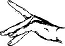
by slowly developing tonic spasms resembling the movement of a worm; they are monotonous, continuous, and often do not stop even during sleep. If the hand is affected by athetosis, the fingers constantly transition from extreme extension to a position of flexion, extreme abduction, and adduction, and these awkward, slow movements can be compared to the play of a polyp's tentacles. A characteristic hand position is also observed where the little finger is strongly abducted, the ring finger is abducted to a lesser degree, the middle finger is slightly flexed, the index finger is extended, and the thumb is extremely extended. All these movements occur with increased muscle tone; thus, if the agonists contract, they meet resistance from the antagonists, which accounts for the slow and difficult nature of the movement. In the intervals between involuntary movements, muscle tone is reduced, and because of this, the impression of a fluctuating spasm (spasmus mobilis) is created. Thus, in athetosis, one observes simultaneously, in the same muscles, now strong hypertonia, now sharp hypotonia. According to modern views, athetosis is a syndrome of damage to the striopallidal system, primarily the neostriatum (putamen + nucleus caudatus). At present, there is an increasing inclination toward the view that these involuntary movements
Figure 1.
are not a symptom of irritation, but the result of the loss of regulatory and inhibitory influences. This is supported by the fact that athetosis continues even when the process causing the athetosis has long since subsided. Among the processes causing such symptomatic athetosis, encephalitis, especially epidemic encephalitis, is most frequently noted. It should be noted that in the latter case, pure athetosis is rarely observed; more often, a combination of various involuntary movements (athetosis, chorea, myoclonus, tremor, etc.) results. Sometimes athetosis can be encountered as a symptom of neostriatum damage in typhus. Athetosis is also observed in such rare diseases as Wilson's disease and pseudosclerosis, and sometimes in brain tumors; hemiathetosis as a symptom of vascular (hemorrhage or blockage) lesions is very rare. If athetosis is added to hemiplegia, it usually develops in the paralyzed limbs significantly later, and moreover, extremely rarely in brain lesions in adults, but more often in childhood. Once athetosis arises, it subsides with difficulty and is little amenable to cure, since the causes evoking it are difficult to eliminate. Symptomatically, bromine, scopolamine, electrization, baths, bandaging, and surgical treatment are applied with almost no success.
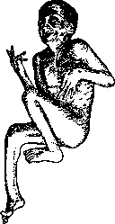
Figure 2.
holding the hand. Often these movements and positions are stereotypical, resembling the atavistic movements of a monkey when it climbs trees. C. and O. Vogt, as well as Foerster, noted that in such patients, movements like those of a newborn remain forever. In complete rest, such involuntary movements may subside, and the musculature may be in a state of hypotonia, but with any irritation, the picture changes sharply: contraction of agonists and antagonists appears, hypertonia arises—the picture of a mobile spasm (spasmus mobilis). Sometimes the picture is colored by "pallidal" rigidity, depending on damage to the globus pallidus. Epilepsy is one of the most frequent complications in athetosis duplex. The mental state may be normal, but more often dementia in a mild form is observed. Reflexes are normal; pathological reflexes are not part of the usual clinical picture of the disease. Athetosis duplex usually appears from the day of birth and is subsequently incurable, although many patients live to an advanced age. In a number of cases, significant improvement has been observed over the years; sometimes it was possible to achieve the development of a number of functions (walking, speech) that were previously absent. In this, one can see the effect and influence of the cerebral cortex on the disturbed innervation of the corpus striatum occurring over the years. In severe cases, Foerster performs surgical treatment, since, according to his observation, the transection of individual motor nerves reduces the tendency toward mass synkinetic movements. Athetosis duplex can manifest in both severe and rudimentary forms, and can present a mixture of choreic, rigid, and spastic states. Thus, athetosis duplex is a collective concept that is not yet clearly enough defined. Rosenthal refers all these cases to symptomatic athetosis, and considers only torsion spasm (see) to be idiopathic, which most authors classify within the athetosis group. With equal justification, extremely rare cases of hereditary progressive bilateral athetosis should be classified as idiopathic athetosis. It is important to note that the disease begins in previously healthy children, often at the age of puberty. Heredity and familial occurrence have been noted by several authors. The disease, in contrast to idiopathic athetosis, progresses.
Related articles
Mentioned in
Cite this page
“Athetosis.” Soviet Medical Encyclopedia. English translation of Bolshaya Meditsinskaya Entsiklopediya, 1st ed. (Moscow, 1928–1936), ed. N. A. Semashko. https://sovietmedicalencyclopedia.pages.dev/article/athetosis/