Extrapyramidal System
Historical document, translated for reference. It reflects medical knowledge of the 1920s–30s and is not medical advice.
Summary
The extrapyramidal system is the oldest phylogenetically motor-tonic mechanism, found even in fish. Its main component is the corpus striatum, but it extends beyond this structure to include other parts like the thalamus, hypothalamus, cerebellum, and mesencephalic formations.
Encyclopedia article (1928–1936)
Extrapyramidal System is the oldest phylogenetically motor-tonic mechanism, already found in fish. The main part of it is the corpus striatum, and therefore, somewhat narrowing the anatomical-physiological substrate, it is sometimes also called the striatal system, striar system, or striopallidal system. But the corpus striatum is only the effector part of this complex reflex mechanism that extends far beyond the corp. striati, and therefore the E. s. as a whole cannot be considered outside the connection with its other parts - the thalamus and hypothalamus, as well as with the cerebellum (striocerebellar system) and mesencephalic formations. The anatomical basis of the E. s. thus consists mainly of basal ganglia, and its center is the corpus striatum (see Basal ganglia), which includes nucl. caudatus (caudate nucleus) and nucl. lenticularis (lenticular nucleus). The latter is divided into putamen and globus pallidus. This nomenclature was replaced by the Vogts (C. and O. Vogt) with a shorter one, namely striatum instead of nucl. caudatus and putamen, and pallidum instead of globus pallidus. Phylogeny. In fish, there is only palaeostriatum (pallidum), which is a collection of cells connected to the thalamus and caudally located nuclei. In amphibians, neostriatum (putamen) also appears. In reptiles, the neostriatal part of the corpus striatum becomes even more developed. In birds, the changes in the corpus striatum consist mainly in an increase in its total mass and in the greater development of connections with various parts of the nervous system. In mammals, sharp changes occur in the structure of the corpus striatum - in addition to an increase in its volume, the internal capsule appearing here divides the neostriatum into two parts - putamen and nucl. caudatus. Ontogenesis. The pallidum is laid down first. According to Kodama, in a 3 cm long human embryo, this can already be clearly seen, somewhat preceding the formation of putamen. In a 4.7 cm embryo, the pallidum divides into two parts. In a 10 cm embryo, the cells of the pallidum are already fully formed, and in a 5-month-old fetus, the first myelinated fibers appear in it. At the 8th month of embryonic life, the myelination of the pallidum almost ends. Putamen lags in its development behind the pallidum. The first differentiation of its cellular structure clearly appears only in a 10 cm long embryo. Myelination begins at the 9th month and ends in extrauterine life. The formation of nucl. caudati begins even later, its first appearance being noted only in a 5 cm long embryo. Its cells develop slowly and reach their full development only by the end of the first year of life. Myelination reaches its maximum at the 11th month of life.
Morphology. The description of the E. s. should more correctly begin with its main, phylogenetically oldest link - the pallidum. By this is meant the former globus pallidus or the two internal segments of the p. lenticularis, which are separated from the outer segment of this nucleus or putamen by means of a plate of myelinated fibers (see figure). The morphological structure of the pallidum is rather primitive in comparison with other parts of the E. s. and amounts to a relatively small collection of gray matter permeated by a dense network of myelinated fibers. Its cytoarchitecture is characterized by the uniformity of the internal structure of cells. In terms of myeloarchitecture, the pallidum also sharply differs from the striatum by its extraordinary richness in nerve fibers, characterizing it as an organ predominantly conductive. Striatum in its morphological structure appears to be a much more complex formation than the pallidum, especially in cytoarchitectonic terms. In any of its sections, its bro
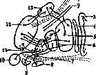
Extrapyramidal system: 1-pedunculus thalami sup.; 2-tractus cortico-spinalis; 3 - insula; 4 - claustrum; 5-putamen; 6-globus pallidus; 7-ansa lenticularis; 8-substantia nigra; 9-nucleus subthalam.; 10-nucleus ruber; 11-pedunc. thalami inf.; 12-thalamus opticus; 13-nucleus caudatus.
one is struck by the huge accumulation of nerve cells. For the structure of the corpus striati and pallidi, see Basal ganglia. According to Vogt, the striopetal fibers, upon entering the striatum, end near small cells with short axons. The latter act on large cells with long axons, which are the source of the striofugal fibers that transmit excitation to the cellular apparatus of the pallidum. In addition to the striatum, the E. s. also includes other basal and mesencephalic formations—Luys' body, the red nucleus, the substantia nigra (corpus Luysi, nucleus ruber, substantia nigra). Of the nuclear formations of the subthalamic area, the largest is the corpus Luysi (see). It is a well-defined, almond-shaped nucleus, located dorsally to the pes pedunculi and substantia nigra. Histologically, when stained for myelin, it appears surrounded by a capsule, the fibers of which connect it to almost all the important formations of the E. s. A very important component of the E. s. is the red nucleus—nucleus ruber (see), located in the tegmentum of the cerebral peduncle at the level of the superior colliculi of the quadrigeminal body. Histologically, it presents the picture of a nuclear formation, not very densely populated with cells, but richly supplied with fibers going in various directions, especially from the superior cerebellar peduncle. According to Foix and Nicolesco, three types of cells are distinguished in the nucleus: 1) large cells, located in the caudal part of the nucleus, 2) medium-sized cells, which constitute the main mass of the nucleus and have connections with other parts of the brain, and 3) small cells, which belong to the proper apparatus of the nucleus. The capsule of the red nucleus is formed by the radiatio of the red nucleus, the radiatio of the tegmentum, and finally by fibers coming from the pallidum. From the caudal part of the nucleus, large cells give rise to one of the main extrapyramidal paths—the bundle of Monakow (tractus rubro-spinalis), which goes to the spinal cord. In the red nucleus, we have a very important motor-coordinative center of the E. s., which receives its excitations from the dentate nucleus of the cerebellum, from the cortex of the frontal lobe, and from the pallidum, with which it stands in close connection, serving as a place of switching of the strio-pallidal apparatus to the underlying mechanisms. A unique mesencephalic node of the E. s. is the substantia nigra. In its cytoarchitectonic aspect, it is divided into two zones: the dorsal, composed of groups of large cells containing melanin, and the ventral, formed by spindle-shaped cells resembling the cells of the pallidum. Its myeloarchitecture is still little studied. Among other formations of the E. s., the substantia nigra sharply differs by the presence of direct connections with the cerebral cortex. Extrapyramidal paths. At the center of the E. s. stand the thalamus and the subthalamic area as indirect and direct sources of impulses. As for the direct connections of the striatum with the cerebral cortex, this question cannot yet be considered finally resolved. A number of authors (Bechterev, Cajal, Economo, Marinesco, Minkowski, etc.) recognize the existence of such connections, while the majority (Vogt, Monakow, Wilson, etc.) deny them, allowing only indirect ones. The main connections of the striatum with the thalamus, subthalamic area, and mesencephalic formations are carried out by means of the following systems. The lenticular loop (ansa lenticularis) is a bundle of striopetal and striofugal fibers, which, crossing the lentiform nucleus, bend along the ventral surface of the pallidum and are directed from the outside inward and from behind forward, ending in the thalamus (or beginning in it for the striopetal fibers), as well as in a number of hypothalamic and mesencephalic formations, especially in the red nucleus. Crossing the internal capsule, the lenticular loop fans out at the border of the hypothalamus into a series of fibers—the anterior ones go to the area of the infundibulum, the middle ones to the thalamus, and the posterior ones, the most numerous, partly also to the thalamus, but mainly to the subthalamic area and to the red nucleus. Forel's thalamic bundle (fasciculus thalamicus Foreli) (H1) originates in the angle formed by the bundle of Vic d'Azyr and the lenticular bundle. It includes fibers of the superior cerebellar peduncle to the thalamus and from the thalamus to the red nucleus. Forel's lenticular bundle (fasciculus lenticularis Foreli) (H2) is a collection of fibers coming mainly from the radiatio of the pallidum, and also partly from the lenticular loop. It contains both striopetal and striofugal conductors. By giving off its posterior fibers to Luys' body, it contributes to the formation of its capsule, while its anterior fibers are closely connected with the periventricular nuclear formations, and its middle fibers with the centers of the hypothalamic area. Fibrae strio-luysianae originate from the radiatio of the pallidum and go along the same path as the previous bundle to Luys' body, also participating in the formation of its capsule. Fibrae strio-thalamicae connect the striatum directly with the thalamus. Commissura Meynerti, according to Déjerine, is an interstriatal system, i.e., connecting both striata with each other. Of the pallidofugal paths, it is necessary to note the tractus pallido-peduncularis—a system of fibers going mainly to the substantia nigra, and the tractus pallido-rubralis, as a further development and continuation of the powerful bundle H3 to the red nucleus. From a group of large cells of the red nucleus begins the fasciculus rubro-spinalis (Monakow's bundle)—one of the main efferent paths of the E. s., going after the decussation in the direction of the spinal cord. Among other descending paths, it is necessary to note the connections of the pallidum through the posterior commissure with the quadrigeminal body, from which the tractus tecto-spinalis begins, going to the spinal cord, as well as the tractus vestibulo-spinalis from the nuclei of Deiters and Bechterev. The main of the indicated paths should be considered the pallidopetal paths from the thalamus to the pallidum of the same side, the paths from the thalamus to the neostriatum, from the neostriatum to the pallidum, and from the latter to the various hypothalamic and mesencephalic formations included in the E. s. At the same time, one cannot ignore other connections of the E. s., in particular with the cerebral cortex, even if indirect ones. Physiology. To form a general idea of the basic functions of the E. s. and of the evolution of the latter, it is necessary to take at least a brief look at the phylogenetic development of these functions in the animal world and on their ontogenesis in man. In fish, the entire motor apparatus is still represented only in the form of the pallium. The latter, receiving impulses from the thalamus, transforms them into motor and tonic ones. In accordance with this simple mechanism, the movements of fish are characterized by the following features: 1) elementariness, 2) constancy, 3) diffuseness of distribution, 4) automatism, 5) rhythm. The movements are smooth, slow, stereotyped, and consist in the sequential contraction of the entire musculature. In reptiles, in connection with the presence, in addition to the pallidum, of an elementary striatum, which plays the role of a brake and regulator of the pallidal apparatus, the movements no longer have a constant character, although they still retain elementariness. In birds, where there is a high development of the striatum, the movements are much more complex and differentiated. In mammals, along with the further evolution of the striatum, the motor cortex and the pyramidal path (see Pyramidal system) appear as a neokinetic system. However, in most higher representatives of mammals (dog, monkey), removal of the cortex does not yet deprive them of motoricity. As Rothmann showed, a dog without a cortex but with the striatum preserved is still capable of independent movements, whereas a dog after removal of the cortex together with the striatum, according to Dresel's data, is already completely incapable of them, although elementary locomotor functions and posture are preserved. In man, removal of the motor cortex causes complete paralysis. The data of ontogenesis in man depict almost the same picture of the development of motoricity as the data of phylogenesis. As Minkowski showed, a 2-5 month embryo already has certain movements of the choreoathetotic type. By the time the child is born, the thalamo-pallidal paths and the pallido-fugal ones are already myelinated, whereas the thalamo-striatal and strio-pallidal ones are not yet fully developed. In accordance with this, the movements of the newborn have clear pallidal features. Only after 5 months, thanks to the participation of the striatum, more complex and differentiated acts appear, for example, the transition from a lying to a sitting position. Even later, the ability to assume and maintain an orthostatic position appears, and finally the ability to walk, which in its differentiated forms is already closely connected with the neokinetic-pyramidal system. The latter, however, by no means takes upon itself the fullness of motor functions. It acquires in man only a dominant significance in motoricity, insofar as it carries out volitional, purposeful, and strictly differentiated movements. Therefore, every complex motor act of man should be considered as a complex act, containing in itself a whole series of components of different origins, of different historical age, and finally of different functional value.
The extrapyramidal system in these motor acts primarily provides a series of kinetic forms of a reactive order, which are instinctive reactions caused by sudden changes in body position or general situation, danger, etc. This includes a whole series of orienting and protective reactions. At the same time, the extrapyramidal system provides a number of auxiliary kinesthetic and automatic components for complex voluntary movements; this is especially true for learned, stereotyped forms of the latter. The close connection of the extrapyramidal system with vegetative centers determines its role in carrying out expressive, mimetic reactions and its lively participation in the emotional-affective life. In its elementary form, expressive movements already appear in the newborn child. Later they become significantly differentiated. Thus arise those automatic changes in tone and reflex movements that manifest themselves in the lively play of facial and eye muscles, in gesturing, which accompany any experience. But inseparably connected with these kinetic functions, the extrapyramidal system performs the statotonic function. Proper tonic innervation of the motor apparatus, normal myostasis are the necessary basis for any complex motor act. To develop maximum dynamics, the motor apparatus with the help of the extrapyramidal system is set in the most favorable kinetic conditions, in conditions of better statotonia and optimal posture. In this case, the body position must be flexible, adapted not only to a given form of movement, but also to a given moment of the developing action. This reflex body position, these postural reflexes are carried out not by the intermediate brain, but mainly by the mesencephalic parts of the extrapyramidal system (a special role here belongs, as Rademaker emphasized, to the red nucleus), which are however closely connected with its central parts. The coordination-tonic function is served by several apparatuses. The main role here is played by the fronto-bridge-cerebellar system, the cerebellum, and the motor-tonic mechanisms of the midbrain and trunk parts. Although the latter work on the basis of proprioceptive impulses, nevertheless they are influenced by the regulating influence of the cerebellum—via paths going to the tectum, i.e., to formations included in the extrapyramidal system, and by the extrapyramidal system itself—via its descending paths. With the help of this complex tonic-coordinating apparatus, the various postural changes in tone, the various body positions are carried out reflexively and automatically. Considering the extrapyramidal system as a whole, we see that, representing a phylogenetically very ancient mechanism, it simultaneously performs kinetic, tonic, and positioning functions. Receiving both nervous and humoral impulses, being in close relations with vegetative centers, it is, as it were, the center of the entire nervous system. Pathology. In accordance with the data presented above, it is quite natural that under pathological conditions we do not encounter a complete splitting of the functions of the extrapyramidal system into purely kinetic and purely tonic ones. In it and here, the ancient connection between motoricity and tone continues to exist. From this point of view, the term 'amiostatic symptom complex' introduced by Strümpell into clinical practice, as not covering the entire sphere of its action, at present can no longer serve as an exhaustive representation of its disorders. In any syndrome with damage to the extrapyramidal system, there are necessarily violations of both of its basic functions. Thus, with damage to the pallidum, a peculiar rigidity of the muscles (pallidal rigidity) occurs, which, in addition to stiffness in the joints, is characterized by cogwheel or step-like movements, persistent spasms (pallidum-spasmus), general stiffness, and amimia. But the matter is not limited to only the violation of tone and myostasis. Along with an increase in plastic tone, there is also a loss of extrapyramidal kinesthetic movements. Among the pallidal syndromes, one should first of all name the paralysis agitans, or Parkinson's disease (see Paralysis agitans and Parkinsonism). The basis of this disease, which has a chronic progressive course, consists of changes mainly in the pallidum (Lewy), which however also extend partly to the striatum (and in severe cases to more diffuse areas). With the same picture of 'parkinsonism', one also encounters in most of the most typical forms of epidemic encephalitis and in other diseases of the extrapyramidal system, e.g., in arteriosclerosis—the so-called 'arteriosclerotic muscular rigidity' of Förster. Parkinsonism was also noted in syphilitic lesions of the pallidary part of the extrapyramidal system. Striatal syndromes, in contrast to pallidal ones, are characterized by hyperkinetic-hypotonic syndromes. One of the typical hyperkinetic striatal syndromes is chorea, especially its chronic progressive form, the so-called chorea Huntingtoni, which is patho-anatomically limited mainly to the lesion of small cells of the striatum. Another characteristic clinical form of striatal syndromes is the so-called double athetosis (see Athetosis, athetose double). To the striatal forms also belongs the one-sided athetosis, which has the most varied etiology. Clinically, this form resembles the previous one, but differs from it in the limitation of hyperkinesia. Patho-anatomically, the basis of the athetotic forms is a process that destroys the large cells of the striatum. The group of striatal diseases also includes torsion spasm and tics, and among the latter also torticollis spastica, which can be considered as a reaction to a narrowly focal lesion of the striatum, in which only a very limited group of muscles suffers (Jakob). To the mixed strio-pallidal syndromes belongs the Hallervorden-Spatz disease (see), characterized by diffuse damage to the medullated fibers (status dysmyelinisatus) in the pallidum and partly in other parts of the extrapyramidal system. Among other extrapyramidal diseases, one should note the so-called hepatolenticular degenerations. These include very close to each other Wilson's disease (see) and Westphal-Strümpell pseudosclerosis (see Westphal-Strümpell disease).
?
*
л
Л
fi
Я
9 § is: Figure 1. Dissociation with interference. The atria and ventricles contract independently of each other (dissociation), with the former being subject to the sinus node and the latter to the Tawara node. The ventricular rhythm is somewhat faster than the atrial rhythm. Individual sinus impulses (2,6) that coincide with the excitable phase of the ventricles cause their premature contraction (interference) and thus shift the automatic ventricular rhythm. In some cases, E. may arise due to the constant work of a second center, as demonstrated by the Vienna authors Kaufmann and Rothberger. Based on a thorough analysis of a series of cases of extrasystolic arrhythmias, mainly of the allorhythmic type, they believe that the existence of two interfering rhythms in the heart is possible, i.e., along with sinus impulses, excitations can rhythmically arise in a new pathologically altered site. They call this additional rhythm 'pararrhythm,' and the irregularity of cardiac activity that results from the functioning of a new center along with the higher one—'parasystole.' A double rhythm can exist only on the condition that each center is protected from external irritations and can work unimpeded. In the opposite case, the sinus node would have to suppress the pathological source of impulses, as happens in normal conditions, or the lower center, having a higher frequency, would become dominant, as in paroxysmal tachycardia, and would displace the normal rhythm. Consequently, the rhythmic work of two centers is possible only in the presence of a 'protective block' around the site of impulse origin. In addition, since only part of the pathological impulses pass to the myocardium, it is obvious that part of them are delayed ('variable block on exit'). The complexity of these assumptions naturally raises objections. Attempts have been made to identify simpler forms of parasystole. Cases have been described that do not require the assumption of 'block on exit,' such as 'dissociation with interference' (Mobitz et al.) (fig. 1) and 'parasystole with simple interference' (Singer, Winterberg). In all the mentioned cases, the proof of pararrhythm is the regular relationships between the intervals separating the extrasystoles (these intervals must be a multiple of a known magnitude—the pararrhythmic period). However, if this magnitude is very small (e.g., less than 0.30 sec), then the calculation becomes very shaky. Moreover, it is unprovable in cases where E. appear strictly regularly at equal time intervals after normal beats. Here heterotopic impulses are closely connected with nomotopic ones. It is clear, for example, that in persistent bigeminy with such a connection, the distances between extrasystoles will be the same, provided the sinus rhythm is regular. In such cases, there is no reason to speak of parasystole; one must assume that the normal impulse somehow causes additional excitation. The mechanism of the occurrence of these irregularities is explained by the theory of 're-entrant waves' (Lewis), based on the experiments of Mines and de Boer. With deterioration of exchange in the cardiac muscle and unevenness of the refractory period in different parts of the myocardium, a single excitation can proceed in stages and make several circular movements, causing two or more systoles. Thus, the mechanism of multiple E., paroxysmal tachycardia, flutter, and fibrillation approaches each other (see Atrial fibrillation). In conclusion, it should be said that the pathogenesis of E. in different cases is apparently different, and that systematic studies of recent years have significantly deepened our understanding of the mechanism of occurrence of various types of this arrhythmia. Symptomatology. Graphic data. Of all disorders of cardiac rhythm, E. belongs to those that most often cause unpleasant subjective sensations. The complaints of patients are very diverse and variable in nature and intensity, ranging from mild, indefinite sensations of palpitations or cardiac arrest to distressing constrictions, blows, and pains in the heart area, radiating to the neck, shoulder, arm, etc. All these phenomena occur in some patients during physical exertion, excitement, or after a heavy meal, while in others—and this is characteristic of E.—at rest, after work, in the evening. Quite often they appear in bed, when falling asleep, and cause the patient to jump up and fear sleep. Sometimes E. are observed only in a certain position (lying, sitting). Whether these phenomena should be attributed exclusively to E. in each individual case is still a question. With careful observation, it can be easily established that in the same patient, E. sometimes causes unpleasant sensations and sometimes passes unnoticed. Obviously, other factors play an important role, such as the state of the psyche and the autonomic nervous system, strengthening or weakening of cardiac activity, vasoconstrictor phenomena, changes in blood pressure, the height of the diaphragm, etc. It is noted that subjective sensations in E. are observed much more often in functional than in organic heart diseases, and that people engaged in intense mental work and leading a restless lifestyle often suffer (in particular, a relatively high percentage of doctors, according to Mandelstam—9%). Sometimes the described typical complaints, associated with arrhythmia, constitute the main or only content of the patient's history, and in the absence of visible damage to the cardiovascular system or with complete compensation of the underlying disease, E. acquires the character of an independent clinical syndrome ('essential E.'). In other cases, arrhythmia does not cause special sensations and is discovered only by chance. Objectively, the recognition of single extrasystoles in most cases is not difficult, both on palpation of the pulse and especially on auscultation of the heart. Against the background of a correct rhythm, a weak premature pulse wave (break) is determined. It can be so weak that it does not reach the periphery (pulse deficit) or at least is not captured on palpation and can be registered only on a sphygmogram. On auscultation, the premature beat corresponds to two additional tones, of which the first is louder than usual, sometimes clapping, and the second is weakened. In very early extrasystoles, the pressure in the left ventricle sometimes increases so insignificantly that it is unable to open the aortic valves (sterile extrasystoles). In this case, only one premature additional tone is heard. If there are noises in the heart, then with premature contractions they may change in strength and in different ways: the systolic noise becomes weaker, the diastolic-presystolic sometimes strengthens, and sometimes weakens. For ventricular extrasystoles, the presence of a compensatory pause and consequently the preservation of the basic rhythm is characteristic. Ventricular extrasystoles do not pass to the atria and do not interfere with their rhythmic contractions. However, the next sinus impulse coming from the atria to the ventricles is absent, as it finds the ventricular muscle in an inexcitable phase (in extrasystole). Only the next impulse passes unimpeded to the ventricles and causes their normal contraction. Due to this, a compensatory pause follows the premature contraction, and the sum of the distances from the normal wave to the extrasystole and from the extrasystole to the next normal wave equals twice the normal cardiac period. For auricular and atrioventricular E., the compensatory pause is shortened or even equals the usual sinus period (in sinus E.). For ventricular and atrioventricular E., another phenomenon is characteristic. During premature contraction of the ventricles, the atria are unable to empty their contents in the usual direction, but drive blood back into the veins. The consequence of the simultaneous systole of the atria and ventricles is a sudden, sharp swelling of the neck veins, visible to the eye and giving a high wave on the phlebogram. For the exact determination of the varieties of E., however, graphic methods must be used, preferably electrocardiography. When analyzing the phlebogram, it is necessary first to recognize the waves of the normal venous pulse, and then establish whether the premature contraction begins with the ventricular wave s. If so, the extrasystole is ventricular; otherwise, it arises in the atrium. For ventricular and atrioventricular E., a large a+c wave is visible on the venous curve, which follows the v wave of the previous contraction or merges with it, and for ventricular extrasystoles, the preservation of the atrial basic rhythm is characteristic. In general, the changes in the venous pulse are diverse depending on the place and time of origin of the extrasystole. It is easier to analyze the nature of the arrhythmia on the electrocardiogram (see the scheme of E. in the article Heart arrhythmia). Auricular E. differ, besides their prematurity, by an atypical (in form and size) P wave and a normal ventricular complex.
When R appears very early, it can be superimposed on the preceding T wave (Fig. 2); sometimes an additional auricular extrasystole occurs. The impulse encounters non-excitable tissue and cannot pass to the ventricles; then one speaks of a blocked extrasystole. Sinus extrasystoles do not differ in form on the electrocardiogram from normal beats. In rare cases, with very early excitations and incomplete restoration of conductivity in the ventricles, the ventricular complex of an auricular extrasystole is atypical and resembles a ventricular extrasystole, from which it differs, however, by the presence of a preceding P wave. In atrioventricular dissociation, the atria and ventricles contract almost simultaneously, since the impulse, originating from the Tawara node, spreads to both sections. On the electrocardiogram, the P wave is negative (Fig. 3) and approaches or merges with the ventricular complex, disappearing within it; the latter has the usual supraventricular form. Ventricular extrasystoles, which occur more frequently than others, give particularly characteristic and distinctive electrocardiograms. They differ, besides their prematurity and absence of a P wave, by their considerable size and atypical form of the ventricular complex (Figs. 4, 5, 6 and 7). The two main types of ventricular extrasystoles (Figs. 4 and 5) are very similar to the dextro- and levogram obtained by cutting the right or left leg of the His bundle. The first consists of a high sharp wave directed upward and a wide gentle wave directed downward (the right-sided type is characteristic of extrasystoles originating from the right ventricle); the second has the reverse form (the left-sided type-in extrasystoles of the left ventricle). The question of the connection between the type of electrocardiogram and the localization of the impulse in one or the other ventricle, however, proved to be more complex than it seemed at first on the basis of experimental data. Irritation of various areas of the myocardium gives various variations of the indicated basic types of electrocardiogram. Furthermore, the form of the electrocardiogram in many cases changes sharply in different leads. Thus, in lead I an extrasystole may have the appearance of a left-sided type, while in leads II and III it may be right-sided and vice versa (discordant type-Fig. 4). At present, it is believed that for determining the place of origin of ventricular extrasystoles, their form in lead III is of greater importance. Therefore, the extrasystole depicted in Fig. 4 should be considered a right-sided, and in Fig. 5 a left-sided ventricular extrasystole. But with respect to the human heart, even this position cannot be considered firmly established. The advantage of the electrocardiogram is also that from the curve it can be seen whether all extrasystoles originate from one place or from different places; polymorphism of extrasystoles indicates polytopic origin of impulses. The magnitude of the waves of the electrocardiogram does not correspond to the force of heart contraction. - In some
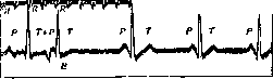
Figure 3. Atrioventricular extrasystole with inverted P wave (E).
electrocardiograms. They differ besides prematurity and absence of a P wave by their considerable size and atypical form of the ventricular complex (Figs. 4, 5, 6 and 7). Two main types of ventricular extrasystoles (Figs. 4 and 5) are very similar to the dextro- and levogram obtained by cutting the right or left leg of the His bundle. The first represents a high sharp wave directed upward and a wide gentle wave directed downward (the right-sided type is characteristic of extrasystoles originating from the right ventricle); the second has the reverse form (the left-sided type-in extrasystoles of the left ventricle). The question of the connection between the type of electrocardiogram and the localization of the impulse in one or the other ventricle however proved to be more complex than it seemed at first on the basis of experimental data. Irritation of various areas of the myocardium gives various variations of the indicated basic types of electrocardiogram. Furthermore the form of the electrocardiogram in many cases sharply changes in different leads. Thus in I lead an extrasystole may have the appearance of a left-sided type while in II and III - right-sided and vice versa (discordant type-Fig. 4). At present it is believed that for determining the place of origin of ventricular extrasystoles their form in III lead is of greater importance. Therefore the extrasystole depicted in Fig. 4 should be considered right-sided and in Fig. 5 left-sided ventricular extrasystole. But with respect to the human heart and this position cannot be considered firmly established. The advantage of the electrocardiogram consists still in that from the curve it is visible whether all extrasystoles originate from one place or from different; polymorphism of extrasystoles indicates polytopic origin of impulses. The magnitude of the waves of the electrocardiogram does not correspond to the force of contraction of the heart. - In some
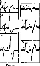
Figure 5.
Figure 4. Ventricular extrasystoles of discordant type; shown is an extrasystole originating from the right ventricle (right-sided type in II and III leads but left-sided type in I lead). Figure 8. Ventricular extrasystoles of concordant type; shown is an extrasystole of the left ventricle giving an atypical form of electrocardiogram of left-sided type in all leads. cases mainly with slow sinus rhythm ventricular extrasystoles do not displace the following normal contraction but end before its appearance (interpolated or interpolated extrasystoles Fig. 6). Such extrasystoles are separated from the subsequent regular contraction not by a compensatory pause but by a shortened interval. Their recognition on the electrocardiogram presents no difficulties. Usually they lengthen the P-R interval of the following systole because the conductivity of the atrioventricular bundle does not have time to completely restore by this time. Early ventricular extrasystoles in rare cases
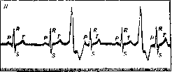
Figure 6. Two interpolated extrasystoles.
can apparently pass in the reverse direction along the His bundle and cause contraction of the atria before the normal systole occurs. Such "reverse" retrograde extrasystoles have been firmly established only experimentally. Allorhythmias are those irregularities of rhythm in which the disorder of the normal mechanism itself repeats with a certain regularity (see Heart arrhythmias). The most frequent and simple form is bigeminy (see) in which each normal beat is accompanied by an extrasystole originating more often from the ventricles less often from the atria (Fig. 7). In other cases one sinus excitation and two extrasystolic excitations alternate regularly (pulsus trigeminus) or one
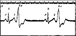
Figure 7. Ventricular extrasystoles of right-sided type with compensatory pause.
sinus and three extrasystolic (pulsus quadrigeminus) etc. More complex allorhythmias are also observed in which whole series of normal and heterotopic excitations alternate in a certain sequence. Such cases represent a transition to paroxysmal tachycardia and are distinguished by the high frequency of the extrasystolic rhythm giving bursts of tachycardia (tachycardie en salves of French authors). Of all allorhythmias bigeminy is practically important. On the pulse curve it gives the regular alternation of a large and small wave the small wave being premature. In this way pulsus bigeminus differs from pulsus alternans. If however extrasystoles appear late the difference is less clear. An apparent bigeminy occurs in the case if every third beat is missing due to partial atrioventricular or sinoauricular dissociation or due to the fact that after two sinus impulses a very early unproductive extrasystole appears. However in this case the paired waves of the arterial pulse are equal in magnitude. The nature of allorhythmia is even easier to recognize from the electrocardiogram. Often pulsus bigeminus simulates bradycardia since the waves of the extrasystole do not reach the periphery-bradysphygmia. On auscultation of the heart palpation of the apical impulse as well as on recording of curves it is easy to ascertain the true character of cardiac activity. Bigeminy can be prolonged or transient often it recurs giving way at times to a normal rhythm tachycardia or chaotically occurring extrasystoles. It is well known that bigeminy appears due to ventricular extrasystoles under the influence of digitalis preparations. In some patients allorhythmia appears with excessive doses or too prolonged treatment; in others with more severe heart damage already after a short time. Generally bigeminy appearing during prolonged treatment is a symptom of intoxication and is an indication to discontinue the drug. It indicates severe damage to the myocardium (Gallavarden's "symptom of alarm"). Extrasystoles can be combined with all other types of arrhythmias most often with sinus and fibrillation. Sinus bradycardia arrhythmia and extrasystoles often occur together which indicates the influence of the vagus nerve (direct or reflex) in the occurrence of these disorders of cardiac activity. In such cases turning off the endings of the n. vagi with the help of atropine can temporarily eliminate the abnormal mechanism. The same result is often obtained with physical exertion (test with movement) or with inhalation of amyl nitrite. On the contrary by reflex irritation of the n. vagi (for example carotid or ocular reflex) in a number of cases it is possible to cause extrasystoles. The combination of extrasystoles with fibrillation is common especially during treatment with digitalis (in approximately */3 of all cases of fibrillation). These are ventricular extrasystoles but they are not accompanied by a compensatory pause due to the absence of sinus rhythm. With more marked increase and irregularity of ventricular activity they can be recognized only on the electrocardiogram.-Close to extrasystoles are heterotopic beats which do not arise on the basis of increased excitability of the cardiac muscle but due to decreased activity of the higher center. They represent manifestations of a slowly and rhythmically working secondary or tertiary center. Individual impulses often occur at the end of long pauses (in sinus bradyarrhythmia) as "escaped beats" (Lewis escaped beats passive heterotopic contractions Wenckebach-Fig. 8) replacing the missing normal excitations (see also Heart arrhythmias). In the case if the sinus node ceases to function
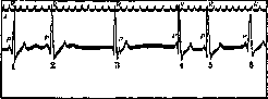
Figure 8. Sinus bradyarrhythmia. "Escaped beats" (.3,4).
to inhibit (in inflammatory changes, sclerosis of vessels or under the influence of the vagus nerve), then the Tawara node can become the leading center and a nodal rhythm is established (nodal rhythm McKenzie). The atria and ventricles contract simultaneously or almost simultaneously (fig. 1). The frequency of the rhythm does not exceed 40-50 beats per minute. The form of the complexes on the electrocardiogram is the same as in atrioventricular extrasystoles (fig. 1 and 8). If, however, the sinus node and a heterotopic (atrioventricular or ventricular) automatic center work with almost the same frequency, then the leading role can repeatedly pass from one to the other and interference of two rhythms occurs (see above). Diagnosis. The clinical picture and the electrocardiogram have great importance. Prognosis. In many cases, the characteristic complaints make it possible to make a presumptive diagnosis based on the history. Extrasystoles can be distinguished from other arrhythmias in most cases already during the direct examination of the patient, best of all during auscultation of the heart. Both single extrasystoles and simple arrhythmias give very characteristic sound phenomena. Palpation of the pulse alone is not sufficient, since the omission of individual pulse beats can be caused by other disorders of the cardiac mechanism (marked sinus arrhythmia, incomplete block). Difficulties arise with very frequent grouped extrasystoles and with extrasystolic tachycardia; here the distinction from paroxysmal atrial fibrillation or sinus arrhythmia without graphic methods (electrocardiogram) is often impossible. Extrasystoles in themselves are not a severe disorder of rhythm and do not have a special effect on the circulation. The presence of extrasystoles, however, necessitates a thorough investigation to determine their cause. If there are no symptoms of organic disease and functional insufficiency of the heart, then the extrasystoles should not be given great importance; practically the heart can be healthy. In any case, the presence of only extrasystoles does not give the right to speak of heart disease and is not a symptom that allows one to distinguish organic disease from functional. In a number of cases, however, extrasystoles acquire the significance of a symptom of damage to the heart muscle or a precursor of circulatory disorders. Thus, during or after an acute infectious disease, extrasystoles, especially with a frequent pulse, indicate pathological changes. Numerous extrasystoles from different foci (polymorphic), differing on the electrocardiogram by their polymorphism, are observed in severe widespread changes in the myocardium. More frequent atrial extrasystoles in mitral stenosis and cardiosclerosis may be a precursor of fibrillation. In hypertension and aortic insufficiency, left ventricular extrasystoles are often a symptom of the beginning weakness of the heart. An unfavorable prognostic significance has digitalis bigeminy, as was said above. Accumulated extrasystoles, forming sometimes attacks of tachycardia, by no means always indicate organic heart disease, and all the more the onset of decompensation. There is no essential difference between atrial and ventricular extrasystoles, although the former are more often observed in functional heart diseases. In general, a correct evaluation of extrasystoles is possible only in connection with the data of the history and a complete objective investigation taking into account socio-domestic and production factors; in individual cases it is necessary to repeat the investigation after certain intervals. Treatment. In the first place stands the treatment of the underlying disease. In the presence of organic heart disease, extrasystoles rarely require special attention. As a rule, extrasystoles must be considered a contraindication to the prescription of digitalis; only in the case if ventricular extrasystoles are accompanied by tachycardia and signs of the onset of heart failure, they cannot be considered a contraindication to the prescription of digitalis, on the contrary, under these conditions the irregularity of the rhythm often disappears with the improvement of the circulation under the influence of digitalis. In any case, extrasystoles or especially bigeminy, appearing during treatment with preparations of the digitalis group, are a symptom of intoxication and an indication to discontinue these remedies. If extrasystoles are the result of reflex or toxic influences, then the elimination of the cause is important (treatment of diseases of the abdominal organs, especially constipation, stomach distension, bloating, etc., restriction of smoking, alcohol, coffee, etc.). In so-called essential extrasystoles, mental calm of the patient and treatment of the nervous system are important (explanation to the patient of the non-seriousness of the suffering, regulation of the way of life, elimination of overwork, rest, light hydrotherapy, nervina, sedativa). If these means are insufficient, quinine recommended by Wenckebach can be prescribed, which has a pronounced ability to reduce the excitability of the heart muscle. The usual course: 3 times a day 0.1-0.2 of any quinine preparation for 10 days with a subsequent break of 8-10 days. For persistent accumulated extrasystoles, quinidine can also be tried in small doses - 0.2-0.3 once or twice a day as needed. Among other means, strychnine and theobromine can be mentioned, which are often combined with quinine in the form of pills. Atropine often eliminates extrasystoles, but mostly only for a short time. In general, we have a whole series of effective remedies (see Heart Arrhythmias), but to judge the therapeutic effect of individual means is usually not easy due to the variability and inconstancy of the arrhythmia. If the extrasystoles do not cause special disorders, then the indicated treatment may be superfluous and it is sufficient for the patient to be explained that the arrhythmia should not inspire fears.
IV. Mandelstam.
Related articles
Mentioned in
- Adiadochokinesia
- Amimia
- Amyotaxia
- Athetosis
- Basal Ganglia
- Clonus
- Fennel (medicinal or Florentine, fennel, Foeniculum)
- Helweg's Tract
- Hemiplegia
- Hyperkinesis
- Hypnophrenia
- Lenticular Degeneration
- Motor Centers
- PARAKINESIAS
- Pyramidal System
- Retro-pulsion
- Subcortical Functions
- Substantia Nigra
- Thalamus Opticus
- Wilson Disease
Cite this page
“Extrapyramidal System.” Soviet Medical Encyclopedia. English translation of Bolshaya Meditsinskaya Entsiklopediya, 1st ed. (Moscow, 1928–1936), ed. N. A. Semashko. https://sovietmedicalencyclopedia.pages.dev/article/extrapyramidal-system/