Atrial Fibrillation
Historical document, translated for reference. It reflects medical knowledge of the 1920s–30s and is not medical advice.
Summary
Atrial fibrillation is a cardiac arrhythmia characterized by irregular and chaotic contractions of the atria, first experimentally induced in the late 19th century. The article discusses its historical recognition, experimental induction, causes, effects on circulation, and associated conditions.
Encyclopedia article (1928–1936)
ATRIAL FIBRILLATION, fibrillation and flutter of the atria and ventricles. 1. Fibrillation of the atria. A disturbance of rhythm which we now call atrial fibrillation (Flimmerarhythmie in German, fibrillation in English) was known long ago. In 1836, Bouillaud described a disease characterized by irregular contractions of the heart, both in strength and rhythm, naming it delirium cordis. Subsequently, this disease was described as arhythmia perpetua, completa, s. absoluta (perpetual, complete, or absolute arrhythmia), pulsus irregularis perpetuus. The impetus for studying this form of rhythm disturbance came from the introduction of graphic methods of research. On the sphygmogram, Hering recorded a complete lack of regularity in the alternation of large and small pulse beats following one another in different sequences, and this disturbance of rhythm was defined by him as pulsus irregularis perpetuus. On the phlebogram, Mackenzie noted the absence of the atrial tooth of the a. This absence could be explained, in his opinion, by two causes: either paralysis of the atria or simultaneous contraction of the atria and ventricles. The latter is possible only when the source of excitation becomes the atrioventricular node. Mackenzie later abandoned the assumption of atrial paralysis and considered that the basis of pulsus irregularis perpetuus is nodal rhythm. The clarification of the localization of the pathological process and the proof of the incorrectness of Mackenzie's views we owe to experimental observations and electrocardiography. At the end of the 19th century, McWilliam, by stimulating the atria of an exposed heart with a strong faradic current in an experiment, brought them into a special state which he defined as fibrillation. In this state, the wall of the atria is as if in diastole and does not contract coordinately. Over the entire surface of the atrial wall, small twitchings and wave-like movements are visible, caused as if by simultaneous and independent contractions of individual muscle fibers. These contractions, causing small fluctuations on the shiny surface of the pericardium and consequently constantly changing reflections in place and time, produce the impression of exactly flickering. After the cessation of faradization, atrial fibrillation continues for some time. Besides faradic current, atrial fibrillation can be induced experimentally by mechanical and chemical irritants. A number of pharmacological substances in certain concentrations (chloroform, barium, etc. and, what is especially important, digitalis) can cause atrial fibrillation. An increase in carbon dioxide content in the blood, an increase in blood pressure, and the administration of thyroxin contribute to the appearance of atrial fibrillation. Stimulation of the vagus nerve and the administration of vagotropic poisons (physostigmine, etc.) not only contribute to the continuation of existing atrial fibrillation but can even cause it. The administration of atropin prevents the onset of fibrillation. Stimulation of the sympathetic nerve reduces the duration of fibrillation after the cessation of atrial stimulation. The appearance of the animal, its age, and the condition of its heart play a large role in the experimental production of fibrillation. For example, the older the animal, the easier it is to cause fibrillation in it. When atrial fibrillation appears in the experiment, the ventricular rhythm immediately changes. Ventricular contractions follow one another with unequal intervals; the strength of individual contractions varies. Fredericq proved that the irregularity in the sequence and strength of ventricular contractions depends on atrial fibrillation. When the bundle of His was cut or compressed, when its conductivity was disturbed, ventricular arrhythmia disappeared and automatic contractions of the ventricles appeared: slow, rhythmic, and equal in strength. In atrial fibrillation, the atria are the source of a large number of impulses to contraction, directed toward the node of Tawara and the bundle of His. If the strength of these impulses is sufficient, then with the complete preservation of the conductivity of the bundle of His, the stimulation will reach the ventricles and they will contract. If, however, the impulse is weaker or the conductivity has not been restored, then the impulse will be blocked, and the ventricles will not contract. Thus, the frequency and sequence of ventricular contractions depend on the following factors, which constantly change during atrial fibrillation: the quantity and strength of contraction impulses coming from the atria, and the state of the conductivity of the bundle of His. This is what determines the irregular alternation of ventricular complexes in fibrillation. As for the reason for the difference in amplitude of pulse waves in atrial fibrillation, it has been clarified by recent works. Korteweg established that the size of the pulse wave in A. F. is determined by the degree of filling of the ventricles and the amount of energy accumulated in them, depending on the duration of the preceding diastole and the strength of the preceding contraction. But experimental and clinical observations showed that the height of the pulse waves in A. F. does not always obey Korteweg's rule and that this disobedience is greater the more severe the pathological changes in the myocardium of the ventricles (Kaufman, Rothberger, Grotele). Kisch as early as 1899 drew attention to the fact that the sphygmogram in the case of paroxysm of absolute arrhythmia described by him is similar to the sphygmogram obtained in a dog when its atria are faradized. But only on the basis of the study of the electrocardiographic picture (Rothberger, Winterberg and Lewis) was the identity established between the arrhythmia in experimentally induced atrial fibrillation and that disturbance of rhythm which was previously defined as pulsus irregularis perpetuus. At the present time, it can be considered unquestionably established that the disturbance of rhythm which was described under the name delirium cordis, arhythmia perpetua, s. absoluta, p. irregularis perpetuus, is a consequence of atrial fibrillation. It is necessary to discard all these obsolete terms and replace them with the term 'atrial fibrillation'. The etiology of clinical cases of fibrillation is extremely diverse. There are no diseases that can be considered as the cause of the occurrence of fibrillation. But there are diseases that are very often accompanied by atrial fibrillation. Most often, fibrillation is observed in lesions of the atrioventricular valve, especially in its narrowing. It usually appears in that period when there is already a significant and long-standing disturbance of compensation. The percentage of cases where fibrillation is associated with lesions of the atrioventricular valves varies, according to data from various authors, from 35 to 50. The next disease in which atrial fibrillation is most often observed is cardiosclerosis. The third disease that is often accompanied by A. F. is Basedow's disease. In addition, atrial fibrillation may occur with prolonged and severe cardiac insufficiency and with lesions of the aortic valves, with syphilitic mesoaortitis and hypertension (essential and nephrosclerotic), but in these diseases A. F. is still observed significantly less often. During the most diverse infectious diseases (most often in typhus and lobar pneumonia), short or more prolonged attacks of fibrillation are sometimes noted. In the clinic, as in the experiment, fibrillation sometimes appears in connection with the administration of vagotropic substances, in particular digitalis. There is no specific pathological picture characteristic of atrial fibrillation. Various changes in the sinus node were very often found: small-cell infiltration, hemorrhages, degenerative processes, etc. Changes in the artery of the sinus node have also been described. These changes were an attempt to explain the appearance of fibrillation. The appearance of atrial fibrillation in lesions of the atrioventricular valves was attributed to the stretching of the cavities of the atria and degenerative changes in their myocardium. However, with the same or even more significant changes in the sinus node and myocardium of the atria, fibrillation may also be absent. On the other hand, it can appear with an intact sinus node and with insignificant changes in the atria. In the experiment (Aver'yanov, Fogel'son and Fedorov), the appearance of fibrillation was never observed after removal of the sinus node. And the existence of A. F. in the form of paroxysms and the restoration of sinus rhythm after prolonged fibrillation under the influence of such a toxic agent as quinine, speaks for the fact that atrial fibrillation is caused not so much by pathological-anatomical changes as by certain biochemical (dystrophic) changes in the myocardium of the atria. The influence of A. F. on the circulation is very significant. The greatest significance of atrial fibrillation for the circulation depends on the fact that atrial fibrillation - as long as the conductivity of the bundle of His is normal - as a rule leads to significant and constant tachycardia. If this concerns a heart that is on the border of insufficiency of its function or not far from it, then this tachycardia, in connection with the unproductive expenditure of myocardial energy on frequent in these cases frustrane contractions, causes more or less severe insufficiency of cardiac function.
Three factors influence circulation in atrial fibrillation: the absence of normal atrial contractions, irregular ventricular activity, and the reduced regulatory role of extracardiac nerves on heart function. Regarding the first factor, its significance is not very great. The amount of blood delivered to the ventricles under physiological conditions by atrial contraction is insignificant. Only in cases of stenosis of the left atrioventricular orifice does the contraction of the hypertrophied left atrium have great importance for filling the left ventricle. In this case, the cessation of atrial contractions apparently causes a significant decrease in blood flow to the heart and a reduction in stroke volume. The greatest importance for circulation in atrial fibrillation is the irregularity in the sequence of ventricular contractions. In a series of prematurely occurring ventricular contractions, their filling is still insufficient. As a result, ventricular contractions may be so weak that they cannot open the aortic valves, and the heart contracts in vain (frustrane Kontraktion). Sometimes the amount of blood ejected by the ventricles is so small that the pulse wave does not reach the periphery. In all these cases, there is a discrepancy between the number of ventricular contractions and the number of pulse beats—the so-called pulse deficit. All this leads to a decrease in minute blood volume. The exclusion of extracardiac nerve regulation of cardiac activity also significantly affects circulation in atrial fibrillation. In the coordination of circulation mechanisms, the regulatory role of extracardiac nerves is of great importance. The clinical picture of atrial fibrillation depends on its form. Three forms of fibrillation are distinguished: 1) tachyarrhythmic (with an increase in heart rate), 2) bradyarrhythmic (with a decrease in heart rate), and 3) paroxysmal form. In the tachyarrhythmic, most common form of fibrillation, patients complain of unpleasant sensations in the heart area, with some complaining of palpitations and others that the heart 'trembles.' Additionally, patients often complain of shortness of breath, dizziness, and internal restlessness. The pulse is rapid; a sharply expressed irregularity in the sequence and strength of pulse waves is noted. In this form of fibrillation, pulse deficit—a discrepancy between the number of heart contractions and the number of pulse beats—is most often observed. On auscultation, there is some intensification and changing resonance of the first tone. Intensification of the first tone is also observed in cases where fibrillation is not associated with stenosis of the atrioventricular orifice. The changing strength of the first tone is explained by the change in the mechanism of valve closure due to the varying rate of increase in intraventricular pressure. The sphygmogram in atrial fibrillation is characterized by irregularity in the sequence and magnitude of pulse waves. On the phlebogram, the P wave is absent. The S waves follow with irregular intervals, their form is usually sharply altered. The electrocardiogram in atrial fibrillation is very characteristic (Fig. 1). The cessation of normal atrial contractions in fibrillation causes the disappearance of the atrial P wave. In fibrillation of the atria, numerous contractions of small areas of muscle occur, which are transmitted to the string and reflected on the electrocardiogram by numerous, varying in size, more or less small oscillations (f). These oscillations appear especially clearly during the S-T and T-P intervals, when the string is at rest. The amplitude of these oscillations varies in different leads. In lead III it is usually maximal, and in lead I minimal, rarely the opposite. Sometimes the magnitude of oscillations in all leads is extremely small; to detect them, it is necessary to resort to special leads of heart action currents. The form of the oscillations sharply changes in the same patient: it can be irregular, fine-waved and pass into a coarse-waved, more regular form, in which the wave approaches the normal size of the atrial P wave. Counting the number of f waves on the electrocardiogram is sometimes very easy, sometimes extremely difficult. The ventricular complexes of the electrocardiogram follow one another with irregular, without any regularity, intervals. Since the impulse to contraction in fibrillation
Phonogramma
J

I
io sei
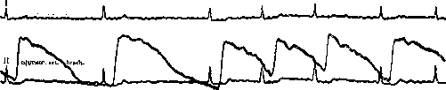
Figure 1. Tachyarrhythmic form of atrial fibrillation. Sphygmogram—waves of varying magnitude, following one another with intervals of varying magnitude. Electrocardiogram—the P wave is absent. Throughout, continuous oscillations of varying waves. The form of the ventricular complex, both its initial and final parts, changes with different contractions. Phonogram—changing strength of the first tone.
Figure 2. Bradycardiac form of atrial fibrillation. Sphygmogram - waves of varying magnitude, following one after another with intervals of varying magnitude. Electrocardiogram - atrial wave is absent. Throughout the entire recording, continuous small oscillations. The form of the ventricular complex is unchanged. Phonocardiogram - changing intensity of the first tone. The absence of the atrial component of the first tone is clearly evident. 009 systoles. Extrasystoles may be identical or varied in form with a constant or changing extrasystolic period. Fibrillation may be combined with complete atrioventricular block. Clinically, such cases proceed as cases of block, and only the electrocardiogram makes it possible to make an accurate diagnosis. On the electrocardiogram, normally formed ventricular complexes follow with regular intervals; the P wave is absent; during diastole, atrial oscillations are clearly visible. - The diagnosis of atrial fibrillation is often made easily on the basis of pulse palpation and auscultation. The appearance of fibrillation in cases of narrowing of the left atrioventricular opening may sometimes be indicated by the disappearance of the characteristic presystolic murmur due to the cessation of atrial contractions. Sometimes the diagnosis of fibrillation presents certain difficulties. The tachycardiac form of fibrillation can easily be confused with grouped extrasystoles. Unlike fibrillation, the intervals between individual extrasystoles in a group are uniform; the group of extrasystoles ends with a compensatory pause. The bradycardiac form of fibrillation can easily be confused with respiratory arrhythmia. The continuing irregularity and changing intensity of heart contractions during breath-holding indicates the presence of fibrillation. An attack of atrial fibrillation can easily be confused with paroxysmal tachycardia. The main differential diagnostic feature is the smaller number of ventricular contractions in fibrillation. Additionally, with prolonged auscultation, especially at rest and after administration of digitalis, the characteristic irregularity of the intervals between individual contractions, which is typical of fibrillation, becomes apparent. Often, however, the diagnosis of fibrillation can only be made on the basis of graphic methods of investigation, especially on the basis of the electrocardiogram. The therapy of atrial fibrillation is based on the same principles as the therapy of atrial flutter (see below).
2. Atrial Flutter. The disturbance of heart rhythm in atrial flutter, with its characteristic significant increase in atrial contractions, was first described by Joly and Ritchie. They also established that the basis of this type of rhythm disturbance is the atrial flutter first obtained experimentally by Mac William. As already mentioned above, Mac William showed that if the atria are subjected to the action of a strong faradic current, they begin to fibrillate. If, however, faradization is performed with a weak current, accelerated rhythmic contractions of the atria are obtained (up to 300-400 contractions per minute). Mac William called this state of the atria flutter. After weakening the strength of the faradic current, fibrillation does not immediately pass into a normal rhythm. Usually there is a transitional period, during which the number of atrial oscillations decreases and more or less correct contractions of the atria of the flutter type (unpure flutter) appear. If during the transitional period the vagus nerve is irritated, flutter again passes into fibrillation. These experimental data show the close connection between fibrillation and atrial flutter. Restoration of conductivity sometimes occurs at regular intervals. The ventricles contract rhythmically at this time. The ratio of atrial to ventricular contractions will depend on the conductivity of the His bundle and will be 1:1; 2:1; 3:1, etc. If restoration of conductivity occurs unevenly, the ventricular rhythm becomes irregular: sometimes 1:1, sometimes 2:1, sometimes 3:1, etc. Irritation of the vagus nerve, if it does not cause the appearance of fibrillation, causes a decrease in conductivity and a reduction in the number of ventricular contractions. The number of atrial contractions is not affected by irritation of the vagus nerve. Irritation of the sympathetic nerve causes intensification and increase of atrial contractions and an increase in the number of ventricular contractions due to improved conductivity. In clinical cases, a regular and irregular form of flutter are also distinguished. Additionally, similar to fibrillation, depending on the ratio of atrial to ventricular contractions, accelerated and slowed forms of flutter are observed. But in flutter, it is even more difficult than in fibrillation to draw a sharp distinction between these two latter forms. In terms of etiology and pathologic anatomy, everything said about fibrillation is applicable to flutter. Flutter is observed in the same diseases as fibrillation, but much less frequently. Fibrillation and flutter very often pass into one another. There is no characteristic patho-anatomical picture of flutter, just as there is none for fibrillation. To explain the occurrence of fibrillation and atrial flutter, a whole series of theories has been proposed. The basis of these conditions, and most authors agree on this, is the same pathologic process, however, the question of the mechanism of the occurrence of this process cannot be considered resolved to this day. The theories proposed to explain flutter and fibrillation can be divided into two main groups. The pathologic process underlying fibrillation and flutter may be caused either by the appearance of additional foci of excitation causing simultaneous dissociated contraction of individual fibers of the atrial musculature, or - in the presence of one focus of excitation - by a change in the sequence of contraction of individual parts of the atrial myocardium. Hering considers the process of occurrence of fibrillation to be analogous to extrasystole. With the appearance of a new focus of excitation in the atria, atrial extrasystoles are obtained. Intensification of the activity of the new focus of excitation leads to the switching off of the sinus node as a source of rhythm and to an acceleration of atrial contractions - tachysystole of the atria. Initially, when the frequency of atrial contractions does not exceed 240, the ventricle responds to each atrial contraction. With increased frequency, some atrial contractions are blocked. This occurs in flutter. In atrial fibrillation, however, there is multiple formation of new foci of excitation in the atria, sending irritations simultaneously and independently of each other. As a result, the coordinated contraction of the atrium as a whole ceases, and only individual muscle fibers contract independently of each other. Hering defines fibrillation as the highest degree of heterotopic formation of excitation. According to Haberland, the numerous foci of excitation are not scattered throughout the atrial musculature, but are concentrated in the atrioventricular node. These foci cause the simultaneous appearance of numerous uncoordinated extrasystolic contractions of individual muscle bundles. However, to detect these contractions, it is necessary to shorten the refractory phase. Without this, the irritation originating from the foci will catch the myocardium in a state of non-excitability, and
Figure 3. Diagram illustrating the course of the wave
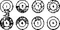
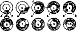
excitation of the muscle ring will not result in fibrillation. The number of supporters of the multiplicity of foci of excitation during fibrillation is small. Most authors at the present time adhere to the view that fibrillation is based on altered activity of the atria with a single source of excitation. Rotberger and Winterberg were the first to express the assumption that multiple foci of excitation are not necessary for fibrillation. Disturbance of atrial activity can be caused by a change in the sequence of contractions of individual parts of the atrial musculature. Subsequently, the occurrence of fibrillation was attributed to the presence of circular movement in the atria. The doctrine of circular movement is based on a number of very interesting experiments (Mayer, Mines). In 1908, Mayer performed the following experiment. He cut out a muscular ring from the swimming bell of a jellyfish, caused temporary compression of the ring by applying a clamp, and then irritated with electric current the portion located in front of the clamped area. The wave of excitation that resulted from the irritation spread in one direction without hindrance, while in the other direction it encountered the clamped area, which prevented conduction of excitation, and died out. If the clamp is removed in a timely manner, the wave of excitation, bypassing the ring, may find the compressed part in the stage of restored excitability. Then conditions are created under which the wave of excitation can repeat its journey around the ring several times. Circular movement is possible only when the duration of the journey around the ring is longer than the refractory phase of the compressed area. Conditions for the occurrence of circular movement can also be created, as shown by Mines on a cut-out muscular ring from a turtle's heart, when this ring is subjected not to a single irritation, but to a series of sequential irritations. With a single irritation (Fig. 3), the waves of excitation spread evenly in both directions, collide, and die out. If, however, irritations follow one another rapidly (Fig. 4), then the refractory phase of the irritated area is shortened and the conduction of excitation is slowed down. In this case, the wave of excitation, on the one hand, may encounter a portion of the ring that is in a relative refractory period, and then the wave of excitation will slowly begin to circle around the ring in the opposite direction. Circular movement will continue as long as the leading part of the wave finds the muscle portion ahead of it in a state of excitability. If, however, on the path of the wave's progression, an additional irritation causes an area to become non-excitable, circular movement will cease. Mines already expressed the assumption that atrial fibrillation is caused by circular movement of excitation waves in the atria. Lewis, together with his collaborators, induced flutter in dogs and by direct derivation from various points of the atria to different galvanometers recorded the passage of the excitation wave and simultaneously recorded the electrocardiogram in lead II. On the basis of his experiments, Lewis believes that the wave of excitation in the atria during flutter does not arise in the sinoatrial node nor in any other specific location of the atrial musculature, but travels along a certain path, describing a circle along the posterior surface of the atria, encompassing the mouths of the inferior and superior vena cava. The path traveled by the excitation wave is the same in all cases of flutter. This explains the regularity of the alternation of atrial oscillations and the similarity of the obtained electrocardiographic curves in all cases of clinical flutter. From this main wave, which is reflected on the electrocardiogram, centrifugal waves extend to the other parts of the atria. The conduction of excitation during flutter is uniformly slowed down and
changes little
even with strengthening of a series of sequential stimuli. The wave of excitation, when it reaches a point in the refractory phase, however, when it spreads in the direction of b. When the wave reaches b (4), it emerges from the refractory phase. The entire time of circular movement, the distance between the head and tail parts of the wave does not change. (From Lewis.) This is accompanied by a change in the shape of the directly recorded atrial oscillations, with pure atrial flutter passing into impure flutter. When the rotation of the main wave intensifies further, obstacles appear not only for the passage of centrifugal waves, but also for the main wave. This is characteristic of atrial fibrillation. In atrial fibrillation, the path of the main wave is shortened, its movements become irregular due to the main wave's tendency to bypass blocked areas. Thus, according to this theory, in atrial fibrillation and flutter we have circular movement of the excitation wave in the atria. In flutter, the main wave has a uniform character and follows a definite path. In fibrillation, however, the main wave, due to obstacles in its path, deviates with a certain tendency to return to the original direction. Two conditions promote circular movement: shortening of the refractory phase and slowing of the passage of excitation. With lengthening of the refractory phase, the excitation wave may find a section of the circular path in a state of non-excitability and die out. The same will happen with acceleration of the passage of excitation. In a ring of a certain diameter, only a wave whose size does not exceed the circumference of the circle can rotate. Between the head and tail of the wave there must be some interval with non-excited, i.e., capable of excitation, muscle tissue. Under the influence of vagus nerve stimulation, the refractory phase of the atria is shortened, and according to some data, the circumference along which the main wave travels in flutter is reduced. Both of these circumstances cause acceleration of the circular movement of the main wave and the transformation of flutter into fibrillation. The cessation of flutter and fibrillation, which sometimes occurs under the influence of vagus nerve stimulation, can also be explained by acceleration of circular movement, bringing the head and tail parts of the wave closer together and causing its extinction. The theory of circular movement has raised a number of well-founded objections. According to Rothberger, it is inexplicable why on the electrocardiogram only the main wave is reflected in flutter, while the centrifugal waves, which cause contraction of the main mass of the atrium, are not reflected. Also completely incomprehensible to Rothberger is the reason for the circular movement to follow a definite path in the absence of an anatomical substrate for this path. Scherf, in his experiments on dogs, showed that when tying the paths along which, according to the theory of circular movement, the main wave travels, experimentally induced atrial flutter and fibrillation do not disappear. Thus, the theory of circular movement cannot be considered proven. Very close to the theory of circular movement is the theory of stepwise contraction of the atria by de Bur. According to de Bur, the impulse originating from the sinus, due to the poor metabolic state of the atria, causes contraction of only a part of the atrial muscle. The contracted part becomes a source of irritation for the next part, and so on. Thus, the contraction of the atria breaks down into a series of stages. Each stage corresponds to a rise on the electrocardiogram. When the number of these stages is small, the amplitude of the atrial waves of the electrocardiogram is relatively large, and their number is reduced; with an increase in the number of stages, the height of the atrial waves decreases, while their number increases. Vagus nerve irritation causes an increase in blocking lines in the atria and an increase in the number of stages. The excitation that caused the stepwise contraction of the atria returns to the starting point. If it finds it out of the refractory period, the sequential stepwise contraction of the atria will be repeated again. The passage of circular movement in fibrillation, according to de Bur, occurs throughout the entire extent of the atria, not in a small ring. The circumstance that fibrillation usually does not extend to the ventricles, de Bur explains by the fact that excitation, reaching the atrioventricular node, quickly reaches the ventricles through the conduction system and causes simultaneous excitation of all their muscle tissue. In its passage, circular movement can capture the atrioventricular node or bypass it. This will cause irregularity of ventricular contractions. The change in the ventricular complex, de Bur explains by different paths of excitation passage in the ventricles, leaving each time separate parts of the ventricular muscle unexcited. Atrial flutter is also caused by slowly occurring circular movement, involving the entire atrium as a whole, not its individual parts. The transition of flutter to fibrillation is caused by the breakdown of atrial contraction during circular movement into separate stages. de Bur's theory has even more unsubstantiated positions than Lewis's theory. Its main postulate, that the contraction of a part of the myocardium can become a source of excitation, is not proven. Also not experimentally proven is the stepwise contraction in fibrillation and the path of circular movement in this condition. Vaquez and Donzelot consider that the mechanism of fibrillation and flutter is not the same. Denying, even under physiological conditions, the connection between atrial and ventricular contractions, these authors believe that in flutter there is an acceleration of atrial automaticity. As for fibrillation, in their opinion, the possibility of circular movement is not excluded. Rothberger believes that the basis of fibrillation and flutter, as well as of extrasystole and paroxysmal tachycardia, lies in the sharply accelerated activity of a limited focus of excitation located in the conduction system. Fibrillation and flutter arise because excitability is not restored simultaneously in all parts of the myocardium.
None of the proposed theories can be considered proven. Only two conditions can be considered firmly established as necessary for the occurrence of fibrillation and flutter: shortening of the refractory phase of the atria and slowing of the passage of excitation in them. Both of these processes are closely related to the activity of extracardiac nerves. Hence - the great importance that extracardiac nerves, especially the vagus, have in the occurrence of fibrillation and flutter. The clinical picture of flutter is not characteristic. The appearance of flutter may not cause any subjective sensations. Sometimes, however, patients complain of weakness and palpitations. In the correct form of flutter (Fig. 5), the rhythm of ventricular contractions is regular. The presence of flutter is indicated by the undulation of the neck veins, caused by the increased number of atrial contractions. Very characteristic is the reaction to muscular tension in flutter. Usually any work causes a gradual increase in ventricular contractions; in flutter, however, the acceleration of contractions is of a jerky nature. This is explained by the deblocking of the conduction system and the transition to a different ratio between atrial and ventricular contractions. Instead, for example, a ratio of 4:1, under the influence of load, a ratio of 2:1 or 3:1 occurs. The jerky increase in the frequency of ventricular contractions and their correct ratio before and after load suggest a correct form of flutter. In the incorrect form of flutter, the only distinguishing feature is the undulation of the neck veins. Only the phlebogram and electrocardiogram are characteristic of flutter. On the phlebogram, instead of one atrial wave a, associated with the ventricular teeth c-v, there are continuously following one another atrial waves a, superimposed on the ventricular teeth. The same is observed on the electrocardiogram. The contraction of the atria is reflected on the phonogram.
Vbracb, "V", "y"
Figure 5. Incorrect form of atrial flutter with a ratio of atrial to ventricular contractions of 2:1 and 3:1. Sphygmogram - waves of unequal magnitude, following at irregular intervals. Electrocardiogram - atrial waves follow each other uniformly, superimpose on the ventricular complex and deform it. On the electrocardiogram, P waves are clearly expressed. These waves usually have a steeply rising ascending limb and a more gently sloping descending limb. Most often, the descending limb of one wave directly passes into the ascending limb of another (Fig. G). Only in individual cases are large intervals noted between individual waves. For flutter in its pure form, the absence of changes in the shape of individual waves and their equal duration is characteristic. As with fibrillation, the amplitude of atrial waves is greatest in leads II and III, while in lead I the height of the waves is less, sometimes negligible. Atrial waves are superimposed on the ventricular complex. With a ratio of atrial to ventricular contractions of 1:1, the electrocardiographic picture is very similar to paroxysmal tachycardia. Even with a ratio of 2:1, the first wave may fall on the R wave, the second on the T wave, and such a picture can also simulate a slower form of paroxysmal tachycardia originating from the atrioventricular node. The number of atrial waves in flutter usually ranges from 200 to 300 per minute. Numbers of oscillations below and above these figures are rare. The stability in the number of oscillations in the same patient is striking. Sometimes for several years their number does not change. Counting atrial oscillations in flutter is much easier than in fibrillation, since their amplitude is significantly greater. Supraventricular ventricular complexes originating in flutter, as in fibrillation, usually have a normal shape. But the atrial waves, having a significantly larger amplitude in flutter, cause a more pronounced deformation of the ventricular complex, and as in fibrillation, the terminal part of the complex (T) is deformed to a much greater extent than the initial part (QRS). The deformation of T is usually so significant that this wave becomes difficult to distinguish. Changes in the conductivity of the His bundle in flutter affect the relationship between atrial and ventricular contractions and the resulting deformation of the ventricular complex due to superimposition. The shape of the ventricular complex therefore takes on a changing character. With a large number of ventricular contractions in flutter, mainly with a ratio of 1:1, as in fibrillation and paroxysmal tachycardia, functional blockade of individual branches and branches of the conduction system can occur. This also causes a sharp deformation of the corresponding ventricular complexes. Otherwise, the clinical picture depends on the disease accompanying the flutter and the state of circulation. The diagnosis of correct form flutter can be made only on the basis of graphic research methods, especially on the basis of the electrocardiogram. But with a ratio of atrial to ventricular contractions of 1:1, it is very difficult to distinguish flutter from paroxysmal tachycardia even on the basis of the electrocardiogram. A characteristic difference for flutter is the tendency to change conductivity and the resulting arrhythmia. The diagnosis of incorrect form flutter is made on the same grounds as fibrillation. Both forms can be distinguished accurately only on the basis of the electrocardiogram. When assessing work capacity and prognosis in atrial flutter and fibrillation, it must be taken into account that the pathological process is localized in the atria. The ventricles are involved in the process secondarily, and assessment of their condition is decisive in the prognosis. When it is possible to record a sphygmogram, it is possible to determine whether the ventricles obey the rule of Korteweg, i.e., to what extent the height of the pulse waves depends on the length of the preceding pause and the height of the preceding wave. Non-compliance with Korteweg's rule "indicates a change in the contractile function of the myocardium. With a good myocardium, patients can sometimes quite long continue their professional activities and even perform heavy physical work. But in cardiac patients with a poor myocardium, atrial flutter and fibrillation is a severe complication and quickly leads to decompensation.
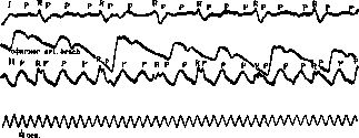
Figure 6. Correct form of atrial flutter with a ratio of atrial to ventricular contractions of 1:3. Sphygmogram - waves of equal magnitude follow at regular intervals. Electrocardiogram - atrial waves follow one another continuously, are superimposed on the ventricular complex and deform it. Phonogram - the strength of the first tone is the same. The absence of the atrial component of the first tone is clearly evident.
In the therapy of fibrillation and flutter, the main roles are played by digitalis and quinine, and especially its isomer quinidine. The significance of digitalis in A. F. lies not in eliminating the arrhythmia, but in eliminating the cardiac insufficiency caused or maintained by it; the mechanism of its action amounts to its vagotropic effect. By irritating the vagus nerve, digitalis causes a deterioration in conductivity between the atria and ventricles. Due to this, a smaller number of impulses for contraction originating from the atria reach the ventricles. The rhythm of ventricular contractions consequently slows down and becomes more regular. At the same time, due to lengthening of the pauses and the direct action of digitalis on the contractile function of the myocardium, the strength of ventricular contractions increases and the pulse deficit decreases or even disappears. All this leads to an improvement in the conditions of blood circulation. Digitalis is therefore especially indicated for tachyarrhythmic forms of fibrillation and flutter. For bradyarrhythmic forms, digitalis should not be given, since here it cannot manifest its main action in A. F.—slowing of the ventricular rhythm. Cardiac insufficiency, caused entirely or in part by A. F., is the most grateful and sensitive object for treatment with digitalis. With marked cardiac insufficiency, one can begin with 0.3 per day of Pulv. foliae Digitalis, if possible titrated. Usually after 3-5 days, slowing of the pulse and establishment of compensation begin. Then the dose of digitalis is reduced by half. If vomiting or bigeminy occurs, the administration of digitalis should be stopped completely. Subsequently, to maintain the pulse at a level of 65-80 beats per minute, small doses (0.03-0.1 per day) are usually sufficient. As in the experiment, intense irritation of the vagus nerve by digitalis can lead to complete cessation of fibrillation and flutter, however, to stop fibrillation by the administration of digitalis alone is very rarely successful. This is more often achieved with flutter, and due to the vagotropic effect of digitalis, the refractory phase of the atria is shortened, flutter passes into fibrillation, and then normal rhythm is restored. The introduction of quinine and especially quinidine into the therapy of fibrillation and flutter played a major role. At present, according to statistical data, with the help of quinidine, in almost 50% of cases of fibrillation, it is possible to restore sinus rhythm for a longer or shorter period. The action of quinine and quinidine is explained differently, depending on the views on the mechanism of origin of fibrillation. Authors who see the cause of fibrillation and flutter in the appearance of additional foci of excitation believe that the cause of quinidine's action is its effect on heterotopic foci of excitation. Quinine and quinidine act suppressively on the automatism of both nomotopic and heterotopic foci of excitation, however, the effect on heterotopic foci is stronger. Therefore, as with extrasystole, the activity of heterotopic foci ceases, and fibrillation disappears. Supporters of circular motion see the cause of quinidine's action in its effect on the refractory phase. Quinidine, according to Lewis, causes lengthening of the refractory phase and a decrease in conductivity of the atrial musculature. Both of these factors act in fibrillation in opposite directions. While slowing the spread of excitation promotes the continuation of fibrillation, lengthening of the refractory phase contributes to its cessation. The ratio of both of these factors determines the action of quinidine in fibrillation. This also explains the inconsistency of quinidine's action. The best results are obtained with quinidine in paroxysmal and tachyarrhythmic forms of fibrillation and flutter, especially when the duration of these rhythm disturbances is not very great. The bradyarrhythmic form gives significantly worse results. Before using quinidine, one should achieve maximum improvement in blood circulation by rest, digitalis, and all other appropriate methods of treatment. Quinidine is given according to a specific scheme. Start with 0.2 × 1 on the first day, 0.2 × 2 on the second day, 0.3 × 2 on the third day, 0.4 × 2 on the fourth day, 0.4 × 3 on the fifth day, and if the patient tolerates quinidine well, then one can give 0.4 × 4 on the sixth day and so on, continuing until a total of 18.0-20.0 of quinidine. Riker recommends giving 0.4 every four hours during the day and night. If the administration of 20.0 of quinidine has no effect, further administration is usually useless. Quinidine treatment is recommended to be combined with injections of 2-4 cm³ of camphor per day and the prescription of diuretin 1.0 × 3 and bromide. During the administration of quinidine, especially when the amount of medication taken reaches 10.0-15.0, patients often feel severe weakness, dizziness, palpitations; occasionally diarrhea and vomiting occur. If marked circulatory disturbance occurs, quinidine treatment must be interrupted. After the rhythm is restored, it is recommended to give quinidine for another 10 days (0.2-0.4 per day). Quinidine treatment should be carried out under hospital conditions with the patient on bed rest. - It is necessary to remember that quinidine is a protoplasmic poison and acts negatively on the entire organism and in particular on the cardiovascular system. Therefore, the administration of quinidine is contraindicated in marked degenerative changes in the myocardium, especially fresh ones, in obstruction of the branches of the coronary arteries and complete atrioventricular block. Quinidine should also not be used in cases where there is reason to suspect the presence of thrombi in the atria, since restoration of normal atrial contraction can cause embolism. Considering all the dangers of quinidine treatment, as well as the fact that the transition to sinus rhythm is sometimes brief, it is necessary to subject each individual case to careful clinical analysis before proceeding to quinidine treatment. Only those cases where fibrillation and flutter appeared not very long ago and where there is no marked circulatory disturbance should be subjected to quinidine treatment. 3. Fibrillation and flutter of the ventricles. Fibrillation and flutter of the atria usually do not pass to the ventricles. The impulse originating from the atria, upon reaching the atrioventricular node, quickly passes through the conduction system to the ventricles and causes excitation of their entire musculature. Experimentally, fibrillation and flutter of the ventricles can be caused by strong mechanical irritation of them, the effect of a strong electric current, and the administration of large doses of various pharmacodynamic agents (chloroform, digitalis, adrenaline, quinine, etc.). Combined irritation of the vagus and sympathetic nerves can also cause ventricular flutter. In humans, ventricular flutter can occur under anesthesia, under the influence of a strong electric current, and with various poisonings, especially digitalis. Cases of sudden death observed after the injection of strophanthin are associated with the appearance of ventricular flutter. A case was observed (Vogel'son and Kabanov) of ventricular flutter following the administration of adrenaline in atrioventricular block, which ended with the restoration of normal ventricular rhythm. Fibrillation and flutter of the ventricles are also observed in obstruction of large branches of the coronary vessels and are the cause of death in these cases. Sudden death due to severe psychic trauma (fear, fright, etc.) is also apparently explained by ventricular flutter. As experimental and clinical observations (Vogel'son) have shown, in the process of dying of the heart, fibrillation and flutter of the ventricles is the terminal act of cardiac activity. Thus, the hypothesis expressed by Hering that ventricular flutter is characteristic only of sudden death is not fully confirmed. The mechanism of origin of fibrillation and flutter of the ventricles is similar to the mechanism of origin of fibrillation and flutter of the atria. And the theories proposed to explain fibrillation and flutter of the atria can also be applied to fibrillation and flutter of the ventricles. The weak and disordered contractions of the ventricles in flutter cannot overcome the pressure in the arterial system and open the semilunar valves. If flutter continues, blood circulation ceases very quickly and death occurs. - When ventricular flutter occurs, the face turns pale and then becomes markedly cyanotic. Unconsciousness quickly sets in. The pulse disappears, breathing stops. Heart tones are not audible. Only with deep palpation in the epigastric region can the characteristic worm-like contractions of the ventricles sometimes be felt. Sometimes clonic and tonic convulsions are observed. A picture characteristic of a Morgagni-Adams-Stokes attack is obtained. And indeed, cases have been described where such attacks were caused by brief ventricular flutter. On the sphygmo-, phlebo-, and phonogram in ventricular flutter, no oscillations are obtained. On the electrocardiogram, instead of ventricular complexes, there is a large number of disordered waves of different shapes and sizes, and in flutter these waves are larger, while in fibrillation they are smaller. Brief attacks of ventricular flutter may end with the restoration of normal ventricular activity, but usually flutter ends in death.
When heart function is restored, consciousness returns, cyanosis disappears, and pulse and breathing appear. The electrocardiogram of the ventricular complex becomes normal. To determine with certainty that this condition and the Morgagni-Adams-Stokes attack are caused by ventricular flutter and fibrillation, it can only be established on the basis of the electrocardiogram. Therapeutic measures for ventricular fibrillation have little effect. In an experiment, a normal rhythm can be restored by intravenous administration of potassium chloride. In humans, it is necessary to immediately resort to measures usually used to restore circulation in collapse (camphor, strychnine, caffeine, etc.). It is possible to attempt cardiac massage or to inject adrenaline directly into the ventricles of the heart.
Related articles
Mentioned in
Cite this page
“Atrial Fibrillation.” Soviet Medical Encyclopedia. English translation of Bolshaya Meditsinskaya Entsiklopediya, 1st ed. (Moscow, 1928–1936), ed. N. A. Semashko. https://sovietmedicalencyclopedia.pages.dev/article/atrial-fibrillation/