Dicrotism
Historical document, translated for reference. It reflects medical knowledge of the 1920s–30s and is not medical advice.
Summary
An article from the 1928-1936 Great Medical Encyclopedia detailing dicrotism, a double pulse wave phenomenon associated with acute febrile diseases and low vascular tone. It covers sphygmographic findings, clinical significance, and various historical theories regarding its physiological origin.
Encyclopedia article (1928–1936)
DICROTISM (from the Greek dis - two and krotos - beat, literally "double beat"), a double pulse beat,
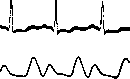
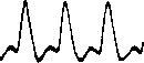
(detected in appropriate cases by simple palpation of the pulse). On the sphygmogram, such a pulse consists fig. 1. Usual dicrotic of two distinct waves—the main and the dicrotic (Figure 1)). A small dicrotic wave is also present on the normal pulse curve, but by palpation, the dicrotic wave of a normal pulse is not detected. A higher pulse dicrotic wave, easily determined by palpation, is observed mainly in acute febrile diseases and is considered a sign of decreased tone of the arterial musculature (Figure 2). In more severe febrile diseases (accompanied by a greater drop in blood pressure and severe tachycardia), the dicrotic wave is already located on the rise line of the next pulse wave—on the anacrote ("super-dicrotic pulse", Fig. 3) or merges with it ("monocrotism"). If the dicrotic wave is weakly expressed, it is located at a high level, on the catacrote of the main wave, relatively close to its apex. The dicrotic wave is small or absent in cases where vascular tone is increased (with high blood pressure) or when the arterial walls are thickened (sclerosed). There is as yet no universally accepted explanation for the origin of the dicrotic rise of the pulse curve. According to one theory, the dicrotic wave is formed due to the fact that part of the blood in the aorta, at the beginning of diastole, due to the slamming of the semilunar valves of the aorta, moves backward and, hitting these valves and being thrown back again, causes a wave directed behind the main wave to the periphery. The experiments of Chauveau and Marey speak in favor of this theory; they showed that the dicrotic rise on the pressure curve taken from the aorta coincides exactly on the blood pressure curve, i
i
i
'
'
" with the protrusion following the slamming of the semilunar valves; the same authors induced artificial aortic valve insufficiency, whereupon the dicrotic wave, which was clearly expressed before this, disappeared. True, these observations relate to the dicrotic notch on the aortogram. On peripheral arteries, the dicrotic wave is, generally speaking, more distant from the main one and more clearly expressed. Therefore, a doubt may even arise as to whether these rises on the curve of the aorta and on the peripheral arteries are of the same nature (Sahli, for example, denies their identity). But the different strength of the main and dicrotic waves can explain the greater lag of the latter from the main wave at the periphery,
vWVWWWWVWW
Figure 3. Super-dicrotic pulse. while the greater delay of the dicrotic wave explains the fact that it is more pronounced here (since on the peripheral sphygmogram it originates from a lower base). According to another theory, the dicrotic wave is of peripheral origin: it is a reverse centripetal wave of blood arising as a result of the collision of the pulse wave with obstacles along the bloodstream. But the reflected wave at the periphery should occur throughout the vascular system, and it is difficult to imagine how a single short and sometimes sharp notch is then created on the sphygmogram. If the distal (behind the sphygmograph) end of the artery is compressed, then, according to this theory, the dicrotic wave should disappear; meanwhile, it does not change in this case. Granstrem's clinical observations also speak in favor of the centrifugal direction of the dicrotic wave. According to a third theory, the wave reflected at the periphery hits the semilunar valves and, being thrown back again, causes the dicrotic wave; thus a centrifugal dicrotic movement is created. Finally, according to the "peripheral heart" theory (M. V. Yanovsky), the dicrotic notch is the result of an active peristaltic contraction of the artery. This latter theory cannot be considered sufficiently substantiated, and in particular, the data presented to prove that the dicrotic wave is a manifestation of the peripheral heart have not been confirmed by corresponding studies. It should be noted that in aortic insufficiency, the dicrotic rise is far from always absent, but only in its severe degree.
Author signatures: A. Myasnikov
Related articles
Cite this page
“Dicrotism.” Soviet Medical Encyclopedia. English translation of Bolshaya Meditsinskaya Entsiklopediya, 1st ed. (Moscow, 1928–1936), ed. N. A. Semashko. https://sovietmedicalencyclopedia.pages.dev/article/dicrotic-pulse/