Blood Pressure
Historical document, translated for reference. It reflects medical knowledge of the 1920s–30s and is not medical advice.
Summary
Blood pressure refers to the pressure exerted by blood on the walls of blood vessels and on the column of blood filling them. This article details the measurement techniques for arterial, capillary, and venous pressure, focusing on historical methods and instruments used in the 1920s-1930s.
Encyclopedia article (1928–1936)
BLOOD PRESSURE, the pressure which blood exerts on the walls of blood vessels (the so-called lateral blood pressure) and on the column of blood which fills the vessel (the so-called terminal blood pressure). Depending on the vessel in which it is measured, blood pressure is differentiated into arterial, capillary, and venous pressure. I. Arterial pressure. Methodology for measuring art. pressure. Methods for measuring arterial pressure. Blood pressure was first measured by Stephen Hales in 1727, by inserting glass tubes into vessels and determining the blood pressure by the height to which the blood rose in them. Accurate measurement of arterial pressure is greatly hindered by constant significant fluctuations, particularly pulse-related ones. Consequently, for accurate measurement of arterial pressure, it is necessary to record it with all these fluctuations over a certain period of time using an appropriate manometer, so that on this recording (curve) its height at each moment of this period can be determined. From such a curve, the magnitude of the average level of arterial pressure over a certain time can be accurately determined using a planimeter. The pressure at the height of the pulse waves is called maximum or systolic pressure; the lowest pressure between the pulse waves is minimum or diastolic pressure, and the difference between maximum and minimum pressure, i.e., the amplitude of the fluctuations of arterial pressure, is pulse pressure. The measurement of arterial pressure is further complicated by the fact that blood in the arteries is in rapid and pulsating motion. Usually, to transmit arterial pressure to the recording instrument - the manometer - a cannula is used, which is inserted into the central end of a transversely cut arterial branch. At this point, the blood flow in this branch ceases, and at the site of this branching, the lateral pressure in the artery from which this branch originates is measured. However, pulse waves continue to penetrate into the branch into which the cannula is inserted, or into the cannula itself, and strike the blind end of the manometer. Here, therefore, a phenomenon similar to the so-called hydraulic hammer develops, significantly distorting the true values of arterial pressure fluctuations and in particular strongly elevating the so-called maximum pressure (Muller, Fabre). This artificial increase in maximum arterial pressure must be taken into account in all measurements of arterial pressure by bloody methods, when a cannula is inserted into the central end of a cut artery, and in all determinations of maximum pressure by clamp methods, since in these methods maximum pressure is determined at the moment of complete or almost complete closure of the lumen of the corresponding artery. The distortion of the magnitude of arterial pressure from hydraulic impact undoubtedly decreases if in the experiment, to transmit arterial pressure to the manometer, T-shaped cannulas are used, which are tied into a cut artery (A. Muller), or a cannula is inserted into an arterial branch so that the end of the cannula is located near the site where the arterial branch originates from its trunk (Frank). In the latter method, an arterial branch with a small diameter compared to the diameter of the trunk should be chosen for measuring arterial pressure. Even with this method and when using a T-shaped cannula, an ideally accurate recording of the lateral blood pressure at a given place in the arterial system is not obtained. These inaccuracies of physiological methods for measuring arterial pressure must always be taken into account when evaluating the results of these measurements. As instruments for recording arterial pressure, various manometers are used. The oldest and most common of them is the simple mercury U-shaped manometer. Being rather inert, it reproduces the pulse fluctuations of arterial pressure in a completely distorted size, but it does provide some idea of the average level of arterial pressure. For this purpose, the mercury manometer is quite suitable and is still commonly used in experiments to record arterial pressure over longer periods of time. For more accurate recording of arterial pressure with all its fluctuations, and pulse fluctuations in particular, a whole series of manometers has been proposed, in which springs, levers, pistons sliding in cylinders, or membranes are used to transmit pressure to the recording instrument. To record the movements of the manometer, various levers are used to record the arterial pressure curve on a rotating drum, or mirrors attached to the end of the lever or to the manometer membrane: the light beam they cast records the movement of the mirror, and thus the pressure fluctuations on a moving strip of photographic paper (photokymography). In a simple mercury manometer, a float with a vertical wire rod is placed on the surface of mercury in the free end of the manometer; to the upper end of this rod a pen is attached, recording the movements of the float on a rotating drum. For accurate recording of arterial pressure fluctuations, all these spring, piston, membrane, and mirror manometers must possess certain qualities: possibly greater sensitivity, possibly a shorter period of their own oscillations and less mass, etc. All points important for the proper construction of such manometers were subjected to detailed theoretical and experimental analysis by Frank (O. Frank), who established the corresponding laws expressed in formulas. At present, in the construction of instruments for recording arterial pressure, these laws are followed, thanks to which modern manometers for measuring arterial pressure have reached considerable perfection. Among them, it is necessary to mention the spring and mirror manometers of O. Frank and the spring and membrane manometers designed by A. Muller. Opening arteries for direct measurement of pressure inside arteries in humans is not applicable in medical practice, and therefore one has to use various indirect methods, with these clamp methods being more or less accessible only to maximum pressure. In all these clamp methods of measurement, the pressure in a given artery is judged by the pressure that needs to be produced on the artery from the outside to obtain certain signs indicating its compression. Usually, clinical measurement of arterial pressure is performed by compressing the artery first with such force that all pulse phenomena disappear peripherally from the site of compression, i.e., with a force exceeding maximum pressure, and then gradually decreasing the pressure until the first appearance of the above-mentioned signs of passage of the pulse wave through the site of compression. The pressure exerted on the artery at this moment is considered to be the corresponding maximum pressure. Only some peripheral arteries are accessible to this method of measuring arterial pressure. For their compression, corresponding pelottes (usually rubber bulbs) (apparatus of Basch and Potain, Sabli) were previously used, but this method is applicable only to arteries that lie on a dense (bony) foundation, are covered only by skin, and are not surrounded by soft parts. The radial artery best satisfies these requirements and is therefore the artery of choice for measuring arterial pressure by these methods, and to a certain extent also the temporal artery. Nevertheless, these methods of measurement are now almost abandoned in practice, since compression of arteries and especially registration of the corresponding pulse phenomena by such methods is technically difficult and imperfect. At present, another method of compressing arteries to determine arterial pressure is generally accepted, namely the cuff method. When using the cuff method of compressing arteries, the brachial artery is used almost exclusively, because only it is located in relation to bony and soft parts in such a way that its compression by a cuff is not hindered. The width of the hollow cuff for compressing the brachial artery should be not less than 12 and not more than 14 cm, because both greater and lesser widths give less accurate data (Recklinghamsen). It would be best in each case to use a cuff of such a width as would be in the correct ratio to the diameter of the arm in the given person. The side of this cuff adjacent to the skin of the subject should be made of elastic rubber, approximately 1/2-1 mm thick. The other side should be of inelastic fabric. Compression of the artery by the cuff is performed by pumping air into it through a suitable rubber tube using a double bulb or other pump. The pressure in the cuff is measured using a mercury or spring manometer showing pressure up to 300 mm Hg. The first pulse waves are most easily determined by palpation (the method of Basch, Potain, Riva-Rocci, etc.) - the so-called palpation method. The appearance (or disappearance) of pulse waves can also be determined by the appearance (or disappearance) of fluctuations of the manometer needle measuring the pressure in the cuff - the so-called oscillation method.
Mercury manometers are unsuitable for this method due to their inertia, so for this purpose, Bourdon, spring, or other manometers sensitive to rapid fluctuations in pressure in the cuff are used (Recklinghausen's, Raspop's, Raga's, etc.). These fluctuations in pressure in the cuff can also be recorded by appropriate instruments (Uskov's, Recklinghausen's, Plesch's, etc.). Finally, the penetration of pulse waves into the segment of the artery under the cuff can be determined by auscultating the resulting sound phenomena - arterial tones (Korotkov's auscultatory method). For this purpose, the end of the stethoscope is placed at the location of the artery below the cuff. To determine maximum arterial pressure at present, the palpation or auscultatory method is used almost exclusively, measuring the pressure in the brachial artery by means of a cuff. It is worth mentioning another method for determining maximum arterial pressure in the finger arteries, proposed by Gartner, as it makes it possible to measure arterial pressure in more peripheral arteries. By placing a small hollow cuff at the base of the finger, it is exsanguinated and air is pumped into the cuff to such an extent that it does not allow blood to enter the finger. Then, by measuring the pressure in the cuff with a mercury manometer, it is lowered until the first appearance of coloration in the finger.
The determination of minimum pressure is somewhat more complex. For this purpose, the greatest fluctuations in either the volume of the brachial artery or the tension of its wall are used, which occur when the cuff exerts pressure on the artery that slightly exceeds the minimum pressure within it. At such pressure in the cuff, the artery in the interval between two pulse waves should collapse to the point where its walls come into contact with each other, and during the passage of a pulse wave, it should expand almost to its normal volume. Simultaneously, the greatest fluctuations in the tension of its wall will occur - from complete relaxation to tension by the full pressure of the pulse wave. If the pressure in the cuff is only slightly below the minimum arterial pressure, the artery between pulse waves will no longer collapse, and the fluctuations in its volume and the tension of its wall depending on the pulse waves will be relatively small. Due to this, when the pressure in the cuff is rapidly lowered below the minimum, there is an abrupt decrease in the fluctuations of the artery's volume and the tension of its wall.
To illustrate the phenomena that occur in the artery when measuring maximum and minimum arterial pressure by the auscultatory and oscillometric methods, a diagram (Fig. 1) of an oscillogram is presented here, as it is obtained when measuring arterial pressure, with the Korotkov sound phenomena schematically depicted alongside it. I phase, II phase, III phase, "Mx", "Mp. - I tones
I tones with noises and noises Figure 1. As already mentioned above, usually arterial pressure is measured by first raising the pressure in the cuff above the maximum and then gradually lowering it. The disadvantage of this method is that the pressure is measured after the veins in the arm have already been compressed to create congestion. But practically, it is easier to catch those transitions that determine the maximum and minimum pressure when the pressure in the cuff is falling rather than rising. As can be seen from the figure, small fluctuations in pressure in the cuff are recorded even at a pressure level exceeding the maximum arterial pressure. This is the result of the impact of pulse waves against the upper edge of the cuff. But at the level of maximum pressure, the fluctuations become distinctly larger, and at the same time, the tones of the so-called first phase of Korotkoff's sound phenomena appear. At a pressure 5-10 mm lower, a palpable pulse also appears in the radial artery. With further lowering of the pressure in the cuff, the fluctuations recorded by the oscillograph gradually become larger, but the tones are soon replaced by noises of the so-called second phase of Korotkoff's sound phenomena. While the tones of the first phase are considered manifestations of the first tensions of the arterial wall by the penetrating tops of pulse waves under the cuff, the noises of the second phase must be considered manifestations of the penetration of pulse waves through the narrowed segment of the artery compressed by the cuff into its distal uncompressed part. With further lowering of the cuff pressure, the fluctuations of the oscillogram gradually and uniformly become even larger, and the noises are again replaced by tones; these tones-the third phase of Korotkoff's sound phenomena-increase in intensity parallel to the increase in the fluctuations of the oscillogram, often acquire considerable sonority, and at the pressure in the cuff immediately preceding the minimum arterial pressure, they reach maximum intensity simultaneously with the maximum magnitude of the oscillogram fluctuations, and then disappear immediately or after a rapidly increasing weakening. The moment of transition from maximum oscillogram fluctuations to their decrease and the moment of the first weakening of the tones of the 3rd phase with decreasing pressure in the cuff determines the minimum arterial pressure. That phase, usually consisting of several weakening tones, which is sometimes captured after the tones of the 3rd phase, is designated as the 4th phase of Korotkoff's sound phenomena. Occasionally, under certain still not fully clear conditions, the tones of the 4th phase are heard at a considerable distance when the pressure in the cuff is lowered even below the minimum arterial pressure, and the transition from the tones of the 3rd phase to the tones of the 4th is indistinct. In such cases, the determination of minimum pressure by the sound method gives incorrect, too low numbers. The determination of maximum pressure is associated with a whole series of inaccuracies. The increase in the measured maximum pressure by clinical methods due to the so-called hydraulic shock has already been mentioned above. As long as there are no clinical methods to determine the magnitude of this hydraulic shock, it must be assumed that it will be the greater the greater the 73^ living force of the pulse wave arising in the aorta will be and the more rigid the arterial wall will be. The living force of the pulse wave arising in the aorta is determined by the amount of blood ejected by the corresponding systole of the left ventricle into the aorta (the so-called Schlagvolumen-systolic volume of blood) and the speed imparted to this mass of blood by the systole. About the pressure that develops when the pulse wave creates a hydraulic shock when measuring arterial pressure in humans by clinical methods, we have an idea only on the basis of measurements by Merke and Muller on two patients, in whom lateral pressure was measured by bloody methods and at times the brachial artery was appropriately compressed below the site of measurement. The increase in maximum pressure due to hydraulic shock in one case was 38 mm Hg, in another case-43. The second source of inaccuracy in determining maximum arterial pressure stems from the fact that in all the methods described for determining maximum pressure, it is measured by the pressure required to stop all pulse phenomena peripherally from the site of compression. This pressure on the artery, necessary for complete or almost complete closure of its lumen, must overcome two obstacles: 1) the blood pressure in the artery and 2) the resistance that the arterial wall offers to compression (AW-Arterienwandspannung). To determine this latter magnitude separately by clinical methods is only possible indirectly and then only approximately. In normal conditions, it apparently amounts to 8-15 mm Hg. Arteriosclerotic changes in the arterial wall, contrary to the generally accepted view, apparently do not have a great influence in terms of increasing AW; the degree of tonic contraction of the arterial muscle has a significant influence on AW; with its strengthening, AW reaches 30-40 mm Hg, with its weakening-2-5. To what extent the minimum pressure measured by clinical methods corresponds to the true minimum lateral pressure in a given artery is not sufficiently clarified. Since the measurement of minimum arterial pressure is made when the artery lumen is closed only between two pulse waves, it must be assumed that the influence of hydraulic shock is less here and therefore the minimum pressure measured by clinical methods is closer to the true value than the maximum arterial pressure measured by these methods. A comparison of the results of arterial pressure measurement obtained simultaneously by bloody methods and by clinical methods on people (in a few experiments) by O. Muller, Merke, and A. Muller nevertheless shows a significant approximation of the numbers obtained by clinical methods to the numbers obtained by bloody methods. Of course, for maximum arterial pressure determined by clinical methods, there remains a significant increase due to hydraulic shock. Arterial pressure under various physiological conditions. Arterial pressure in large arteries falls little and gradually, and only in the precapillaries does it decrease sharply. An idea of the change in maximum and minimum arterial pressure separately from the aorta to the capillaries is given by a comparison of the results of arterial pressure measurements in various arteries made by Klisiecki. From these data, it is seen that towards the periphery, maximum arterial pressure decreases, while minimum pressure gradually somewhat increases. Correspondingly to this, the amplitude of pressure pulse fluctuations gradually decreases towards the periphery and in arteries with a diameter of 0.2-2.3 mm it disappears. If in previous measurements (Spengler, Volkmann, etc.) in individual experiments, a somewhat higher maximum pressure was sometimes obtained in an arterial branch than in the proximal trunk, this is explained partly by the fact that hydraulic shock in more distal arteries gives a higher pressure rise than in more central ones (see the results of Merke, Muller, Fabre), and partly by the fact that arterial pressure in peripheral arteries fluctuates within wide limits due to temporary local vaso-motor influences. Therefore, in individual measurements, higher numbers for arterial pressure can be obtained in more peripheral arteries compared to central ones.
age
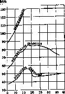
Figure 3. Changes in arterial pressure in youth and middle age. As for the level of arterial pressure in various animals, the curves of Klisetsky provide an idea of the arterial pressure in dogs. In horses, the average arterial pressure is 155 mm, in guinea pigs -75 mm Hg. As these examples show, there is no proportionality between the size, resp. weight of the body, and arterial pressure in various animals. This incidentally confirms that basic principle of hemodynamics that the level of arterial pressure by itself does not determine the intensity of the circulation, as it is measured for example by the amount of blood ejected by the heart per unit of time. (A reservoir located at a very great height does not give a greater amount of water per unit of time if the pipeline is narrow.) Factors determining the level of arterial pressure. In humans, the level of arterial pressure depends on sex and age (see table and fig. 3-6, as well as below - blood pressure in children). The indicated arterial pressure is the average for a given sex, age, etc.). Deviations from these average values within 10-20 mm Hg must still be considered normal. Changes in arterial pressure in old age (according to Richter). This fact of the coincidence of increased arterial pressure in women with menopause is the most convincing example of that influence which internal secretion has on blood pressure. During pregnancy, starting from the 5th month, a slight increase in arterial pressure is observed (on average no more than 10 mm Hg). Childbirth is accompanied by a significant rise in arterial pressure, but after its completion the pressure quickly drops 10-20 mm Hg below the original level; this drop sometimes reaches even 50 mm Hg. In old age, a decrease in minimal arterial pressure and an increase in the amplitude of pulse fluctuations of arterial pressure should be noted, apparently due to sclerosis of the aorta (see below). (On the influence of constitution on arterial pressure see table, as well as the curves of Alvarez-Zimmermann - fig. 7.) Influence of constitution on arterial pressure (according to Larimore). Constitution Amount Max. pressure Min. pressure Pulse pressure Surface of body in m2 M. F. M. F. M. F. M. F. M. F. 1. Sthenics ...... 65 62 126.3 118.4 78.8 73.4 47.5 45.0 1.81 1.539 2. Hyposthenics .... 68 55 116.3 115.6 71.7 72.3 44.6 43.3 1.61 1.66 3. Asthenics ...... 51 106.8 63.3 68.2 43.5 37.5 1.67 1.5J6[ Age 4 n ё™ 5
__________ women Figure 4. Changes in maximum arterial pressure by age (according to Saller). There are none. Worthy of note is the increase in pressure pulse fluctuations during the period of puberty (fig. 3) and the sharp rise in arterial pressure in women after 40-45 years. The greater a person's height and weight, the higher their average arterial pressure. On the influence of climate, conditions and time of year on arterial pressure - see Hypotension. Fluctuations in arterial pressure during the day in normal conditions do not usually exceed 10- --------ви- - я 75 o 70- § з: 65- -1 м в г [ г + _1 к - 1 Figure 13 16 19 22 25 28 31 34 87 40 43 46 49 52 55 58 61 64 67 age ----------- women Figure 5. Changes in minimum arterial pressure by age (according to Saller). years, but they differ in a certain regularity. Arterial pressure reaches its lowest level during the deepest sleep, by morning and during the day it gradually increases, reaching a maximum in the afternoon and evening hours. After food intake, max. arterial pressure also increases by mm 10, while min. arterial pressure does not increase, but even falls, as a result of which the amplitude of pressure pulse fluctuations clearly increases. This increase in arterial pressure with simultaneous acceleration of the pulse is a manifestation of the increase in minute blood volume after food intake (see Circulation, physiology). In the standing position, pressure (maximal-119, minimal-80) is slightly higher than in the lying position (115 and 72). On the influence of muscle work on arterial pressure, the experiments of Liljestrand and Stenstrom (Liljestrand, Stenstrom) give a good idea (see table).
6. Changes in pulse pressure by age (according to Saller). 20 mm Hg, but they differ in a certain regularity. Arterial pressure reaches its lowest level during the deepest sleep, by morning and during the day it gradually increases, reaching a maximum in the afternoon and evening hours. After food intake, max. arterial pressure also increases by mm 10, while min. arterial pressure does not increase, but even falls, as a result of which the amplitude of pressure pulse fluctuations clearly increases. This increase in arterial pressure with simultaneous acceleration of the pulse is a manifestation of the increase in minute blood volume after food intake (see Circulation, physiology). In the standing position, pressure (maximal-119, minimal-80) is slightly higher than in the lying position (115 and 72). On the influence of muscle work on arterial pressure, the experiments of Liljestrand and Stenstrom (Liljestrand, Stenstrom) give a good idea (see table). ются резкие колебания артериального давления (Gries-bach) и чаще, чем среди других профессий, встречается повышенное артериальное давление (Гельман). Наклонность к гипертонии отмечается еще y рабочих-пивоваров и рабочих анилинового (красочного) производства. Умственная работа также вызывает повышение артериального давления, но в значительно меньшей степени, чем физическая. B общем и она дает Влияние работы мышц на артериальное давление (по Liljestrand'y и Stenstrom'y). Поло же ние Cp. скорость в м в мин. Систолич. давление в мм Hg Стоя.................. . Bo время ходьбы..........} Bo время бега ............) 78,3 95,3 И3.0 184,5 236,6 264,3
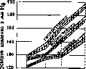
возраст These changes in arterial pressure during muscular work are determined by the increase in minute blood volume. Since the degree of decrease in total resistance to outflow of blood from central arteries to periphery lags behind the degree of increase in minute volume, arterial pressure increases, and pulse pressure increases the most. The more familiar the physical movement, resp. the better the training, the smaller the increase in average pressure level, but the greater the increase in pulse pressure. Чем привычнее физ. движение, resp. чем лучше тренировка, тем повышение среднего уров-давления меньше, но тем боль-Рисунок 7. Изменение арте-ше увеличение пуль-риального давления y rnR(Xrn ттЯРТ1рттист Чя-женщин различных кон- LUbU1(J давлении, oa ституций.
ВИСИТ ЭТО OT ТОГО, что чем больше тренировка, тем меньше при данной физ. работе учащение пульса и тем больше нарастание систолического объема крови (Schlagvolu-men). Вызванное физ. работой повышение артериального давления после прекращения работы y здоровых людей быстро-в течение нескольких минут-проходит и притом тем скорее, чем работа была легче. После продолжительной и тяжелой работы вслед за повышением артериального давления непосредственно следует период его понижения.-Значительное влияние на изменение артериального давления вследствие физ. работы имеют те внешние условия, при к-рых происходит работа. При высокой внешней t° (напр. в условиях работы литейщиков) физ. работа вызывает особенно сильное понижение минимального артериального давления и вследствие этого очень резкое увеличение пульсового давления. B качестве примера приведены на рис. 8 диаграммы изменений артериального давления y рабочего-вы-нимадьщика литейного цеха после 8 и 16 мин. тяжелой работы вблизи печи (Стож-кова-Гольдфарб). Из имеющихся пока немногочисленных наблюдений над влиянием различных профессий на артериальное давление вытекает, что как-раз y литейщиков отмеча- 124,6 132,5 134,2 142,3 161,5 168,6 164,0 Диасто-лич.давление в мм Hg Пульсовое давление в мм Hg 95,0 94,1 98,6 97,5 101,7 102,4 35,8 37,5 40,1 43,7 64,0 66,9 61,6 Частота пульса 73,8 88,8 92,4 94,8 127,2 141,3 153,4 Потребление 02 (в см3) в мин. 270 1.010 1.330 1.860 3.120 3.390 3.650 тем большее повышение, чем она менее привычна и чем труднее. Ho особенно резко повышается артер. давление под влиянием всякого псих, возбуждения и в'особенности такого, к-poe сопровождается ощущениями отрицательного свойства (гнев, страх, и т. п.). Это влияние псих, моментов необходимо иметь в виду при каждом измерении артериального давления, т. к. самый процесс измерения, в особенности если он для данного лица непривычен, вызывает большее или меньшее волнение и благодаря этому некоторое повышение артериального давления. Влияние сравнительно небольших изменений атмосферного давления на артериальное давление отчетливо отмечается только непосредственно после его изменения, причем как понижение, так и повышение его вызывают подъем артериального давления. При значительном понижении атмосферного давления, например на больших горных высотах (4.000 - 5.000 м), наблюдается наклонность к стойкому повышению артериального давления, достигающему уровня на 40-50 мм Hg выше нормы. Физич. работа на больших высотах вызывает более резкий подъем артериального давления, чем та же работа на уровне моря. Особенно выражена наклонность к повышению артериального давления на больших высотах y пожилых людей. Это повышение артериального дав"-ления на горных высотах есть результат недостатка O2, так как исчезает под влиянием вдыхания 02(Loewy). Между артериальным давлением. давлением спинномозговой жидкости и внутриглазным давлением при физиол. и пат.
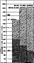
Рисунок 8.
under certain conditions, a certain parallelism can be noted in the sense that an increase in arterial pressure is accompanied by a certain rise in pressure of the cerebrospinal fluid and intraocular pressure. On the other hand, an increase in intracranial pressure usually causes an increase in arterial pressure in the initial period. Blood pressure is the transformed energy of heart contractions; it in turn is converted into the energy of blood movement and into the energy of tension of arterial walls; the latter during the diastole of the ventricles is again converted into blood pressure and its movement. Blood pressure is an essential intermediate phase in the transformation of a significant part of the energy of heart contractions into the energy of blood movement. Consequently, a certain level of arterial pressure is an essential condition for adequate blood circulation. - Due to the intermittent nature of the pumping work of the heart, constant fluctuations in blood pressure and rhythmic alternation of the said energy transformations occur in the arterial system. In addition to pulse waves, which depend on the work of the heart and are called first-order waves (about them - see Pulse), second-order waves are also observed in the arteries. These include fluctuations in arterial pressure synchronous with respiration. These fluctuations occur as follows: at the beginning of inspiration, arterial pressure falls and reaches its minimum at the very beginning of inspiration, after which follows a rise that continues throughout the remaining part of inspiration and reaches its greatest height at the beginning of expiration. Then in the remaining part of expiration, pressure falls, to again reach its minimum at the beginning of inspiration. Consequently, inspiration corresponds mainly to an increase in arterial pressure, expiration to a decrease, but these respiratory fluctuations in arterial pressure lag somewhat behind the respiratory waves, and this lag is the stronger the faster the respiratory rhythm. Respiratory waves of arterial pressure are the result of the fusion of a whole series of arterial pressure waves arising in various ways in connection with the phases of respiration. A distinction is made between 1) respiratory fluctuations of arterial pressure of mechanical origin. They are of two kinds: a) as a result of those displacements of blood that occur during respiratory movements of the chest due to a decrease in pressure on the heart, aorta and large veins during inspiration and due to an increase in this pressure during expiration, and b) as a result of suction of blood into the chest cavity during inspiration (with simultaneous compression of veins in the abdominal cavity) and as a result of a delay in the entry of blood into the chest cavity during expiration. The mechanical influences of the second category have a decisive influence on the character of respiratory waves of arterial pressure and cause the lag of these waves compared to the phases of respiration. 2) Respiratory fluctuations of arterial pressure of nervous origin: a) Traube-Hering waves; they arise as a result of the spread of periodic irritations of the respiratory center by irradiation to the vasomotor center; these waves have the opposite character to the respiratory waves of arterial pressure of mechanical origin and, being weaker than the latter, apparently contribute to some weakening of them; b) Fredericq waves depend on the effect on arterial pressure of acceleration of the heart rate during inspiration and its slowing down during expiration (see Pulse). Third-order waves, or Siegmund Mayer waves. These fluctuations in arterial pressure are independent of both cardiac and respiratory functions. They are observed with a frequency of 6-9 per minute, may be quite regular, but also completely irregular both in height and in length. The height of these waves in rabbits can reach 40 mm Hg. These waves are often obtained in a pronounced form also on plethysmograms of humans. They are regarded as the result of independent fluctuations in the tone of vasomotor centers, causing periodic contractions of vessels in extensive areas of the body and thus fluctuations in arterial pressure. Significance of changes in arterial pressure for judging blood circulation. The level of arterial pressure is determined by the amount of blood ejected by the heart per unit of time into the aorta, and by the resistance which the movement of blood encounters, mainly on the periphery of the arterial system in the form of friction. The decisive significance for the magnitude of this friction is the lumen of small precapillary arteries in the aggregate (see Blood circulation, physiology). Consequently, the level of arterial pressure is determined by the ratio between the degree of narrowing of peripheral arteries and the work of the heart, measured by the minute volume of blood. The level of arterial pressure is regulated by the vasomotor nervous system and is established by it depending on the need to adapt blood circulation to the demands of organs and tissues in the sense of greater or lesser blood supply to them, determined by their functional state. However, an increase and decrease in blood pressure are not the main way by which the organism uses to change blood circulation and blood supply to organs. On the contrary, by changing the distribution of blood and the speed of blood circulation, the organism rather strives to maintain as normal a level of arterial pressure as possible. Thus, for example, during enhanced muscular work, the amount of blood ejected by the heart into the aorta per unit of time increases up to 10 times, while arterial pressure increases only by 7/9. - In the vasomotor nervous system - see Blood circulation, Vasomotors and Vegetative nervous system. - Besides the vasomotor nervous system, the function of regulating arterial pressure is also attributed to the system of endocrine glands, from which the medulla of the adrenal glands, hypophysis, ovaries and other glands secrete, as numerous experiments and clinical observations show, secretions that undoubtedly affect the tone of arterial musculature. On the basis of these same observations, the impression is gained that the correct relationship of the functions of these various glands maintains the normal level of arterial pressure. But as long as the prevailing view is that the regulation of arterial pressure by these secretions nevertheless occurs with the mediation of the vasomotor nervous system; the latter, like the entire vegetative nervous system with the endocrine glands, is connected into a complex but functionally unified apparatus regulating vegetative functions, including arterial pressure. In particular, under normal conditions, the vasomotor nervous apparatus coordinates the work of the heart and arterial pressure above all for the purpose of adequate blood supply to tissues. Therefore, with a primary strengthening of the tone of the musculature of peripheral arteries, i.e., with their narrowing in extensive areas of the body, arterial pressure rises, since the work of the heart is strengthened, in order to by compensatory acceleration of the current through narrowed peripheral arteries to ensure adequate blood supply to tissues. With weakening of the tone of the musculature of peripheral arteries, i.e., with their dilation, arterial pressure falls, and also by increasing and strengthening the work of the heart as much as possible, the necessary level of arterial pressure for adequate current of blood in capillaries is maintained. But since strengthening of the work of the heart is possible only with sufficient blood supply, and with significant dilation of vessels in extensive areas of the body - particularly in the area of the abdominal viscera - the blood supply to the heart decreases, then often under these conditions arterial pressure nevertheless falls, and a picture of insufficiency of blood circulation develops due to vascular insufficiency as a primary phenomenon and cardiac insufficiency as a secondary one. With strengthening of the work of the heart, if the vasomotor nervous apparatus works normally, no increase in arterial pressure occurs, since the peripheral vessels dilate to a corresponding degree and the current of blood is strengthened. With weakening of cardiac activity, i.e., with the heart ejecting per unit of time into the aorta an insufficient amount of blood, arterial pressure with a normally working vasomotor apparatus does not fall, since it (the vasomotor apparatus) by strengthening the tone of arterial musculature strives to maintain the level of arterial pressure necessary for adequate blood supply to the most vital organs, mainly the heart and brain. Since blood from arterioles enters capillaries, theoretically it is quite possible for arterial pressure to rise also as a result of narrowing of the lumen of all capillaries in the aggregate. Some authors assume such an origin of an increase in arterial pressure in certain pathological conditions and in particular in acute nephritis. They consider the latter not as a local disease of the kidneys, but as a disease of the capillaries of the entire body, leading to an increase in resistance to the current of blood in them (Frank, Kylin). This point of view is not yet generally accepted, and the possibility of an increase in arterial pressure due to changes in capillaries has not been clarified. In comparison with the lumen of arterioles and capillaries, other factors that could influence the magnitude of friction and
BLOOD PRESSURE
746 Consequently, they have no significant importance, as they can easily be compensated for by changes in the lumen of small vessels. Thus, for example, the viscosity of blood, with properly regulating lumen of small vessels in the vasomotor apparatus, has no influence on blood pressure. The level of arterial pressure gives no possibility to judge the useful work of the heart, the speed of circulation, the degree of blood supply to tissues, and the minute volume of blood in particular. The amount of blood ejected by the heart per unit of time into the aorta can be large even with low pressure (e.g., in anemia) and small with high pressure (e.g., in hypertension with insufficient cardiac activity). Conversely, with high arterial pressure, the minute volume of blood can be both large (e.g., during heavy physical work) and small with low arterial pressure (e.g., in vascular insufficiency). A high level of arterial pressure only gives the right to judge that the left ventricle with each contraction ejects a certain amount of blood into the aorta, but it is completely impossible to judge this amount based on the level of maximum and minimum arterial pressure separately. Some idea of this amount is rather given by the pulse pressure, since its magnitude undoubtedly depends on this systolic volume. But since the magnitude of pulse pressure is also determined by a second factor, namely the elasticity of the walls of central arteries, it is possible to judge the magnitude of the systolic volume by pulse pressure only, taking into account this second factor, which however is far from easy. Nevertheless, high maximum arterial pressure and large pulse pressure give the right to assume a heart muscle contracting with considerable force; but we have no right to assume that such a muscle is completely healthy, since an altered heart muscle, but working with all its reserve force, can for a certain time maintain high arterial pressure. If high arterial pressure persists for several weeks, one can assume the presence of hypertrophy of the left ventricle. All attempts to extract from measurement data (maximum, minimum and pulse pressure) and pulse frequency more precise indications of the functional state of the circulatory organs by means of formulas (Strassburger, Erlanger, Nikolaev, Kabanov, etc.) have not been practically justified and are theoretically incorrect, since for the reasons stated above blood pressure and pulse frequency give no right to judge the sufficiency or insufficiency of circulation. The level of minimum arterial pressure depends mainly on the speed of outflow of blood from the arterial system. This speed of outflow of blood is determined primarily by the width of the lumen of small arteries. Consequently, minimum arterial pressure gives some right to judge the degree of tonic contraction of the peripheral arterial musculature. When evaluating the significance of pathological changes in arterial pressure, it is necessary first of all to take into account the influence of heart rate on it. It has an influence on arterial pressure in the sense that maximum arterial pressure decreases with increased pulse rate, minimum pressure increases, and pulse pressure decreases. With bradycardia, opposite changes in these values are observed. The influence of bradycardia on arterial pressure is especially characteristic in complete heart block, when maximum pressure is elevated, minimum pressure is lowered, and pulse pressure is increased to a significant degree. The indicated influence of pulse rate on maximum, minimum, and pulse pressure of course takes place only under otherwise equal conditions and in particular, if the amount of blood ejected by the heart into the aorta per unit of time does not change. A certain height of the blood pressure level is usually attributed to the so-called physio-constants, i.e., to such constant physiological phenomena that are necessary for the proper function and life of the entire organism. In this respect, blood pressure is compared with body temperature. This comparison is quite appropriate, since indeed significant deviations of arterial pressure from the normal level, as well as significant changes in body temperature, are incompatible with life. And just as the organism relatively easily tolerates significant increases in body temperature, it easily tolerates significant increases in arterial pressure, even increases twice the normal, whereas corresponding decreases in arterial pressure, as well as significant decreases in body temperature, are not observed or are observed only shortly before death. In general, however, arterial pressure under different physiological and pathological conditions fluctuates and often even very significantly and for long periods. For long-term changes in arterial pressure, both in the direction of increase and decrease, see Hypertension and Hypotension; for changes in arterial pressure in arteriosclerosis, see Arteriosclerosis; in nephritis, see Nephritis. Pathological changes in arterial pressure. Short-term increases in arterial pressure are observed with strong painful sensations, asphyxia, lead colic, during tabetic crises, in some cases of angina during the attacks themselves (angina pectoris vasomotorica), during the so-called vascular crises of Paly (see Crises), with increased intracranial pressure, under the influence of adrenaline injections, etc. Short-term decreases in arterial pressure are observed in humans with vascular insufficiency, collapse (see Circulation, pathology), after significant blood loss, with acute weakening of cardiac activity. All the indicated acute increases in arterial pressure are the result of rapid strengthening of the tone of the arterial musculature, mainly of central origin. In acute decreases in arterial pressure, as they are observed in the most pronounced form in collapse, there is, on the contrary, a decrease in the tone of the arterial musculature, mainly in the area of the abdominal viscera. At this time, dilation of the capillaries of the same area, caused by the influence of substances acting like histamine directly on capillaries, may participate or even have main importance; in the dilation of vessels, plexuses of small veins also participate; the so-called reserve blood reservoirs (see Circulation) are filled. Large blood losses apparently lower arterial pressure not by direct decrease in the mass of blood, but by causing a paretic state of the vasomotor centers through disruption of their blood supply. Blood losses up to 1/2 liter in humans usually do not cause a decrease in arterial pressure. In general, even relatively large changes in the total blood volume, as shown by experimental observations, do not significantly change arterial pressure, as long as the vasoconstrictive nervous apparatus reacts normally. Thanks to it, the vascular system, by changing the tonic contraction of its musculature, quickly and without allowing blood pressure to decrease or increase, changes its capacity. From clinical facts proving the ability of the circulatory system to adapt without changing arterial pressure to fluctuations in the total blood volume, one can point to polycythemia and the algid period of cholera, which as a rule are not accompanied by significant changes in arterial pressure. Drinking large amounts of fluid with a normally functioning vasomotor apparatus also does not cause significant changes in arterial pressure. But in hypertensive conditions, when the vasomotor apparatus does not react normally, drinking 1 liter of water, e.g., during the so-called water test for determining functional capacity of the kidneys, often causes very strong increases in arterial pressure. With valvular heart diseases, with arteriosclerosis of the heart, arterial pressure is on average somewhat below normal. If with these diseases cardiac insufficiency develops, then pressure does not change or slightly increases. This latter phenomenon was also noted by Sali (Hochdruckstauung). With improvement of cardiac activity, arterial pressure in these cases somewhat decreases. The absence of a decrease in arterial pressure or even some its increase despite insufficient work of the heart, i.e., despite the decrease in minute volume of blood, is explained, as already indicated, by the corresponding vasomotor reaction. A sharp decrease in arterial pressure in diseases of the heart itself is usually observed only shortly before death or with the addition of vascular insufficiency, e.g., with the addition of some infectious process. In this respect, among diseases of the heart itself, the only exceptions are acutely developing infarctions of the heart muscle due to thrombosis of the coronary arteries; their development is accompanied by a sharp decrease in arterial pressure, not corresponding to the increase in temperature that is usually observed in these cases. Edema do not influence the level of arterial pressure. Ascites, in general accumulation of fluid in the abdominal cavity and increase in intra-abdominal pressure undoubtedly cause an increase in arterial pressure. With compensated valvular heart diseases, pulse pressure partly reveals typical
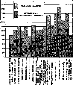
Figure 9. Diagram of changes in arterial pressure in various pathological conditions (in mm Hg). Changes having diagnostic significance. Thus, in pure mitral stenosis, pulse pressure as a rule is decreased, and the degree of this decrease, all other conditions being equal, is proportional to the degree of stenosis. In aortic insufficiency, on the contrary, an increase in pulse pressure is highly characteristic due to a lowering of minimal arterial pressure. This increase in pulse pressure is usually more pronounced in aortic insufficiency of endocarditic origin than in aortic insufficiency of syphilitic or arteriosclerotic origin. The increase in pulse pressure in aortic insufficiency is explained by the fact that the systolic volume of blood is increased and the outflow of blood from the aorta is intensified due to the backflow of a portion of blood from the aorta back into the left ventricle. The degree of increase in pulse pressure in aortic insufficiency also makes it possible to a certain extent to evaluate the degree of insufficiency (fig. 9). Vascular insufficiency (see Blood circulation, pathology) is accompanied by a fall in arterial pressure. In its most pronounced form, as already indicated, it is observed in collapse and in anaphylactic shock. In the clinic, vascular insufficiency is observed as a rule in infectious diseases; in abdominal and typhus fevers and in severe influenza it is most pronounced and gives the characteristic lowering of arterial pressure in these diseases during the period of most severe condition and during the period of temperature decrease. At the same time, pulse pressure is also decreased. At the beginning of infectious diseases, arterial pressure may even be somewhat elevated, especially during chills. Among chronic infectious diseases, tuberculosis is distinguished by a tendency to lower arterial pressure. This lowering usually corresponds to the degree of pulse acceleration. Among diseases of the internal secretion glands, Addison's disease gives a characteristic lowering as a manifestation of insufficiency of the chromaffin system and the consequent loss of the stimulating effect of adrenaline on the vasomotor nervous system. Diseases of the thyroid gland, myxedema and Basedow's disease also give characteristic changes in arterial pressure. For myxedema, a certain lowering of arterial pressure with decreased pulse pressure is typical, while for hyperthyreosis, on the contrary, there is a tendency to increase arterial pressure and especially to increase pulse pressure. These changes in arterial pressure in hypo- and hyperthyreosis are fully explained by those changes in blood circulation which accompany the corresponding disturbances of thyroid function: in hyperthyreosis, accelerated blood circulation with increased minute volume due to both tachycardia and increased systolic volume, while in hypothyreosis, on the contrary, slowing of blood circulation. In severe anemias, certain changes in arterial pressure are also as a rule observed - an increase in pulse pressure with a tendency to lower the average level of arterial pressure (F. B. Greenberg). These changes are also explained by the increased speed of blood circulation due to increased systolic volume with a tendency to lower vascular tone.
Blood pressure in the pulmonary circle is considerably lower than in the systemic; the average B. p. in the pulmonary artery corresponds approximately to 1/3-1/4 of the aortic pressure; in the dog it in numerous determinations fluctuated around 15-20 mm Hg, differing significantly less in constancy than the arterial pressure in the systemic circle. The maximum pressure in the pulmonary artery in the dog averages 43.3 mm Hg, the minimum - 11.9 (Wiggers), pulse pressure - 24 mm Hg. Pulse pressure in the pulmonary artery is also characterized by considerable lability. The magnitude of B. p. in the pulmonary artery depending on the phases of respiration in the dog during natural breathing fluctuates in such a way that at the beginning of inspiration a decrease in pressure occurs, which at the end of inspiration is replaced by an increase; during expiration pressure rises, and only at the end of expiration or in the respiratory pause does a decrease in pressure begin again. Capillary pressure - see Capillaries, physiology.
P. Venous pressure. Venous pressure is measured in experiments on animals either by means of a tube tied into a vein or a cannula thick enough to be inserted into a vein without disturbing the blood flow in the vein. Usually a water manometer is used, since venous pressure is so low and its fluctuations are so small that they cannot be accurately measured by mercury manometers. More often for the same reason venous pressure is expressed in mm of water (to convert to mercury column it is necessary to divide by 13.5). In humans venous pressure is measured either by direct - bloody - or indirect methods. The first method, proposed by Moritz and Tabora, is quite analogous to the experimental method of determining venous pressure in animals. As there, it is necessary to pay special attention to the fact that the place in the vein where venous pressure is measured is exactly at the level of the right atrium; this is necessary to exclude the influence of hydrostatic pressure, which at the generally low venous pressure has the most essential influence on the height of the latter. In humans venous pressure is usually measured by the bloody method in the veins of the bend of the elbow.
Non-bloody, or indirect clinical methods of measuring venous pressure are based on the principle of determining the external pressure necessary to cause collapse of a superficial vein. The most common method consists in hermetically attaching over a superficial vein of the arm a bottomless capsule with a glass cover and increasing the pressure in this capsule until the vein collapses. The pressure in the capsule is measured by a water manometer attached to it.
The following numbers give an idea of the height of venous pressure in the dog (see table). Venous pressure in the dog (according to Burton-Opitz). mm Hg
femoral dext. 0.4
saphena sin. 0.4, brachial dext. 0.4, portae 0.4, mesenterica 0.4, gastro-lienalis 0.4
renalis .... 5,4 7,4 3,9 8,9 14,7 10,1 10,9 V. facialis sin. . . » jug. ext. sin. . . . . . dextr. . . 0,1 . . . cav. sup. (peripheral segment) . . . 1,4 . . . cav. sup. (central part) .......2,8 . . . fem. sin.......5,4 When measuring venous pressure by normal and more perfect methods, the following numbers have been obtained (see table). In the veins of the lower extremities, in the recumbent position, the venous pressure does not differ significantly from the venous pressure in the veins of the elbow bend; in the standing position it increases considerably, but regarding the degree of this increase, the data are inconsistent. With complete rest and other equal conditions, the venous pressure in the same individual is almost constant. Age and sex do not have a significant influence on venous pressure. Muscular work as a rule increases it, and this increase can reach 100%. The negative pressure inside the chest cavity, so-called Donders' pressure, has a significant influence on venous pressure. As a result of opening the chest cavity in Burton-Opitz's experiment, the pressure in the superior vena cava of the dog increased from -30 mm water to +32.4, i.e., by 62.4 mm H2O. Correspondingly, respiratory movements cause significant fluctuations in venous pressure, and the magnitude of these fluctuations is directly proportional to the depth and speed of respiratory movements. But these respiratory fluctuations in venous pressure are expressed only in the central veins; here they reach 70 mm H2O, and during inspiration, negative pressure is obtained in these veins, which, as is known, can cause aspiration of air into the vena cava when it is opened. In the veins of the lower extremities, the respiratory fluctuations in venous pressure are of exactly the opposite character to those observed in the upper half of the body, i.e., in the veins of the lower extremities, venous pressure increases during inspiration and decreases during expiration. This is explained by the fact that the inferior vena cava during inspiration is under increased intra-abdominal pressure. Toward the periphery, the respiratory fluctuations in venous pressure are relatively quickly smoothed out, and in the cubital vein with normal breathing through a wide cannula (more than 0.5 mm in diameter) they do not exceed 0.5-2-4 mm H2O, and with the strongest breathing-5-8. If in normal conditions an enhanced inspiratory movement cannot cause a corresponding decrease in venous pressure in the veins of the elbow bend, then prolonged difficulties in the entry of air into the lungs, as we observe in emphysema of the lungs, pleural adhesions, rigidity of the chest cavity, kyphoscoliosis, etc., by lowering Donders' pressure, can significantly lower the venous pressure measured in peripheral veins. This factor, which significantly affects the level of venous pressure, must be taken into account when evaluating other pathological deviations of venous pressure (Kroetz). In general, the level of venous pressure is determined primarily by the difference between the level of blood pressure in the capillaries and the level of blood pressure in the right atrium. The level of blood pressure in the capillaries depends on the central arterial pressure and on the lumen of the precapillary arteries and capillaries or on the total number of open capillaries. This last factor - the degree of opening of precapillaries and capillaries, varying in different organs and parts depending on their functional state, can cause different levels of venous pressure in the corresponding peripheral veins. During muscular work, to this factor locally increasing venous pressure, is added the influence on it of the mechanism of muscular contractions and venous valves. The level of pressure in the right atrium depends on the ratio between the influx of venous blood to the heart and its performance capacity, measured by the amount of blood it can transfer per unit time further into the pulmonary artery. Another basic factor determining the level of venous pressure is the degree of tonic contraction of the venous musculature. The less this tone, the greater the total lumen of all veins and the lower, under other equal conditions, will be the venous pressure. Apparently the venous system, especially at the periphery, possessing numerous and richly developed networks, has the ability to vary its capacity within very wide limits, and accordingly can also vary the amount of blood contained in it. Part of these venous plexuses can apparently to a certain degree be disconnected from the main venous branches and trunks by narrowing the corresponding branches. This too, by changing the amount of circulating blood, should influence the blood pressure in general and venous in particular. The tone of the venous musculature, as well as the arterial musculature, is determined by the chemical composition of the blood, in particular by the CO2 content in it (Henderson), and partly by the vasomotor innervation. All these numerous and complex factors determining the level of venous pressure are not yet amenable to sufficiently precise accounting, and therefore the pathogenesis of the changes in venous pressure observed in various pathological conditions is not always amenable to precise analysis. It has been most definitely established that insufficiency of cardiac activity is accompanied by an increase in venous pressure. This increase goes more or less in parallel with other manifestations of blood stasis in the great circulation, enlargement of the liver, congestive renal phenomena, edema, etc. It is most sharply expressed in patients with mitral valve defects, during decompensation of which venous pressure rises to 200-312 mm H2O; followed by arteriosclerotic cardiosclerosis and insufficiency of the aortic valves. With compensation, mitral defects give for venous pressure an average number-103, cardiosclerosis-72, hypertension-78, syphilitic aortitis-70, aortic insufficiency-66 mm H2O. A fact deserving special attention is that in heart diseases the level of venous pressure in each case, as a rule, under other equal conditions changes strictly in proportion to the degree of decompensation. Curves (Pavlovskaya and Soboleva) (figure 10) give an idea of these fluctuations and the corresponding changes in other manifestations of cardiac insufficiency. Eyster points out the practical significance of measuring venous pressure for establishing more precise indications for bloodletting in severe cardiac insufficiency and for controlling the effect of this therapeutic method. Bloodletting of 350-600 cm3 in suitable cases of cardiac insufficiency gives a direct decrease in venous pressure by 100-200 mm H2O. The influence on venous pressure of vascular insufficiency is studied less in detail (see Circulation, pathology). But on the basis of available experimental observations and clinical data, for pure vascular insufficiency, a fall in arterial pressure and a fall in venous pressure are particularly characteristic. Thus, in histamine shock (see Circulation, pathology) and in severe acute infectious diseases, a fall in venous pressure is as a rule observed; at the end of typhoid fever, for example, venous pressure usually falls to 20-40 mm water column (Waldman).
g.
Lang. III. Blood pressure in children. Arterial blood pressure in children generally follows the same conditions as in adults; - its height fluctuates at lower figures, but the fluctuations themselves are more significant, which is explained by the influence of the nervous system, increased reflex excitability of the vascular musculature, further - the influence of hormones of the internal secretion glands, which are in the period of growth and development, and especially - changes in the anatomical and functional relationships between the heart and the vascular system during the child's growth period. The gradual increase in arterial blood pressure with the child's growth may depend partly on the increase in the strength of the heart muscle (Benjamin). In determining arterial blood pressure in children, generally the same technique and apparatus are used as in adults. As for Korotkov's auscultatory method, it is fully applicable to children, and with skill and some patience, it can give sufficiently reliable figures for systolic blood pressure - even at the very earliest age; however, in regard to diastolic blood pressure in very young children, this is not always possible. In infants, a cuff 41/2-6 cm wide is used, in older children - a wider one, but not wider than 12 cm. The state of excitement of the child - crying, weeping movements - produce a short-term but rather significant increase in arterial blood pressure, while the intake of food, as well as the transition from a sitting position to a recumbent one, do not have any special effect; during sleep, blood pressure is usually somewhat lowered. To avoid errors, several determinations of blood pressure should be made in succession and the average taken: it has been noted that in subsequent measurements, the figures obtained are somewhat lower. Arterial blood pressure in newborns fluctuates, according to Seitz's data, between 75 and 100 mm Hg; according to Neu, it averages 90 mm (Gartner's apparatus); on average it equals 80 mm Hg. Blood pressure in premature infants is even lower; according to Trumpp, it does not exceed 60-70 mm Hg. With age, arterial blood pressure increases. This increase, proceeding unevenly, is most intense in the first year of life: during this period, the increment is the same as in the subsequent 12 years of life. Besides infancy, the greatest intensity of blood pressure increase falls on the age from 6 to 10 years. According to Popov's data, arterial blood pressure increases on average by 2 mm monthly during the first year, reaching 100 mm by its end. Bauchwitz, Trumpp, Feer, Bruning, Rominger and others give an average figure of 80 mm up to 6 months of age and 90 mm up to 2 years. Systematic studies of arterial blood pressure in healthy infants gave the following average figures (according to Korotkov): from 3 to 6 months - 84.7 mm Hg, from 6 to 12 months - 92.5 mm Hg, from 1 to 2 years - 97.5 mm Hg. The amplitude of fluctuations is considerable; thus, blood pressure at the age of 3 to 6 months fluctuates between 73 and 100 mm; from 6 to 12 months between 81 and 100 mm, from 1 year to 2 years - between 85 and 105 mm (Sokolov and Rosenthal). The figures for blood pressure in older children can be taken from the summary data of Potten, Nobecourt and others, obtained with Potten's apparatus: arterial blood pressure in cm Hg Age K- cm g . . . l . . . » ... 7-9 8-10 9-12 9-12 L . , 12-14 » . . 14-16 » . . 10-14 11-15 13-15 Arterial blood pressure increases not only parallel to age, but also parallel to growth and weight. In this respect, the comparisons of Nirnheim (E. Nirnheim) are interesting, who determined blood pressure with the Recklinghausen apparatus with a saline manometer. z ^ =1 то =1 3 i a m ЫЗ я ьс Age нК o o o н« « нч 100-105 15-20 7 » ...... 106-110 но 21-25 121.5 8 » ...... 111-115 26-30 126.5 9 » ...... 116-120 31-35 133.7 10 » ...... 124.9 121-125 36-40 11 » ...... 129.5 126-130 125.5 41-53 151.6 12 » ...... 131-135 133.5 | 13-14 ...... 142.5 136-140 141-145 136.6 136.6 L46-150 148.4 ' All these data refer to a. brachialis. When measuring blood pressure in the finger arteries with Gertner's apparatus, smaller figures were found: in infancy - from 64 to 82 mm, from 1 year to 16 years - 82-108 mm . There are formulas for calculating the average height of arterial blood pressure for a given age; thus, Katzenberger gives the formula (up to 13 years of age): 80 + 2x, where x=number of years. This formula, however, does not give sufficiently accurate figures, especially for early age. According to Popov, blood pressure of children up to 1 year can be determined by the formula: 76 + 2w (w=number of months of life). Maslov gives the formula: 100 + x, where x=number of years. As for the minimal, diastolic blood pressure, by the end of the year it averages 49 mm, reaching 88 mm by 15-16 years (Maslov). Its determination in small children is associated with technical difficulties and is not always reliable. It can be considered that in healthy infants it does not undergo large fluctuations and is approximately 35-45 mm (Rosenthal's data by Korotkov's method).- Pulse pressure in early age is relatively large - approximately 50- 60 mm; with age it decreases. Arterial blood pressure undergoes lowering or elevation in the most diverse diseases; thus, sharp drops in blood pressure are described with significant dehydration of the body, with jaundice, anaphylactic shock, etc.; low blood pressure during diphtheria, indicating myocardial damage, serves as a poor prognostic sign. An increase in blood pressure occurs in pneumonias, meningitis, etc.; it is particularly persistent in chronic nephritis, in acute glomerulonephritis, in scarlet fever, where this phenomenon can be one of the early signs of kidney damage. It should be emphasized, however, that with the same disease, blood pressure may be elevated, normal, and even lowered depending on how much those mechanisms on which blood pressure depends are involved in the suffering; for example, in a child with pneumonia, blood pressure is often elevated; but if (as is often the case in the so-called cardiac form of pneumonia) there is simultaneously damage to the heart muscle or paralysis of the precapillary network, then blood pressure may be lowered. However, certain pathological conditions are characterized by persistent changes in blood pressure; for example, in physically and mentally retarded children, blood pressure is lowered (Strokov); low blood pressure figures are apparently characteristic of lymphatic children; in neurotics and spasmophiliacs, it is, on the contrary, rather above normal. Similarly, in children with exudative diathesis, with and without eczema, blood pressure often gives persistent elevations.- Venous pressure in small children has not yet been studied. Capillary pressure (according to Frontali; Kylin's apparatus) for infancy fluctuates between 100 and 160 mm of water, from 2 to 10 years - between 90 and 160 mm. A. Sokolov.
Related articles
Mentioned in
Cite this page
“Blood Pressure.” Soviet Medical Encyclopedia. English translation of Bolshaya Meditsinskaya Entsiklopediya, 1st ed. (Moscow, 1928–1936), ed. N. A. Semashko. https://sovietmedicalencyclopedia.pages.dev/article/blood-pressure/