Blood Circulation
Historical document, translated for reference. It reflects medical knowledge of the 1920s–30s and is not medical advice.
Summary
This article from the 1928–1936 Great Medical Encyclopedia examines the physiology and pathology of blood circulation, detailing the cardiovascular system's structure, the driving forces of blood flow, and regulatory mechanisms as understood in early 20th-century medicine.
Encyclopedia article (1928–1936)
BLOOD CIRCULATION. Contents: I. Physiology. Plan of the structure of the blood circulation system....... 543 Driving forces of blood circulation............ 545 Movement of blood in vessels........ 546 Velocity of blood circulation................. 549 Minute volume of the heart.......... 553 Velocity of the blood circuit...... 557 Regulation of blood circulation and distribution of blood in individual organs.......... 559 II. Pathology. Circulatory insufficiency of cardiac origin ................ 566 Circulatory insufficiency of vascular origin ................ 575 Circulatory disorders in connection with pathological states of other organs ............. 582 I. Physiology. Blood circulation is one of the methods of connection 1) between individual parts of more complex animal organisms and 2) between the external environment and the tissues of the organism through the mediation of the organs of respiration, digestion, and excretion. The means of connection is the blood, and the pathways of communication are blood vessels forming a closed circuit; through this circuit, blood circulates under the influence of the work of engines, the most important of which is the heart. The functions of blood circulation are: 1) delivery of nutrient substances from the places of their entry into the organism to the organs or tissues consuming or storing them, resp. from places of storage to places of consumption; 2) transport of metabolic products from the places of their formation to the places of excretion or further processing; 3) transport of various types of white blood cells, which under certain conditions pass into tissues to perform various functions; 4) delivery of hormones; 5) various protective substances, and 6) heat (Bethe). Blood circulation, which performs all these functions, is found in higher animals—vertebrates, whereas lower animals have various other forms of transport. These forms represent either stages on the path of development of the complex blood circulation system of higher animals or various devices of another type serving the same purpose. History of the question. Our modern concepts of blood circulation in higher animals have developed gradually over the past two millennia. Among the scientists who contributed to this development, the greatest credit belongs to W. Harvey, who was the first to give (about 300 years ago) a correct description of the movement of blood. Harvey's discovery was preceded by the works of M. Servet and the Italians Leonardo da Vinci, Colombo, Cesalpino, and Fabricius ab Aquapendente. In Harvey's doctrine of blood circulation, only the anatomical proof of the communication between veins and arteries was lacking. It was soon provided by the description of capillaries and the blood flow in them by Malpighi and Leeuwenhoek (Malpighi, 1661; Leeuwenhoek, 1686). A correct concept of arterial pressure in higher animals was given only in 1733 by Stephen Hales. Plan of the structure of the blood circulation system. The entire apparatus of blood circulation in higher animals consists of few parts that differ from each other in structure and function, but together constitute a closed circular path along which blood continuously circulates. This circular movement of blood is caused, maintained, and directed by the pressure difference created by the work of the heart, which has the function of a forcing pump inserted into a complex system of tubes. The tubes with the smallest lumen and thinnest walls, penetrating almost all organs in huge numbers (blood capillaries), are the most important part of the blood circulation system, where between the blood and tissues, as well as between the blood and atmospheric air, the exchange of respiratory gases and substances necessary for tissue nutrition, products of their metabolism, etc., is carried out. Other tubes of larger diameter carry blood from one capillary system to another; some of these tubes—veins—collect blood from the capillaries of the entire organism except the lungs, and uniting into two large vessels, the so-called venae cavae, carry it into the right atrium; from it, the blood enters the right ventricle and is pumped by it through the pulmonary artery and its branches into the pulmonary capillaries. Blood along this entire path—from the capillaries of all organs to the capillaries of the lungs—is venous, i.e., blood that has given up a significant part of its oxygen to the tissues in the capillaries of the organs, consequently relatively poor in O2, but has accepted CO2 from the tissues and is therefore relatively rich in it. In the pulmonary capillaries, the arterialization of blood takes place, i.e., the absorption of O2 by the blood from the atmospheric air and the release of excess CO2 to it. From the pulmonary capillaries, arterial water—i.e., blood saturated with O2 and having given up a significant part of CO2—flows through the pulmonary veins into the left atrium and thence into the left ventricle. The latter pumps it through the aorta and arteries back into the capillaries of the entire organism (see figure on page 390). In higher animals, the blood circulation system is usually divided into the so-called lesser or pulmonary circle and the so-called greater circle. It is more precise to speak of the lesser and greater half-circles, since the pulmonary circle and the greater circle, communicating with each other in the right and left heart, only together form the common closed circle of blood circulation. Two pumps are inserted along the path of the blood: the right and left heart. The pulmonary half-circle of blood circulation, consisting of the pulmonary artery, pulmonary capillaries, and pulmonary veins, is short and, in terms of branching and vessel distribution, is built relatively simply; it has a special purpose—the exchange of blood gases with the atmospheric air by means of respiratory organs. The organ performing this function—the lungs—is the only organ through the capillaries of which the entire circulating mass of blood flows entirely. In mammals, the lesser half-circle of blood circulation has a definite engine—the right heart. The greater half-circle of blood circulation has a more complex structure: its purpose is to perform all the above-listed functions of the blood circulation system except for the exchange of blood gases with atmospheric air. In the greater half-circle, one can distinguish a whole series of constituent parts (half-circles or loops), each of which begins in the aorta and ends in the superior or inferior vena cava and consists of corresponding arteries, capillaries, and veins. The shortest such half-circle is the system of coronary vessels of the heart itself, beginning in the aorta near its mouth and ending in the right atrium and all heart cavities by the Thebesian veins. Each of the organs of the greater circle of blood circulation thus always receives only a part of that blood which flows through the lungs and is expelled by the left ventricle into the aorta. In terms of blood supply, they are all located side by side. The only partial exception is the liver, which is supplied with blood not only directly from the arterial system (through the hepatic artery), but also by blood that has already flowed through the spleen, stomach, and intestine (through the portal vein). Thus, in this loop, blood flows through two capillary networks: first through the network of the spleen and gastrointestinal tract, and then through the network of the liver. Such a structure protects the blood circulating in the rest of the organism from sharp fluctuations in composition that would otherwise result from the direct entry into the general bloodstream of nutrient and other materials absorbed in the intestine. In the liver, as is known, these materials undergo processing and storage or are excreted by bile back into the intestine. (The special significance of the spleen for blood circulation will be discussed later.) The lungs and liver, according to the stated scheme of construction of the blood circulation system, receive mainly venous blood. But this venous blood serves a special function: in the lungs—for its arterialization by gas exchange with atmospheric air, and in the liver—for the filtration processing just mentioned. Just like the liver, the lungs, in addition to venous blood (through the pulmonary artery), also receive arterial blood (through the bronchial arteries). Driving forces of blood circulation. The heart is the main engine of blood circulation. It acts mainly as a forcing pump; its function as a suction pump has not been finally clarified. In addition to the heart, there is undoubtedly a whole series of other auxiliary engines. The main ones are located where they are most needed, i.e., in the venous system. The fact is that the living force imparted to the blood by the heart is spent in a significant part on overcoming obstacles in the sphere of small pre-capillary arteries and capillaries, and therefore the outflow of blood from the capillaries to the heart needs additional motive forces. As such an auxiliary engine of blood flow in veins, first, the contraction of skeletal musculature acts, expressing blood from the veins in the direction of the right atrium with the assistance of venous valves. This auxiliary engine works automatically—specifically when the blood supply to the muscles increases, i.e., precisely during their work. The second very important auxiliary engine is the suction force of the thoracic cavity, which increases with each inspiration and directly contributes to the inflow of blood to the heart through the superior vena cava. Simultaneously, during inspiration, due to the contraction of the diaphragm, intra-abdominal pressure increases and pressure on the liver increases particularly. As a result, blood rushes from it into the inferior vena cava and the thoracic cavity, where pressure decreases at the same time.
The acceleration of blood flow by peristaltic contractions of the arterial musculature in response to their stretching by the pulse wave, assumed by some authors, should be considered as yet unproven in higher vertebrates. Likewise, such contractions of capillaries and veins that could play the role of additional engines of blood flow have not yet been established in them. Movement of blood in vessels. Movement of blood in arteries. Due to their structure, arteries possess the most important functional qualities: extensibility, elasticity, and contractility. As experiment shows, in a system of tubes, a pulsating (i.e., rhythmically intensifying) fluid flow is converted into a uniform one under the condition of extensibility and elasticity of the tube walls, at a sufficient length thereof, and under the condition of the presence of a certain resistance to the outflow of fluid from the entire system. All these conditions are realized in the arterial system thanks to the elastic properties of the arterial walls and due to the fact that the tonic contraction of their musculature creates a certain obstacle on the periphery for the outflow of blood into the capillaries. Thus, the conversion of a pulsating blood flow in the arterial system into a uniform flow at its end is the most important function of the elastic properties of the arterial walls. But out of the pulsating flow, a uniform one is obtained thanks to the elastic properties of the arterial walls only in the capillaries, whereas in the arteries there is a pulsating flow. As direct experiment also shows, with elastic walls, a rhythmically intensifying fluid flow through a system of tubes at an equal mean pressure gives a better effect in the sense of the amount of fluid flowing per unit time than a uniform flow (Fleisch). The correctness of this observation was also confirmed by theoretical calculation. Consequently, with a blood flow pulsating due to the work of the heart in the arteries, the elasticity of their walls facilitates the work of the heart; it also converts the pulsating flow into a uniform one by the time of transition into the capillaries. The significance for blood circulation of the elastic properties of the arterial walls also follows from the following considerations: if the arterial walls were rigid, the heart with each contraction would have to move the entire mass of blood in the arteries as a whole. Thanks to the elasticity of the arteries, the heart ejects its systolic volume of blood into the central arteries, mainly into the aorta. The walls of these vessels, stretching, easily perceive the systolic volume of blood, but, stretching, they at the same time accumulate the energy of cardiac contractions and then, contracting by virtue of their elasticity during ventricular diastole, convert the accumulated energy of the heart back into the movement of blood. Thus, heart systoles cause pressure waves and flow acceleration waves in the arterial system (see Pulse), which run through all arteries and maintain the blood flow. Among the central arteries, the aorta plays the main role in the function of accumulating the heart's energy; it should be regarded as a special regulating reservoir (Windkessel). This function of it corresponds to the circumstance that the lumen (more precisely, the cross-sectional area of the lumen) of the aorta in an adult human is somewhat larger than the sum of the lumens of the branches extending from it, whereas subsequently during the branching of the arteries, the sum of the branch lumens is always larger than the lumen of the trunk from which they depart. Due to this, the blood pressure does not drop along the length of the aorta and is equally high at the mouths of all its branches (Hürthle, R. Thoma). The capacity of the entire circulatory system in total (including reserve reservoirs—see below) is determined by a constant greater or lesser tonic contraction of the contractile elements of the walls of the heart, arteries, veins, and capillaries. The degree of this tonic contraction for each of these parts of the circulatory system is such as to maintain in it the proper level of pressure necessary to support the proper blood flow. Blood pressure in the arteries is the converted energy of cardiac contractions, accumulating due to that difficulty of blood outflow from the arteries which is caused and regulated by the tonic contraction of the arterial musculature, mainly pre-capillary arteries. The energy of arterial pressure is converted into the movement of blood through the entire circle of blood circulation. The highest pressure in the circulatory system is created at the beginning of the arterial system. By the proper ratio of heart work and tonic contraction of the arterial musculature, such a level of blood pressure is constantly maintained in the arterial system as is necessary for the proper flow of blood through the capillaries and veins. But the arterial musculature has yet another most important function. The fact is that the function of various organs and tissues varies in quantitative and partly in qualitative respects within the widest limits depending on the needs of the whole organism. To perform the corresponding work by organs, their proper blood supply is necessary; with their increased work, it must increase accordingly. Thus, for example, during increased work, skeletal muscles receive an amount of blood per unit time up to 10 times exceeding their blood supply at rest. In order to guarantee to all organs the possibility of maximum work if necessary, their blood supply would have to either be constantly maximal or have the ability to quickly change and adapt to the activity of one or another organ. In the organism of higher animals, the second, incomparably more economical principle is implemented. The blood supply of organs and tissues must therefore change in accordance with their function: this is an indispensable condition for their normal working capacity. From this stems the necessity for the blood circulation system of a purposeful distribution of the blood at its disposal and directing it to one or another organ in such an amount as they need at a given moment to perform the work they carry out in the interests of the whole organism. The simplest way to increase the blood supply of a working organ would seem to be the dilation of the arteries supplying it with blood; but in view of the fact that the organism has a definite limited amount of blood at its disposal, such a dilation of the supplying arteries of the working organ for the purpose of its increased blood supply would lead to a drop in central arterial pressure, and thanks to this, the influx of blood to the working organ would not only not be increased, but could even decrease. From this it follows that the increased blood supply of one organ is possible only with the simultaneous restriction of the blood influx to others, i.e., simultaneously with the dilation of the supplying arteries of the working organ, the supplying arteries and capillaries of other organs must be narrowed, since the total capacity of the entire circulatory system must be preserved, and the level of blood pressure necessary for sufficient blood circulation must be retained in it. Thus, during the work of one or another larger organ, a redistribution of blood throughout the entire organism must always occur. This redistribution of blood is carried out first of all by the arteries. By changing the degree of tonic contraction of their musculature, the main arterial trunks and their branches direct the blood entering the aorta into various organs in such an amount as they need at a given time to maintain life and perform the work assigned to them. At the same time, the small arteries, capillaries, and veins of those organs that are working and are therefore subject to increased blood supply expand accordingly, and at the same time the tonic contraction of the musculature in the arteries of other, non-working organs increases. In aggregate, the tone of all arterial musculature is established in such a way as to keep the central arterial pressure at the proper level. The main role in the distribution of blood by the arteries belongs to the smaller peripheral pre-capillary arteries, which are especially rich in musculature. But the walls of the central arteries are also very rich in musculature (it is about 50% in the wall of the aorta and carotid arteries), and apparently the distribution of blood among organs begins already in the large arterial trunks. It is possible that this function of the arteries is facilitated by the marginal muscular ridges described by Dragendorff, which are present on the inner surface of the arterial wall at the sites of branches. Capillary blood circulation. The most important role of capillaries in blood circulation has already been defined above (see also Capillaries). To perform the function of the exchange of respiratory gases and various substances between blood and tissues, three conditions are necessary: 1) the largest possible surface of capillary walls and the possibility of changing the surface depending on the work or rest of one or another organ; 2) the greatest possible thinness of capillary walls; 3) the proper time of contact of blood with the capillary wall. The first two conditions are ensured by the structure of capillaries (see). The third condition for the fulfillment of their function by capillaries (the proper time of contact of blood with the capillary wall) is achieved by a corresponding slowdown of blood flow in the capillaries. In this regard, it must be noted that blood flows most slowly precisely in the capillaries of those organs where particularly intensive exchange of various products between blood and cells takes place: in the capillaries of the liver and in the intertubular capillaries of the kidneys.
This particularly slow blood flow is achieved thanks to the fact that the greater part of the blood flows through these capillaries after it has already overcome one capillary system. Venous blood circulation. The function of veins is considered to be the return transport of blood from the capillaries to the heart; until very recently, very little attention was paid to their role in the blood circulation of the entire organism. Their crucial role in regulating the supply of blood to the heart and in influencing changes in the circulating and reserve blood volume is now becoming clear. Undoubtedly, the proper capacity of the venous system is of essential importance for blood circulation and this capacity is determined, like the capacity of the arterial system, by tonic contraction of the vascular musculature. But since blood pressure in veins is very low compared to arteries, this musculature in veins is correspondingly less developed. The capacity of the venous system is estimated to be 3-4 times greater than that of the arterial system and in all probability varies to a much greater extent than that of the arteries. Since the work of the heart in terms of the amount of blood pumped by it per unit of time depends primarily on the amount of blood that flows through the venae cavae to the heart, the proper performance of their function by the veins—specifically the supply of blood to the heart—is of paramount importance for blood circulation. This supply will depend primarily on the proper tone of the musculature of the venous system; a decrease in it must cause an increase in the capacity of all veins, which will lead not only to a slowing of the blood flow in the veins themselves, but, taking into account the large capacity of the venous system, also to a constant retention in it of a significant part of the blood and, by virtue of this, to a decrease in the filling of the lesser circulation and of the arteries and capillaries of the greater circulation; as a result, a general deterioration of blood supply is obtained.-In some places, arteries and veins are connected not only by capillaries, but also by so-called shunt channels described by Hoyer (see Blood vessels). Velocity of blood circulation. Velocity of blood flow. The fundamental phenomenon by means of which the function of blood circulation is carried out is movement, the flow of blood in the vessels. Therefore, the velocity of blood flow is that magnitude which is of the greatest importance for both physiology and pathology of blood circulation. With respect to the velocity of blood flow in the circulatory system, the following concepts can be distinguished: 1) the speed of movement of a single blood particle at a given point in the circulatory system; 2) the amount of blood flowing per unit of time through the cross-section of the lumen of a given vessel; 3) the so-called minute volume of blood, i.e., the amount of blood flowing in 1 minute through the cross-sectional area of the lumen of the vascular pathway at one or another point of the general circulatory loop (e.g., through the cross-section of the mouth of the aorta or pulmonary artery or through the cross-section of all pulmonary capillaries combined or through the general cross-section of all capillaries of the greater circulation), excluding the liver and renal glomeruli; 4) the velocity of circulation of one or another blood particle; it is determined by the time required for this blood particle to return to its starting point after completing a path along the entire circulatory loop; and 5) the blood supply of one or another organ, i.e., the amount of blood flowing per unit of time through the area of one or another cross-section or combination of sections of arteries, veins, or capillaries of a given organ. The velocity of blood flow in various sections of the circulatory system is extremely diverse. It is determined according to Poiseuille's law by the difference in blood pressure between two points in the direction of the axis of the vessel and by the resistance that friction offers to the movement of blood (see Hemodynamics).-It is necessary, however, to draw attention to the fact that the resistance to blood flow in the arteries, starting from the aorta and ending with the precapillaries, increases, although the cross-section of the total lumen of the vascular bed in this direction increases significantly. This gradual increase in the total lumen in itself should facilitate blood flow, since, other things being equal, the obstacle to fluid flow through tubes decreases with an increase in their lumen. But to a greater extent than the lumen increases as the arteries branch into an ever-increasing number of branches and into ever smaller branches, the resistance increases due to the enormous increase in the area of contact of the blood with the vascular walls and the consequent increase in friction.--An idea of the increase in the area of contact of the blood with the vascular walls as the aorta branches into arteries, arterioles, and capillaries is given by Krogh's calculation (see Capillaries). Both of these factors taken together (the change in the lumen and the increase in the area of contact of the blood with the vessel walls) create in the arterial system at its periphery that resistance to blood flow which is necessary to maintain the proper pressure level and the corresponding blood supply to organs. The organism uses changes in the size of the arterial lumen to alter blood pressure and change the blood supply of organs and tissues. Thus, the arterial system is a system of tubes that are branches of one main trunk, the aorta, and the cross-section of the total lumen (or the sum of the lumens) of these tubes gradually increases toward the periphery. Corresponding to this increase in the total lumen toward the periphery, blood pressure and the velocity of blood flow also decrease, since, other things being equal, when a fluid flows through a system of tubes, the lateral pressure of the fluid and the velocity of its flow decrease in proportion to the expansion of the lumen of the tubes. But the increase in the total lumen, and therefore the slowing of blood flow and the drop in arterial pressure throughout the arterial system, occurs to a relatively small degree. Only in the precapillary arterioles does the blood pressure drop sharply, and upon their transition into capillaries, where a very sharp expansion of the entire blood bed occurs over a very short distance, the blood flow slows down correspondingly and strongly. However, the notion that the main resistance to blood flow is encountered in the capillaries is incorrect. As corresponding blood pressure measurements show, its greatest drop occurs in the small arteries; in the capillaries, the pressure is already very low, it does not exceed 10 mm Hg (see Capillaries) and drops along the capillaries by only 1–2 mm. This is explained by the fact that the capillaries, compared to the arterioles nourishing them, represent a colossal expansion of the lumen of the blood bed. This expansion compensatorily and excessively reduces that increase in resistance to blood flow which occurs in the capillaries due to increased friction caused by the increase in the area of contact of the blood with the vascular walls. Resistance to blood flow in the capillaries is furthermore small due to their very short length (0.2–0.5 mm). The curves on page 737 (Fig. 2) give a schematic representation of the correlation between the change in the area of the total cross-section of the circulatory system, the velocity of blood flow, and blood pressure throughout the vascular system. They are compiled on the basis of Schleier's curves obtained by him by measuring an injection preparation of the blood supply system of the mesenteric artery of a dog with the addition of a schematic curve of changes in the velocity of blood flow. To measure the velocity of blood flow in one or another place of the arterial and venous systems, apparatuses built on the principle of the so-called Ludwig's clock are used, or methods utilizing Hering's principle (see below), or devices built on the pendulum principle (e.g., Chauveau's hemodromograph), or on the principle of Pitot tubes (Cybulski's photo-hemotachometer). Using the latter apparatus, Klisiecki obtained the following figures on a dog in 1929 (see Table 1). The values for the velocity of blood flow in a dog obtained by previous authors are generally lower. For example, Chuyevsky obtained 149–385 mm per 1 sec. for the carotid arteries of a dog (Grützner's clock). According to previous authors, the pulse oscillation of the flow velocity in arteries Table 1. Periods of heart contraction Velocity of blood flow (in mm) in 1 sec. In upper end of femoral art. and in carotid art. In lower end of femoral art. In art. saph. (branch of femoral art.) lumen diam. 0.5–1.2 mm In ram. plant. art. saph. diam. 0.2–0.38 mm During diastole ... During systole ... During dicrotism ... Fluctuations from respiratory waves 3rd order oscil. ... 500 545 570 50 100 600 650 660 20–70 100 10–45 10–45 30–70 135 135.5–138 4–7 is significantly greater; for example, according to Cybulski, in the femoral artery of a dog, systole is 356 mm, dicrotism 300 mm, diastole 177 mm per 1 sec. Chauveau, Bertolus, and Laroyenne determined 520 mm for systole and 150 mm per 1 sec. for diastole in the carotid artery of a horse, and established an acceleration during eating of up to 1,200 mm for diastole and up to 1,570 mm for systole. In the human aorta, the velocity of blood flow was determined on the basis of a calculation from the diameter of the aorta and the amount of blood pumped by the heart per unit of time into the aorta (see above); 144–216 mm per 1 sec. was found. This calculation cannot be considered accurate. In the above-cited data of Klisiecki, attention is drawn to the greater velocity of blood flow in the branches of the aorta than in the aorta itself.
This is partly explained perhaps by the fact that the sum of the cross-sectional areas of the branches of the aorta, according to Thoma's data (at least in humans), as already mentioned, is somewhat smaller than the cross-sectional area of the aorta, whereas with further branching of the arteries, the sum of their lumens progressively and significantly increases. In the arterioles, the blood velocity sharply decreases. Gürtle found it, for example, to be equal to 5 mm per 1 second with an arteriole diameter of 0.03 mm. In the capillaries, the blood flow, as is necessary for the performance of the function of gas exchange and other dissolved substances between the blood and tissues, is uniform and even slower. On average, the speed of blood flow in the capillaries is 0.5 mm per 1 second, fluctuating within fairly wide limits primarily depending on the diameter of the capillaries (from 0.2 to 1.7 mm per 1 second). The flow velocity in the capillaries is determined directly under the microscope, respectively by microphotographic means. The blood flow in the veins compared to the capillaries accelerates again, because their total lumen compared to that of the capillaries decreases significantly. But since the total lumen of the veins still remains greater than the lumen of the aorta, the blood flow in them is significantly slower than in the aorta. The highest blood flow velocity was found (Burton-Opitz) in a dog in the external jugular vein, namely 147 mm with a lumen of 5.5 mm; for the femoral vein of a dog, he also found 63 mm with a lumen of 4.4 mm, in the mesenteric vein (lumen 6.5 mm) - 85 mm. Frank found in the same dog that blood makes the journey from the femoral vein to the right atrium in 16-18 seconds; in humans (according to Blumgart and Weiss) (see below) blood from the antecubital vein reaches the right atrium in 6.7 seconds, i.e., it flows with a velocity of 60 mm in 1 second. Minute volume of blood. To enhance blood circulation in a working organ, as indicated above, the arteries supplying it expand with a corresponding constriction of the arteries of non-working organs. Due to this, an increased amount of blood flows through the working organ. This change in blood distribution in itself should not be accompanied by a change in the general speed of blood circulation and an increased workload on the heart in the sense of increasing the amount of blood pumped by it per unit of time. But the increase in the blood supply to the working organ achieved solely by this change in the lumen of the corresponding vessels will be relatively small. For a more substantial increase in the blood supply to the working organ, it is necessary not only to redistribute the blood entering the aorta per unit of time, but also to increase its quantity. This can be achieved only by increasing the minute volume with an acceleration of blood flow through the heart, through the pulmonary and systemic circulations, and in the systemic circulation the accelerated blood flow should go predominantly, due to appropriate expansion or constriction of vessels, through that loop or those loops which supply blood to the working organs. There is no doubt that a change in the speed of blood flow, or more precisely the speed of its circulation, is the main method used by the organism to enhance the blood supply to a particular system or a particular organ during its increased work. To measure changes in the entire blood circulation under the influence of various physiological and pathological conditions and influences, the determination of the so-called cardiac minute volume of blood is currently predominantly used. Therefore, the question of the minute volume of blood is of paramount importance for the doctrine of blood circulation. To determine the minute volume of blood pumped by the atria or ventricles, one can use in animal experiments the plethysmographic method, determining the decrease in the volume of a given section of the heart (both atria together, a single ventricle in amphibians, both ventricles together in warm-blooded animals) at each systole and multiplying this value (systolic volume of blood) by the number of heart contractions per minute. For the experimental determination of the minute volume of blood pumped by the left ventricle into the aorta, one can also use the Ludwig or Tigerstedt clock and other physiologists. Both methods do not take into account the amount of blood flowing through the coronary arteries into the heart itself. In addition, these methods are of course associated with rather gross violations of the normal conditions of blood circulation and especially the work of the heart. Therefore, the principle of determining the minute volume proposed by Fick in 1870 is of particular importance. If the content of O2 (or CO2) in the blood entering the pulmonary capillaries, i.e., in the mixed venous blood, and the content of O2 (or CO2) in the blood flowing out of the lungs are known, and if the amount of O2 absorbed from the inhaled air (or CO2 released in the lungs from the blood) per unit of time is known, it is easy to calculate the amount of blood that flowed through the lung in the same unit of time. By determining the difference in the content of O2 or CO2 in venous and arterial blood, it is determined what amount of O2 was absorbed by each cm3 of blood that passed through the lungs. The amount of O2 absorbed per unit of time is divided by this value, and the amount of cm3 of blood that flowed through the lungs in a given unit of time is obtained. A condition for the correctness of the values obtained by this method for the minute volume is the absence of significant absorption of O2 and release of CO2 by the pulmonary tissue. Only after the final refutation of Bohr's doctrine on the significant participation of the lungs themselves in the process of combustion did Fick's principle find wide application. To determine the gas content in mixed venous blood in animal experiments, it was first extracted from the right atrium; then Pflüger proposed determining the gas tension in venous blood by their tension in alveolar air (see Pulmonary catheters). Subsequently, numerous methods were developed for determining the tension of O2 and CO2 in venous and arterial blood by determining them in alveolar air. All these methods require active participation from the subject, and the necessary deep respiratory movements can themselves change the blood circulation. Therefore, an essential progress in the methodology for determining the minute volume of blood in humans should be considered the principle first proposed by Bornstein. The subject inhales some gas that is indifferent to humans in appropriate amounts; the amount of this gas disappearing from the alveolar air, i.e., absorbed from it by the blood during breath-holding, is determined; the duration of breath-holding in any case must be less than the duration of one blood circulation cycle (see below). Knowing, in addition, what amount of this gas is absorbed by a given volume of blood under given conditions, the minute volume of the heart can be calculated. Bornstein used nitrogen, Krogh and Lindhard used easily diffusible N2O, Marshall and Grollman also used easily diffusible ethylene. The best result is apparently given by acetylene proposed for the same purpose by Grollman. This author also improved and at the same time simplified the entire methodology for determining the minute volume using this gas. Another method proposed by Henderson and Haggard, the so-called iodo-ethyl method, based on the review of the majority of scientists who tested it, cannot be recognized as correct. Very recently, Lauter began determining the minute volume of blood in humans by obtaining mixed venous blood directly by puncture of the right heart through the chest wall (!), and arterial blood by puncture of a peripheral artery. The most accurate and numerous determinations of minute volume in healthy humans were made by Grollman using the acetylene method. 50 healthy students aged 20-30 years at complete rest and on an empty stomach gave an average minute volume of 3.88 liters; calculated per 1 m2 of body surface - 2.21 liters, with observed deviations from the average value corresponding to 6.4% on average. The amount of blood pumped by the left (or right) ventricle at each systole at rest averages 60 cm3. Using the above-mentioned method, Lauter determined the minute volume of blood at a norm equal to 4.5 liters, but the number of subjects examined by him is not indicated. With age, according to Grollman, the minute volume of blood decreases somewhat; he found no difference between men and women. Other authors found a lower volume in women than in men (Liljestrand and Stenström); but their data are absolute and not reduced to a unit of body surface or weight. In women, according to Collet and Liljestrand, the minute volume in the premenstrual period increases, during menstruation it decreases compared to the average level. During pregnancy, Gammeltoft found an increase in minute volume. External temperature affects the minute volume in the sense of increasing it under the influence of heat and decreasing it under the influence of cold. Body position, according to the studies of Collet and Liljestrand, has a fairly significant effect on the minute volume - it decreases in the sitting position, and even more so in the standing position compared to the horizontal position.
Grollman, however, was unable to establish a significant influence of body position on the minute volume. Food intake increases the minute volume; according to Grollman, a heavy meal by 1-2 liters (for about 3 hours), a light meal by 0.5-0.9 liters (for 1 hour). Protein food increases the minute volume more than carbohydrate and fat food. An intake of 1-1.5 liters of water increases the minute volume by no more than 26%, and usually only by about 10%. Grollman also found an increase in minute volume under the influence of psychic excitation by 0.1-0.9 liters. The sharpest increase in minute volume is observed under the influence of muscular work. During heavy work, it can reach 35 liters, i.e., increase by 6-10 times. In trained individuals, the increase in blood minute volume during physical work is less than in untrained ones. The systolic volume increases during physical work up to 200 cm3. The magnitude of the minute volume is determined by the systolic volume, i.e., the amount of blood that the ventricle ejects with each of its systoles, and the frequency of heart contractions. During heavy muscular work, the increase in minute volume occurs both due to an increase in systolic volume and due to an increase in the frequency of heart contractions. In general, in persons accustomed to this muscular work and in persons generally trained, the increase in minute volume predominates due to an increase in systolic volume, while in persons untrained and unaccustomed to physical work, it predominates due to an increase in the heart rate. The filling of the ventricles occurs mainly at the beginning of diastole, and this predominance of ventricular filling at the very beginning of their diastole is stronger the more blood flows to the heart through the venae cavae. With a properly functioning blood circulation system, the blood flow to the heart during work is also correspondingly enhanced. This factor—the blood flow to the heart—generally has a decisive significance for the magnitude of the minute volume. If it increases, respectively if the pressure in the atria increases, the heart automatically begins to fill and empty more frequently (Bainbridge's reflex) and during each diastole and systole to take up and eject an increased volume of blood. As for the relationship between the minute volume of blood and arterial pressure, under physiological conditions, arterial pressure does not exhibit such strong fluctuations as are observed on the part of the minute volume. With an increase in minute volume, e.g., during muscular work, a certain rise in arterial pressure is observed, but it is significantly less than that which would be expected if the resistance at the periphery of the vascular system did not change. Thus, for example, Liljestrand and Zander determined an increase in minute volume by 480% during muscular work in a healthy person, and an increase in mean arterial pressure by only 61%; consequently, the resistance at the periphery under these conditions drops sharply and in this case constitutes only 28% compared to the resistance at rest. This drop in resistance occurs due to the dilation of arteries, veins, and capillaries mainly in the working organ. Thus, the bulk of the blood ejected by the heart per unit of time flows through the working organ, returns to the heart, is driven through the lungs for arterialization, and is again directed by the left heart and arteries to the same working organ. What amount of blood flows through other organs during the enhanced work of a single organ? There is reason to assume that it is significantly reduced compared to the amount that flows through them during the general rest of the organism. An exception is, of course, the lungs, respiratory musculature, heart, and (in all probability) the corresponding sections of the nervous system. In them, when one or another organ works, the blood supply undoubtedly also increases. With each enhanced work of a larger organ or system, especially with enhanced muscular work, blood circulation also increases significantly in the integuments, and consequently heat dissipation as well. Under conditions when enhanced blood circulation is required in the largest sections of the circulatory system, it would be very advantageous for the organism to have at its disposal a larger volume of blood than usual; indeed, the total volume of blood circulating in the circulatory system is not constantly the same (Barcroft). It has been established that it can change in the sense that a larger or smaller part of the entire mass of blood is delayed in certain sections of the vascular system, forming a sort of reserve, and the organism puts this reserve into circulation in those very cases when an increase in the mass of circulating blood is required to enhance the blood supply of a particular system, for example, during heavy muscular work involving a significant number of muscles, at high external temperature (enhanced blood supply to the skin) the amount of circulating blood also increases at the expense of the reserve blood. Outside of those conditions when the organism requires enhanced blood circulation, a significant part of the blood is transferred to the corresponding reservoirs (economy of heart work). We have a completely concrete idea of only one such reservoir—the spleen. Up to 10-12% of the entire mass of blood can accumulate in it and, if necessary, be re-joined to the circulating blood (Barcroft). But there is reason to assume that in other venous, and possibly also capillary areas of the circulatory system, there are devices for detaining a part of the blood. It is assumed that the liver, the vessels of the stomach and intestines, and the subpapillary venous plexuses of the skin possess such a reservoir function. One can imagine that in these organs, the reservoirs in which blood is delayed are those networks formed by small veins (and capillaries). Blood flows through these plexuses only along individual branches, whereas in the majority of loops it stagnates due to the greater or lesser narrowing of the afferent and efferent branches. According to observations on humans, the volume of circulating blood can change in the same person by 1-2 liters or more (Eppinger, Wollheim). Nevertheless, the increase and decrease of the total volume of circulating blood appear to be only quantitatively secondary methods of increasing blood circulation, if one compares these numbers with those fluctuations that have been established, as set forth above, regarding the minute volume of blood. Velocity of blood circulation. Alongside the minute volume, for the physiology and pathology of blood circulation from the point of view of assessing its rapidity, the so-called velocity of blood circulation is of essential interest, measured by the time required for a blood particle to return to one or another initial point after completing a path along the entire circle of blood circulation. The velocity of blood circulation for various blood particles must be different depending on which path a given particle takes in the lesser and greater circle. The duration of circulation will be the smallest for that particle which goes in the lungs along the shortest loop of the pulmonary vessels, and in the greater semicircle along the shortest loop of the coronary vessels of the heart, and the largest for that particle which goes along the longest loop of the pulmonary semicircle and through the vessels of the toes. The duration of circulation of such a particle that enters the vessels of the spleen and from there into the portal vein and liver or is carried into the bone marrow will be even greater in all probability, because in these organs and in the portal system in general, the blood flow is the slowest. Thus, the velocity of circulation of a blood particle will depend on the different lengths of the loops of the lesser and greater circle through which it goes, and on the different rapidity of the blood flow in these loops. In addition, to some extent, the velocity of circulation of a given particle of blood will also be influenced by the circumstance of whether this blood particle will pass predominantly closer to the vessel wall or in the so-called axial flow. Thus, the velocity of blood circulation for its various particles and for various branches of the blood path will be very different. Practically, the measurement of blood circulation is carried out in such a way that a substance is introduced into the blood (in humans, usually into the subcutaneous vein at the elbow bend), the appearance of which in another specific place of the circulatory path can be easily and quickly determined. In animals, Ed. Hering used potassium ferrocyanide for this purpose; later in humans, Koch (E. Koch) used fluorescein. The most perfect method must be considered the method developed by Blumgart and Weiss, who inject a radioactive solution into the cubital vein and determine the appearance of "radium C" in one or another place of the circulatory path using a modified Wilson chamber. This method, however, requires relatively complex equipment. Very simple and apparently sufficiently accurate (and therefore especially suitable for clinical purposes) is another method proposed also by Blumgart—the introduction of histamine into the cubital vein and the determination of the time of its appearance in the small vessels of the facial skin by the reddening of the latter.—For various animals, the duration of blood circulation from the external jugular vein of one side to that of the other side was established by Hering and Vierordt. Table 2. Cf. duration.
Average heart rate (in 1 min.) Object of blood circulation experiment (in 1 sec.) circulation Guinea pig 7.5 Rabbit . . . 7.79 28.5 Cat .... 6.69 26.8 Dog .... 16.7 26.7 Goat..... 14.1 Horse . . . 31.5 28.8 Man, according to Koch .... 20.9 25.1 In this table, attention is drawn to the fact that the number of heart contractions during one circulation is almost equal in various animals and in man. But, as special experiments have shown, there is no complete correspondence between the acceleration of the cardiac rhythm and the time of a single revolution of the blood in one and the same individual. Nor is there complete correspondence between the minute volume of blood and the speed of blood circulation along a definite path in the same individual under various conditions, e.g., at rest, during digestion, and during muscular work. On the basis of the above, this is quite understandable, since the minute volume, for example during digestion, changes due to the intensification of blood circulation through the abdominal organs, whereas blood circulation through the muscles and skin does not change or even decreases. Blumgart's radium method 589 determines the duration of the blood path in man from the cubital vein to the atrium to be 6.7 seconds, for the path from the right atrium to the arteries of the forearm - 10.8 seconds; for the pulmonary path itself, i.e., the path from the pulmonary artery to the left atrium, Blumgart calculates the duration to be 6.8 seconds. For the entire path measured by Blumgart (from the vein of one arm to the artery of the other), the duration at rest averages 18 seconds; for the path of blood from the cubital vein to the vessels of the face according to the histamine method - 23 seconds. The difference of 5 seconds is explained by the fact that in the histamine method the reaction is obtained upon the penetration of histamine into the capillaries through the smallest vessels, where the blood flow slows down faster, whereas in the radium method the moment of appearance of the radioactive substance in a large artery is determined. - Age, according to Koch, has a noticeable effect on the speed of blood circulation: for persons aged 15-19 he found an average of 18.4 seconds, for 30-40 year olds - 21.0 seconds, and for 70-80 year olds - 22.6 seconds. Blumgart also found a greater speed of blood circulation at a young age, but could no longer find further slowing down at an older age. Regulation of blood circulation and distribution of blood to individual organs. As indicated above, blood circulation in higher animals is organized on the principle of the most economical distribution of blood and its direction to one or another organ depending on the need of each of them at a given moment for blood supply; this need is determined by the work that a given organ must perform at a given time in the interests of the entire organism. The direct - mechanical - regulation of this distribution and direction of blood is performed by the arteries. What regulates the almost constant changes in the degree of contraction of blood vessels in one or another region for the rational distribution of blood? Undoubtedly, there are also purely local influences in the bodies of higher animals, e.g., chemical or physico-chemical changes in tissue juices caused by the work of the organ, which regulate the blood flow in small vessels. The role of these local factors and mechanisms is limited to such changes in blood circulation that do not disrupt the blood supply to other organs or regions of the organism. But since all more significant changes in the blood supply of a larger organ, in view of the relatively small amount of blood available to the organism, must result in changes in the blood supply of other organs and tissues, the main regulation of blood circulation in the interests of the whole organism is carried out by a special organ - the vasomotor nerve apparatus, which is part of the autonomic nervous system and is closely connected by its centers with other parts of this system. Vasomotor centers regulate, firstly, the distribution of blood in the aforementioned sense and, secondly, simultaneously the level of central arterial pressure [see Autonomic nervous system - vasomotor nerves, Vasomotors, Hypertension]. It is necessary to specifically point out that the tone of the venous musculature is also under the influence of vasomotor centers (Gollwitzer-Meier), which in this way apparently regulate the inflow of venous blood to the heart. Henderson, however, suggests a special tonic effect of CO2 on the venous system acting in the same sense. It can be assumed that the irritations causing the redistribution of blood necessary to supply a starting-to-work organ with blood are produced by metabolic products obtained in larger quantities in the corresponding organ during its work. These metabolic products include first of all CO2, and it has been precisely established that even the slightest decrease in the normal, weakly alkaline reaction of the blood due to a certain excess of CO2 is sufficient to locally cause the expansion of blood vessels and increased blood supply to the tissue. Based on the study of Atzler and Lehmann, it can be assumed that normally the vessels, under the influence of the weakly alkaline reaction of the blood, are in a state of a certain tonic contraction. A decrease in this alkalinity reduces, and an increase enhances, the contraction. The contraction or weakening of vessels is influenced in this case (at least under physiological conditions) exclusively by the concentration of hydrogen ions. It is necessary to draw attention here to the fact that the CO2 of the blood acts on the vasomotor centers in precisely the opposite sense with respect to its influence on peripheral vessels, i.e., an increase in its concentration in the blood has an enhancing effect on the tone of the vasoconstrictor center and in this way leads to a general increase in blood pressure (e.g., during asphyxia). At a certain dose of inhaled CO2, an increase in minute volume is obtained, possibly due to an enhancement of the tone of the venous musculature and an improvement thanks to this of the inflow of blood to the heart. All these facts indicate that CO2 - this most important metabolic product - plays a major role in the regulation of blood distribution. It is assumed that other metabolic products regulate blood supply in the same sense, i.e., in the sense of enhancing blood circulation. The impression is created that there is automatic regulation of blood circulation in the respect that metabolic products, the accumulation of which in tissues can disrupt their function, cause increased blood circulation in these tissues and are thereby removed from them. But such a concept explains only the local influence of metabolic products on blood circulation. How does an increase in metabolism in a working organ cause a redistribution of blood supply throughout the organism? One might think that the same metabolic products reach the vasomotor centers via the bloodstream and act on them directly. But the adaptation of blood circulation in the sense of enhancing the blood supply of the working organ and the redistribution of blood supply in general occurs so quickly that the transmission of an impulse via the bloodstream to the vasomotor centers is out of the question. One can assume the irritation of the peripheral endings of centripetal nerves embedded in the walls of the smallest vessels by the same metabolic products and the transmission of irritation to the corresponding centers via the nervous system. With regard to the increased blood supply to the abdominal viscera during digestion, a similar mechanism can be assumed. But the peripheral endings of centripetal nerves transmitting impulses for the redistribution of blood supply to the vasomotor centers are apparently sensitive not only to chemical, resp. physico-chemical irritations, but also to a variety of others. In any case, they are also sensitive to thermal irritations, since it is known that a change in external temperature has a great influence on the redistribution of blood. In general, there is reason to assume that the paths and methods of action from the periphery on the vasomotor centers are extremely numerous and diverse. The realization of changes in blood circulation in the sense of the redistribution of blood supply to organs occurs through the mediation of vasoconstrictor and vasodilator influences that are closely interconnected. The change in blood circulation can also be carried out by local influences, and apparently the closer to the capillaries, the greater and greater role these local influences play. It is known in particular with respect to capillaries, based on the work of Ebbecke, Krogh and others, that they are distinguished by considerable independence with respect to various influences causing a change in lumen. Thus, they react differently than arteries to a change in the concentration of hydrogen ions, to histamine, urethane, etc. One or another irritation, e.g., thermal, can, as Ebbecke has shown, cause opposite changes in the lumen of arteries and capillaries. Such independence of capillaries is quite understandable if one keeps in mind that they perform a completely special function compared to arteries. Whereas arteries and veins represent only conducting and outflow paths, capillaries are the organ where exchange between tissues and blood is carried out, and therefore it is quite understandable that under many conditions they must react completely differently than arteries and veins.
One can imagine, for example, that in order to carry out the exchange of difficultly diffusible substances between the blood and tissues, a significant slowing of the flow in the capillaries is necessary. For this purpose, the corresponding afferent arterioles must be constricted and the capillaries dilated. There is no doubt that the cardiovascular system is one of those systems whose function is in the closest connection with the autonomic nervous system and in the greatest dependence upon it. And given the closest connection that exists between the autonomic nervous system and the system of incretory organs, those most important relationships that exist between the blood circulation system and certain hormones, e.g., adrenaline (see), are fully understandable. All that has been stated about the regulation of blood distribution and blood supply gives an idea of the considerable complexity of this regulation. Although we still have a far from complete idea of the function of the entire apparatus regulating the distribution of blood, it must nevertheless be recognized that the vasomotor nervous system plays a dominant role in this apparatus. An idea of how blood is distributed in the organism during the most important and frequent functional states is provided by the varying degree to which the vessels of various regions are supplied with vasoconstrictor nerves. The skin and the region innervated by the splanchnic nerves are supplied most abundantly; the vessels of the central nervous system, heart, and lungs are supplied least of all. As for the latter two organs, they must work continuously to a greater or lesser extent, and the increased work of other organs or systems must always be accompanied by an intensification of their function as well. Therefore, strong vasoconstrictor nerves are not needed for them. As for the brain, its blood supply depends, apparently, also exclusively on the central arterial pressure. During mental work, the blood supply to the brain increases (Mosso, Weber), while simultaneously the volume of the extremities and even of the ears decreases (plethysmographic determinations). From these observations it follows that the enhanced blood supply to the brain under these conditions is achieved by the contraction of vessels in other regions of the body. The abundant supply of vasoconstrictors to the skin is understandable from the standpoint of the importance of this organ for thermoregulation. Particular importance is attached to the rich supply of vasoconstrictor nerves to the vessels innervated by the splanchnic nerves. The work of the digestive apparatus can be slowed down without harm to the organism if the intensive work of other organs and systems is necessary in the interest of the whole organism. The part of the circulatory system innervated by the splanchnic nerves contains 45% of the entire blood mass when the body is at rest (Gürtler), and if there is a need for an increased blood supply to other organs, a significant part (about 3/4) of this blood mass is transferred at the disposal of other sections of the circulatory system through the enhanced contraction of the abdominal viscera vessels for the purpose of an increased blood supply to the working organ. The most common such case is increased muscular work. Distribution of organs and tissues by blood. From all the foregoing, it follows that the amount of blood flowing through one or another organ per unit of time is an extremely variable quantity depending not only on the state of rest and work of the given organ, but also on the corresponding state of almost all other larger organs. But with the general rest of the organism, the magnitude of the blood supply to each organ, resp. each tissue, is apparently a more or less constant magnitude. The table compiled by Schleier on the basis of determinations by various authors of the blood supply to the organs of a dog gives some idea of the distribution of blood in its organism and of the specific magnitude of the blood supply to various organs (Table 3). Large Table 3. Distribution of arterial blood among individual organs in a dog weighing 13 kg (according to Gürtler). Organ Organ weight as a percentage of body weight Lungs . . . . . . . . . . . . . . . . . . Resting musculature . . . . . . . . . . Brain . . . . . . . . . . . . . . . . . . Kidneys . . . . . . . . . . . . . . . . Intestine . . . . . . . . . . . . . . . . Heart . . . . . . . . . . . . . . . . . . Liver when filled through art. hepat. . . . . . . . . . . . . . . . . Stomach . . . . . . . . . . . . . . . . Spleen . . . . . . . . . . . . . . . . . Pancreas . . . . . . . . . . . . . . . Adrenals . . . . . . . . . . . . . . . Salivary glands . . . . . . . . . . . Thyroid gland . . . . . . . . . . . . Skeleton . . . . . . . . . . . . . . . . Skin . . . . . . . . . . . . . . . . . . Other organs **** . . . . . . . . . . Blood . . . . . . . . . . . . . . . . . Total . . . . . . . . Absolute organ weight (in g) 173 3,770 130 104 410 145 19,5 1,3 14,3 1,3 3,472 2,177 967 1,000 Pressure in pulmonary art. or in aorta in mm Hg 23 95 95 95 95 95 95 95 95 95 95 95 95 95 95 95 Absolute blood volume in cm3/sec. 33 6,7 2,3 2,5 1,9 1,6 1,6 0,66 0,66 0,23 0,10 0,16 0,12 10,0 3,88* 0,6 33,0 Volume as a percentage of aortic blood 100 21,6 7,4 8,0 6,1 5,1 5,1 2,0 2,0 0,08 0,03 0,05 0,04 32,2 11,7 1,8 Specific blood supply* 0,6098 0,01332 0,01853 0,004203 0,008609 0,003384 0,00340 0,008127 0,01039 0,06052 0,008324** 0,06934 0,002215 * Amount of blood flowing through 100 g of organ for 1 sec. and under a pressure of 1 cm H2O. ** At rest. *** Determined on the assumption that the specific blood supply of the skin is equal to the blood supply of resting skeletal musculature—0.00138. **** Genital organs and sense organs; adipose and connective tissue. A large part of these data was obtained by measuring the blood flow in the corresponding artery or vein using devices of the Ludwig clock type. These numbers therefore differ only in very approximate accuracy. Some values are even very doubtful, such as, for example, the value of the blood supply to the bones. In these tables, the lungs occupy a completely special place, differing from all other organs in that the entire circulating mass of blood flows through them. The pressure in the pulmonary artery of a dog is 1/5–1/4 of the pressure in the aorta; consequently, the resistance in the pulmonary vessels must be correspondingly less than in the rest of the vascular system. This corresponds to a significantly shorter pulmonary circulatory path and an insignificant width of the pulmonary precapillary arterioles. The pulmonary capillaries are no wider than the capillaries of other organs, but the width of the precapillaries in particular is the most important factor in the resistance to blood flow through the organ in general (see above). Blood particles pass through the entire pulmonary circulatory path in a dog in 3–4 sec. (Stewart), and in a human in 6.8 sec. (Blumgart). The special significance of the vascular system innervated by the splanchnic nerves has already been pointed out. In this regard, the fact that the hepatic veins are relatively richly supplied with smooth muscle, which in the region of the main hepatic veins of the dog and man even forms something akin to sphincters (Simonds-Mautner), also deserves attention. One can imagine that this is a special apparatus regulating the blood flow through the liver and its influx to the heart both from the liver and from the veins of the portal system. It is also worth noting the extremely rich blood supply, in comparison with other organs, of the endocrine glands studied in this direction—the thyroid gland and the adrenals. In the table, they stand in first place after the lungs. Regarding the blood supply to other organs, it is necessary to point out yet another factor having a significant influence on the blood supply to organs: this is the length of the circulatory path of a given organ or the length of that loop which its vascular system forms. Since for all loops of the greater circulation (half-circulation) the difference in blood pressure at the beginning (in the aorta) and at the end (in the venae cavae) is the same, the speed of blood flow through these loops will be greater the shorter the loop (Hess). This ensures, at least in part, the particularly abundant blood supply to the heart muscle, brain, kidneys, etc. The influence of hydrostatic pressure on blood circulation in man could be imagined as significant, taking into account the vertical position that he predominantly occupies, and the arterial pressure he possesses, which exceeds only slightly the hydrostatic pressure of a blood column with a height equal to the distance between the heart and the sole. However, in fact, this influence is not particularly great even in the lower extremities. The fact is that in a system of closed tubes arranged in a large part vertically, the impediment to blood flow caused by gravity when blood flows from bottom to top is compensated by that acceleration of blood flow which, due to gravity, occurs in that limb of the same vascular loop in which blood flows from top to bottom. In loops of vessels consisting of arteries, capillaries, and veins arranged vertically, blood pressure, despite the narrow lumen of the capillaries, is undoubtedly transmitted from the arterial limb to the venous one and back. When fluid flows through closed tubes first upward and then downward (in the aorta, vascular loops of the head), an acceleration of the flow is obtained due to the action of the siphon.
Generally, experiments with corresponding models and theoretical calculations show that the vertical position of the body would not cause any difficulties for the blood flow due to the influence of hydrostatic pressure if the walls of the blood vessels were rigid. With the elasticity of the vessels, however, the influence of gravity on the blood column somewhat disrupts blood circulation by altering the distribution of blood, since the elastic walls of vessels located below the heart have to withstand increased pressure (hydrostatic + blood pressure). As a result, they undergo some additional stretching, while on the walls of vessels located above the heart, the blood pressure decreases accordingly, and they undergo less stretching. Due precisely to this influence on the elastic walls of the blood vessels, hydrostatic pressure has a certain effect on blood circulation, causing some displacement of blood upon a change in position. The organism counteracts this influence of hydrostatic pressure on blood distribution by a corresponding change in the tone of the musculature of the arteries and veins. The stronger development of muscle tissue in the veins of the lower extremities in humans (Soborov) must be regarded as a manifestation of this compensatory function of the vascular musculature. In humans, the adaptability of vessels to changes in body position and the vasomotor compensation for the influence of hydrostatic pressure on blood circulation are better developed than in animals (with the exception of monkeys). Still, in a diseased human organism, a change in body position can cause significant blood displacement and lead to severe consequences (fainting, etc.). As Mosso established, the difference in the blood content of the lower extremities between vertical and horizontal positions can reach 100 cm3. Atzler and Herbst determined in healthy individuals an increase in leg volume under the influence of a sitting position for 3/4 of an hour by an average of 1.11%, and in convalescents by 3.47-4.71%. P. Pathology. The most important general pathological disorders of blood circulation boil down to general slowing of blood circulation and incorrect distribution of blood. General slowing of blood circulation leads to circulatory insufficiency, i.e., to a state in which the amount of blood flowing per unit of time through organs and tissues does not satisfy their need for blood supply at a given time. Circulatory insufficiency is observed daily, on the one hand in various diseases of the heart itself, blood vessels, and the nervous apparatus directing their function, and on the other hand as a result of the influence of general diseases on the cardiovascular system (e.g., severe general acute infectious diseases and diseases of other organs and systems). A general slowing of blood circulation can be precisely established by determining 1) the minute volume of blood and 2) the velocity of blood circulation. Modern methods for determining the minute volume of blood in humans are still very imperfect, and their application is particularly difficult in cardiac patients suffering from dyspnea (see data by Siebeck below). In view of this, the number of determinations of minute volume in cardiac patients is still very small, and their results are not in complete agreement with each other. Thus, Plesch and Eppinger and his coworkers obtained conflicting data, and the latter was even inclined to consider a certain acceleration of blood circulation correct in a certain phase of developing circulatory insufficiency. Definite data in the sense of a decrease in minute volume in heart diseases with greater or lesser decompensation were obtained by Lundsgaard, as well as a number of other researchers. The methodology of these researchers is also far from flawless, but since they determined the minute volume of blood under normal conditions as well, their results at least have the significance of relative values. Thus, Lundsgaard (Krogh and Lindhard method) determined the minute volume of blood for normal men to be 5.3 l, for several men with valvular heart disease—an average of 3.2 l; Meakins, Dautrebande, and Fetter (Meakins and Davies method) found the minute volume of blood for normal women to be 5.5-7 l, and in four women with mitral stenosis (in the period of periodically appearing decompensation) determined it to be 3.31, 2.88, 4.37, and 3.47 l. Lauter and Baumann always found a decrease in minute volume in heart failure. In full agreement with these data are the results of determining the velocity of blood circulation in circulatory insufficiency, performed by Koch, Blumgart, Weiss, Myasnikov, and Tetelbaum. Koch found under normal conditions 12-26 sec., in patients with compensated valvular defects—12-36 sec., in circulatory insufficiency—30-63 sec.; Blumgart and Weiss under normal conditions (radium method)—12-24 sec., on average—17-19 sec., in heart failure due to rheumatic and syphilitic valvular defects—30-40 sec., in arteriosclerosis (and arteriosclerotic heart lesions)—30-50 sec. Myasnikov and Tetelbaum using the histamine method found under normal conditions—on average 22.2 sec., in valvular defects and arteriosclerosis without phenomena of heart failure—22.6 sec., in heart failure—36.3 seconds. Circulatory insufficiency of cardiac origin. The most frequent cause of insufficiency, resp. slowing of blood circulation, must be considered insufficiency of the heart muscle work, most often as a result of those changes that develop as a result of coronary artery sclerosis, excessive heart load during mechanical difficulties of its work due to valvular defects, hypertensive states, etc., and as a result of various inflammatory and toxic influences on the myocardium. A poorly working heart is unable to transfer the necessary amount of blood from the veins of the systemic circulation into the pulmonary vessels and from them into the aorta to maintain the proper speed of the entire blood circulation. The first manifestation of the weakening of the heart ventricles is their inability during systole to transfer into the aorta, resp. pulmonary artery, all the blood received during diastole. In such cases, a certain part of the blood remains in the ventricles by the end of systole, which they should have transferred into the aorta, resp. pulmonary artery. Despite the presence of this residual blood in the ventricular cavity by the beginning of diastole, it nevertheless takes in the same amount of blood during diastole as before after complete emptying, and therefore expands. The increased diastolic stretching of the ventricles leads to their increased contraction; thanks to this, even with an expanded heart, normal blood circulation can still be maintained for a certain time. But with significant constant or increasing difficulty in the heart's work, its working capacity decreases, and its reserve forces are ultimately depleted. As a result, blood is delayed (stagnates) in the overlying sections of the circulatory system. If predominantly the left ventricle works insufficiently, blood stagnates first in the left atrium and in the lesser circulation, if the right works insufficiently—in the right atrium and in the systemic circulation. With a normal heart muscle, the overfilling of the venae cavae and atria with blood, resp. an increase in pressure in them, causes an acceleration of heart contractions and an increase in the systolic volume of blood (Bainbridge reflex); with a weakened heart muscle, the overfilling of the venae cavae and atria with blood also causes, as a rule, as a result of the same reflex, an acceleration of the heart rhythm. By this acceleration of its rhythm, the heart to some extent compensates for the decrease in the amount of blood ejected at each systole, but this acceleration of heart contractions, if significant and prolonged, in turn weakens the heart muscle. Blood stagnation in the lesser circulation is accompanied by an increase in pressure in it, manifested by an intensification of the 2nd tone of the pulmonary artery, a slight increase in lung volume, limitation of their respiratory excursions, and other consequences of blood stagnation in the lungs (see). Stagnation in the veins of the systemic circulation is manifested by their swelling, which is especially noticeable in superficially located veins, particularly the jugular veins of the neck, and a corresponding increase in venous pressure, as well as a congested liver, kidneys, edema, etc. In capillaries, stagnation is manifested by a characteristic expansion of the venous knee of capillaries (see Capillaroscopy). With insufficient heart work in the sense of transferring an insufficient amount of blood from the venous system into the aorta, with the overfilling of the venous system as a result and a rise in pressure in it, one might expect a drop in pressure in the arteries due to their insufficient filling. In fact, blood pressure in the arteries drops in heart failure only if this insufficiency reaches extreme limits (usually shortly before death) or if the insufficiency of the vasomotor apparatus joins in. Frequently, in heart failure (e.g., in valvular defects), even a certain increase in arterial pressure is observed (Hochdruckstauung Sahli).
This is explained by the fact that the vasomotor apparatus establishes a tonic contraction of the arterial musculature in such a way as to preserve the level of arterial pressure necessary to maintain blood circulation in general and for sufficient blood circulation of the organs most important for life—the brain and the heart in particular. The exact pathway by which irritation of the vasomotor centers occurs in this process is unclear. Perhaps an enhanced irritation of these centers by CO2 plays a role here, the content of which in the arterial blood in severe heart failure is sometimes increased. According to the foregoing, in blood circulation failure due to heart failure, the amount of blood in the arteries is decreased, and in the veins it is increased. As for the total volume of circulating blood, under these conditions—specifically in heart failure (according to Plesch, Griesbach, Rowntree and G. Brown, Hitzenberger, and partly according to Wollheim)—the total volume of circulating blood is increased, and this increase in volume equally concerns both red blood cells and plasma. Thus, it should be considered more probable that the so-called blood reservoirs are emptied in heart failure, and an increased amount of blood circulates in the circulatory system. But the largest part of it is located in the stretched veins, overflowing them and slowly flowing through them; in the arteries, however, the amount of blood is reduced, or the total lumen of the arteries at the periphery is reduced. In any case (and this is the most essential thing), the blood supply to the organs—the amount of blood flowing through the capillaries per unit of time—is reduced in heart failure due to the slowing of blood circulation. The question of the total volume of circulating blood in heart failure cannot yet be considered completely elucidated, especially since our ideas about the so-called blood reservoirs, excluding the spleen, are still not precise enough. What influence an increase or decrease in the volume of circulating blood will have on blood circulation in cardiac activity failure is also not yet entirely clear. It can be assumed that the activity of the heart will be hindered all the more, the larger the volume of circulating blood. But the work of the heart and the blood supply to the tissues are determined by the cardiac output. The cardiac output, in turn, depends not so much on the volume of circulating blood as on the speed of blood circulation. Depending on the speed of blood circulation, the cardiac output can be completely identical with both a larger and a smaller volume of circulating blood. In any case, the indications of Eppinger and Wollheim deserve attention, that the improvement of blood circulation in heart failure, occurring under the influence of successful treatment with digitalis group preparations, is accompanied by a decrease in the volume of circulating blood. Adrenaline, camphor, caffeine, and strychnine, on the contrary, increase the volume of circulating blood. Blood circulation failure due to heart failure is accompanied by definite changes in gas exchange and the chemism of blood and tissues. Basal metabolism in heart failure is often somewhat elevated (Grafe, Eppinger, and others). This increase is generally the stronger, the more pronounced the blood circulation failure and the stronger the dyspnea. Whether this increase in gas exchange depends only on the increased work of the respiratory musculature and the more restless state of patients is not yet entirely clear. Most authors lean toward the opinion that other factors also play a role in the origin of the increase in basal metabolism in heart failure (see below). In any case, muscular work in blood circulation failure causes, as a rule, a significantly greater absorption of O2 than in normal blood circulation. In blood circulation failure, the increase in O2 absorption under the influence of muscular work occurs not during the muscular work itself, but after it and is distinguished by a significantly greater duration; during the work itself, the absorption of O2 is even, as a rule, significantly lower than in normal blood circulation (Minin, Lewy). According to the given examples (from the work of Eppinger—Fig. 1 and 2), under normal conditions, the entire increase in O2 absorption due to a certain muscular work, the so-called "requirement" of Hill, corresponded to 2,028 cm3 of O2, and a part of this value which falls on the time after work (the so-called "debt" of Hill) constituted 565 cm3,
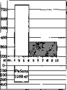
Figure 1. Normal. Increase in oxygen absorption during and after a certain muscular work—a total of 2,028 cm3. Of this amount, the time after the end of work (shaded) accounts for 565 cm3 = 27.8% (according to Eppinger).
that is, 27.8% of the requirements. In heart failure, the same work causes a requirement equal to 6,110 cm3 of O2, and a debt of 4,830, that is, 79%. Eppinger associates the increase in O2 consumption observed in blood circulation failure and the change in the degree and order of amplification of O2 absorption in cardiac patients under the influence of muscular work with a second, very constant phenomenon in blood circulation failure—with an increase of lactic acid in the blood (according to Romberg, up to 30–40 mg% versus 13–17 mg% in norm) and with a sharper increase in its content in the blood of cardiac patients under the influence of muscular work compared to healthy ones.
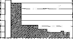
Figure 2. Cardiac defect. Increased oxygen uptake during and after a certain muscular work - 6,110 cm3. Of this amount, the time after the end of work (shaded) accounts for 4,830 cm3 = 79% (according to Eppinger). In circulatory insufficiency, the resynthesis of lactic acid into glycogen in the muscles (and, in all probability, also in all other tissues) is hindered, and only 1/2 or even only 1/3 (instead of 4/5 in the norm) is converted back into glycogen, while the remaining part accumulates in the muscles, passes into the blood, and is only gradually burned, insofar as it is not excreted by the kidneys and sweat glands. An idea of the degree and duration of the increase in the lactic acid content in the blood under the influence of muscular work in heart failure compared to the norm can be given by the following experiments of Eppinger. Table 4. Norm and heart defects. Amount of lactic acid in the blood in mg% before work. Work and its duration. Amount of lactic acid in the blood in mg% after work. In parentheses - number of minutes after the end of work when blood was taken. Heart defects standing on the verge of compensation and decompensation... Burning of excess blood lactic acid in the norm occurs in a large part in the liver. Since all functions of the liver in circulatory insufficiency are more or less impaired, including the oxidation of lactic acid, the excessive accumulation of the latter in the blood in heart failure could also be explained as a consequence of the impairment of the corresponding liver function in this condition. It can also be assumed that the accumulation of lactic acid in the body will be facilitated by the decrease in the excretory function of the kidneys characteristic of heart failure. Undoubtedly, the functional insufficiency of these organs in circulatory insufficiency will contribute to the accumulation of lactic acid in the blood of the respective patient. However, Dresel and Himmelweit proved that the resynthesis of lactic acid into glycogen during work is disrupted under the influence of circulatory insufficiency already in the muscles themselves. They determined the lactic acid content of venous blood flowing from the arm before and during work performed by this arm, and in heart failure, compared to the norm, they found a much sharper and more prolonged increase in lactic acid in the outflowing blood. Insufficient resynthesis of lactic acid into glycogen in the muscles during work in circulatory insufficiency can be considered a consequence of insufficient supply of O2 to the muscle during its work, since that abundant irrigation of the muscle with arterial blood, which is carried out in the norm, does not occur (due to a lack of blood flowing per unit of time from the arteries into the capillaries) by the opening of a mass of new capillaries for the blood flow during work (see physiological part). That insufficient blood supply to the muscle during its work indeed leads to excessive accumulation of lactic acid was proved by Eppinger in experiments on dogs in which, during shock caused by the injection of peptone with a sharp drop in arterial and venous pressure and a decrease in the minute volume and the amount of circulating blood, an increase in the lactic acid content in the blood was observed under the influence of muscular work, many times exceeding that determined in the same animals under the influence of the same work, but with normal blood circulation. Eppinger suggests that the above-mentioned increase in O2 uptake in cardiac patients is explained by its enhanced consumption for burning the excess lactic acid accumulating in the body. One can imagine that with normal blood circulation, precisely thanks to sufficient irrigation of working muscles with blood, O2 is absorbed during the work itself in a larger amount than with insufficient blood circulation. The abundant supply of working muscles with oxygen ensures normal sufficient resynthesis of lactic acid into glycogen. In circulatory insufficiency, due to a lack of O2, resynthesis occurs on a reduced scale, and therefore an excessive amount of lactic acid accumulates during work. Long after the cessation of work, enhanced blood supply and respiration must occur to deliver the O2 necessary in an increased amount for burning the excess lactic acid, as well as for removing it and its combustion products from the body. It must be borne in mind that burning lactic acid requires a significantly greater amount of O2 than resynthesizing it into glycogen. The constant excess of lactic acid in the body may also explain the increased consumption of O2 in circulatory insufficiency both at rest and during activity. It must be borne in mind that in cardiac patients, the intensification of respiratory movements due to dyspnea and the forced position often associated with it is essentially a muscular work constantly increased compared to the norm. This theory gives the right to consider the primary phenomenon in circulatory insufficiency to be the insufficient blood supply to the muscles during work, while the secondary phenomenon is the accumulation of lactic acid in the tissues and in the blood. But this accumulation of lactic acid in turn requires, as just indicated, an enhanced blood supply to the tissues, and this necessitates enhanced blood circulation and respiration; the latter, in turn, implies intensified muscular movements. Thus, a circulus vitiosus of pathological processes apparently develops, which is generally so often the basis of severe diseases. In addition, the accumulation of lactic acid in circulatory insufficiency causes a disturbance of the acid-base balance in the tissues and in the blood, and in particular leads to the enhanced use of reserve alkalinity with the displacement of CO2 from the corresponding buffer compounds and its removal from the body through the lungs. As a result, in circulatory insufficiency, acidosis occurs in the sense of an increased content of organic non-volatile acids in the blood and tissues and a decrease in the blood's ability to bind CO2; however, the pH of the blood does not change due to its buffer properties. The increased acidity of the blood and tissues can be a source of disruption of a whole series of other functions. Against this concept of the role of lactic acid in the pathogenesis of those disorders in the body that accompany circulatory insufficiency, there are some objections. Thus, it is pointed out that there is no sufficient correspondence between the increase in the lactic acid content in the blood and the degree of circulatory insufficiency (Jahn), nor is there any in the norm between the amount of lactic acid in the blood after muscular work and the degree of increase in O2 uptake (Gollwitzer-Meier), nor between the amount of lactic acid in the blood and the degree of increase in O2 uptake in heart failure (Jervell, Jahn). Therefore, to explain the increase in basal metabolism in circulatory insufficiency, other causes besides the accumulation of lactic acid are assumed. The whole doctrine of these chemical processes in circulatory insufficiency, developed mainly by Eppinger and his school, is currently still only in the phase of initial development; however, its great importance for the pathology of blood circulation is already beyond doubt. From Eppinger's doctrine, however, his initial view on the primary role of metabolic changes in the muscles in heart failure cannot be accepted: the insufficiency of blood supply to working muscles due to insufficiency of cardiac activity or due to other causes causing circulatory insufficiency must be recognized as primary. Nor can Eppinger's view on the acceleration of blood circulation in certain phases of circulatory insufficiency and his doctrine of the acceleration of blood circulation during attacks of cardiac asthma be accepted. These views contradict all our doctrine of circulatory insufficiency based on clinical and experimental observations. They cannot be accepted all the more because the corresponding results were obtained using methods whose application to cardiac patients with labored and altered respiration is fraught with numerous possibilities of error. This is fully confirmed, for example, by the fact that Lauter, using a more direct method of obtaining arterial and venous blood—puncture of the artery and puncture of the right heart—to determine the minute volume of blood, obtained in three cases of cardiac asthma during the attack itself a significant decrease in minute volume. But all this does not detract from the importance for the development of a correct understanding of heart failure of the metabolic changes clarified by this doctrine in it. The exchange of O2 and CO2 between the blood and tissues and the blood and alveolar air is one of the most important functions of blood circulation.
When circulation is slowed, besides the insufficient supply of oxygen to tissues and the resulting disruption of chemical processes in tissues (which was partly discussed just now), one should also expect an increased absorption of oxygen from the blood by tissues, as a result of which the venous blood in circulatory insufficiency becomes poorer in O2 than under normal conditions. Instead of the normal 65%, the oxygen saturation of venous blood can drop to 30%. The increased absorption of O2 from arterial blood by tissues (increased utilization of blood O2) in circulatory insufficiency is analogous to the same phenomenon in muscles during work. It compensates to a certain extent for the insufficiency of blood supply, but to a far from sufficient degree when the decrease in blood supply is more or less significant. The given figures show how much the content of reduced hemoglobin in venous blood increases during circulatory insufficiency. This partly explains cyanosis in circulatory insufficiency. But since in heart failure there is also an overfilling of small subpapillary venous plexuses due to blood stagnation in the veins, cyanosis in heart failure with blood stagnation in the systemic circulation will be particularly strongly expressed. The increased utilization of O2 in blood flowing through capillaries has, however, certain limits, because with a decrease in the O2 content in the blood, its pressure in it progressively and increasingly drops and approaches that in the tissues; as a result of this, the further transition of O2 from the blood into the tissues soon becomes impossible. Furthermore, with the very frequent presence of edema in heart failure, the increased utilization of blood O2 by tissues during slowed circulation gives way to a deterioration of this utilization (Harrison, Pilcher). The CO2 content in mixed venous blood (in the blood of the right heart) in heart failure, according to available data, does not present regular changes. In any case, in the lungs, venous blood in cardiac patients is in most cases subjected to quite sufficient arterialization, and therefore arterial blood in them may not differ from the norm in terms of O2 and CO2 content. Regarding the O2 content in arterial blood, there are contradictory indications. Eppinger did not observe a decrease in the O2 content of arterial blood in a single case, even in severe cardiac patients. Kroetz, however, in moderately severe decompensation in patients with valvular defects and hypertension, found in 8 cases out of 16, using arterial puncture to obtain blood, a decrease in blood O2 saturation (92–89% instead of the normal 96%). One should not lose sight of the fact that the normal O2 content certainly does not ensure a sufficient supply of O2 to the tissues if the blood supply—that is, the amount of blood delivered to the tissues per unit of time—is insufficient. As for the carbon dioxide content in arterial blood, in heart failure it is often not only not elevated, but even lowered. This decrease occurs apparently partly due to the aforementioned decrease in the blood's capacity in circulatory insufficiency to bind CO2, and partly due to increased breathing resulting from the irritation of the respiratory center as a result of the impairment of its blood supply. However, in severe cardiac patients, significant breathing difficulties easily develop due to a decrease in the distensibility of the alveolar walls—initially of a functional character (due to the overfilling of pulmonary capillaries as a result of blood stagnation), and then of an organic character [due to the development of connective tissue (Basch's lung stiffness)]. In addition, bronchitis often develops on the basis of stagnation, and the effect of respiratory movements is reduced during their excessive acceleration due to dyspnea, etc. (Siebeck). As a result of these breathing difficulties in severe cardiac patients, gas exchange in the lungs may be reduced to such an extent that the O2 content in the arterial blood becomes insufficient, and the release of CO2 is even hindered; but the dyspnea of cardiac patients in any case does not depend on an increased CO2 content in the blood or depends on it only in a part of the cases, since the CO2 content in the arterial blood in circulatory insufficiency, as just indicated, is as a rule not only not elevated, but even lowered. Dyspnea in heart failure is the result of irritation of the respiratory center partly by arterial blood altered in its composition in the sense of an increased content of other acidic compounds, in particular lactic acid, and partly as a result of chemical changes of the same direction in the tissue of the respiratory center itself due to its insufficient blood supply. Figures 3 and 4 provide a clear illustration of the relationship between respiratory function and circulatory function during muscular work in circulatory insufficiency compared to the norm. Circulatory insufficiency of vascular origin. Besides diseases of the heart itself, the cause of circulatory insufficiency can also be such diseases of other parts of the circulatory apparatus that lead to a disruption of its entire work. On the part of the arteries, changes in the distensibility and elasticity of their walls must primarily cause circulatory insufficiency, judging by the great importance that the functions associated with these qualities of the arterial walls have for the entire circulation.
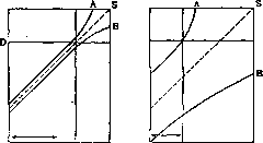
Figure 3.
Figure 4.
Figures 3 and 4. Relationship between blood circulation and respiration during work in a healthy person (Fig. 3) and a person with circulatory insufficiency (Fig. 4). The ordinate shows the increase of these values in % compared to the figures at rest, the abscissa shows the work performed. In both individuals, metabolism (S) increases during work. In a healthy person, respiration (A) and blood circulation (B) increase uniformly up to a certain point. Then B rises more slowly, while A rises faster (excessive ventilation). In circulatory insufficiency, B is already below normal at rest, and A is above it. During work, B and S increase uniformly, and excessive ventilation begins earlier and reaches a greater degree. The horizontal line (D) indicates the point at which the sensation of air hunger (dyspnea) appears. (According to J. H. Means.) Arteriosclerosis (see) is of the greatest importance in this respect. It should be noted that the impairment of function of one part of the circulatory apparatus is compensated by the work of other parts, but among these parts, in terms of the ability to compensate for the functional impairment of others, the heart stands in first place. In arteriosclerosis, as a rule, a compensatory strengthening of cardiac activity also occurs, and complete circulatory insufficiency in arteriosclerosis most often occurs only when insufficiency of the compensatorily overworked heart develops. - It is not yet possible to distinguish pathological impairment of the function of the arterial musculature from impairment of the function of the vasomotor nervous apparatus, since the pathogenesis of the main types of pathological changes in the functions of the arterial musculature that have an effect on blood circulation throughout the body (hypertension and hypotension) has not yet been elucidated. How hypertension (see) leads to circulatory insufficiency follows from what is set forth in the corresponding chapter on the pathogenesis of this pathological state. In hypertension, the narrowing of peripheral, mainly precapillary arteries due to increased tonic contraction of the arterial musculature impedes the blood supply to the tissues. This narrowing can be compensated only by accelerating the blood flow through the narrowed vessels. To achieve this acceleration, it is necessary to establish arterial pressure at a higher level; this is possible only with increased work of the heart, in particular the left ventricle. This increased work leads to its hypertrophy. With adequate heart work, arterial pressure is maintained at the high level necessary for such an acceleration of blood flow through the precapillary arteries as is required to maintain an adequate blood supply to the tissues. If the heart, as a result of overwork, weakens, it ceases to transfer a sufficient amount of blood into the aorta, as a result of which the minute volume of blood decreases and, consequently, the blood supply to the tissues decreases. On the other hand, insufficient heart work leads to blood stagnation in the superior parts of the circulatory system and to an increase in venous pressure. Thus, circulatory insufficiency in hypertension ultimately develops as a result of cardiac insufficiency as well. The heart, undergoing hypertrophy and intensifying its work, is able to compensate for a given impairment of arterial function for the time being. Regarding such pathological impairments of the functions of capillaries and veins that would lead to circulatory insufficiency in the whole body, our knowledge is even less perfect. As follows definitely from modern concepts of blood circulation, set forth in the section on the physiology of blood circulation, cardiac activity is the main and almost the only source of the energy that carries out blood circulation. But the work of the heart, in turn, depends entirely on the inflow of blood to it and is largely determined by it (Starling). The inflow of blood to the heart, although it occurs to a large extent at the expense of the energy of cardiac work, is nevertheless completely dependent on the function of the vascular system, in other words, on whether the latter correctly or incorrectly returns blood back to the heart. The enormous importance in this respect of the "periphery" of the circulatory system, i.e., blood vessels, is not yet sufficiently appreciated by everyone. This insufficient appreciation partly explains the tendency to attribute new functions to the vascular system, such as the function of a "peripheral heart." As already indicated in the physiological part, we do not yet have sufficient grounds to recognize the function of generating energy for the movement of blood in vessels. In this respect, the heart, as stated above, plays a special role in the circulatory system. But this does not contradict the fact that all parts of the circulatory apparatus together constitute a single functional whole, one part of which cannot work without another, and the formulation of the question of which is more important—the function of the heart or of the vessels—is therefore, of course, completely incorrect. Regarding the pathology of circulation, it must be recognized that those circulatory disorders that depend on incorrect or insufficient heart function are comparatively well known and studied, whereas with regard to those pathological states of the entire circulation that depend on impairment of the vascular system's function, our knowledge is still in the period of initial development. For clarity, it is necessary to contrast circulatory insufficiency due to insufficientia cordis with circulatory insufficiency due to insufficiency of vascular activity. Vascular insufficiency in this case should be understood in the sense of unsatisfactory supply of blood to the heart, i.e., insufficient delivery of blood to the heart. Even a fully functioning heart, with insufficient blood supply, is unable to transfer a sufficient amount of blood into the lungs for its arterialization and into the arteries to ensure proper blood supply to organs and tissues per unit of time. Both in insufficientia cordis and in vascular insufficiency (in the sense just indicated), the blood flow is slowed down, the minute volume of blood is reduced, tissues and organs receive an insufficient amount of blood per unit of time; but in insufficientia cordis, blood is delivered to the heart in sufficient quantity, yet the heart is unable to transfer it into the arterial system in the amount necessary. Blood therefore stagnates in the superior parts of the circulatory system and, in particular, in the veins of the systemic circulation. As a result, venous pressure is elevated in cardiac insufficiency. But arterial pressure, as a rule, does not decrease in this case (see below). In vascular insufficiency, blood is retained in the peripheral vessels, and both venous and arterial pressure necessarily decrease. The basis of vascular insufficiency is a decrease in the tone of the contractile elements of the vascular walls throughout the entire vascular system or in one or another of its large sections. In the physiological part of the article, the special significance is indicated that belongs, with respect to the distribution of blood, to the vascular region innervated by the celiac nerve—a region corresponding in a large part of it to the portal vein system. The special significance of this region and its exceptional role stand out with particular distinctness under conditions of pathological changes in vascular tone. This section of the vascular system reacts stronger than others as a rule to influences that decrease and increase vascular tone, and due to its large capacity, a decrease in the tonic contraction of its musculature leads to the accumulation of a significant part of the blood in it and to a corresponding decrease in the inflow of blood to the heart and, consequently, to a reduced flow of blood into the arterial system, i.e., to the same thing to which cardiac insufficiency also leads. But between circulatory insufficiency caused by insufficientia cordis on the one hand and circulatory insufficiency due to vascular insufficiency on the other, there is a substantial difference. In cardiac insufficiency, an insufficient amount of blood enters the arterial system per unit of time, but thanks to the normal function of the vascular system, arterial pressure is maintained at the proper level, and a purposeful distribution of this insufficient amount of blood takes place in the sense of supplying it first of all to the organs most vital for life—the brain and the heart. In vascular insufficiency, however, when an insufficient amount of blood enters the aorta, arterial pressure cannot be maintained at the necessary level due to that same vascular insufficiency; therefore, there is no rational distribution of blood, and the heart and brain suffer particularly from insufficient blood supply. Due to this, cardiac insufficiency and impairment of the functions of the central nervous system, which is especially sensitive to all impairments of blood supply, as a rule join vascular insufficiency. This difference in circulatory impairment in cardiac and vascular insufficiency explains, among other things, the fact that even in severe cardiac insufficiency, phenomena of brain anemia are relatively rarely observed, whereas in severe vascular insufficiency, as we observe it in its most pronounced form in collapse (see), these phenomena are frequently observed and can lead to loss of consciousness. The unsatisfactory blood supply to the brain, which is characteristic of vascular insufficiency, partly explains, in all probability, the fact that in vascular insufficiency, unlike cardiac insufficiency, patients prefer a horizontal position.
It should be noted that the pallor of the integuments, which is especially characteristic of vascular failure, manifests as a result of their blood depletion, and a small and soft pulse manifests as a result of inadequate filling of the arterial system. The fact that cyanosis in vascular failure, compared to cardiac failure, is usually significantly weaker is explained by the fact that of the two factors determining the cyanotic coloration of the integuments—the reduced O2 content in venous blood and the engorgement of the small veins of the integuments with venous blood—the second factor, which apparently has the greatest importance in the origin of cyanosis, disappears in vascular failure. The less pronounced and less constant dyspnea in vascular failure can be explained by the simultaneous depression of both the vasomotor and respiratory centers. From the standpoint of the stated concept of the pathogenesis of vascular failure, it is entirely understandable that under appropriate conditions, neither cardiac enlargement, nor hepatic enlargement, nor engorgement of superficial veins, nor any congestive phenomena whatsoever on the part of the organs of the greater and lesser circulations are observed. Unfortunately, with regard to vascular failure in humans, there is almost no direct, more precise data on changes in those main quantities that characterize blood circulation, i.e., the minute volume of blood, the velocity of blood circulation, the amount of circulating blood, O2 absorption, resp. basal metabolism, O2 and CO2 content in the blood, lactic acid, etc. Even regarding venous pressure, there is insufficient data. Our concepts of vascular failure in corresponding pathological conditions are based only on its indicated clinical manifestations, on observations of the action of certain remedies used for its treatment, and finally on experimental observations. The latter consist of 1) observing and analyzing the effect on blood circulation of such factors that cause a picture of vascular failure in humans, e.g., bacterial toxins, or 2) studying pathological conditions in animals similar to vascular failure in humans, caused by certain substances, e.g., histamine and peptone shock. The picture of vascular failure is most often observed in acute infectious diseases. Romberg and his coworkers deserve the credit for experimentally proving that bacterial toxins, by depressing the function of the vasomotor centers, cause a decrease in the tone of the vascular musculature, predominantly of the abdominal visceral vessels. In addition to vascular failure of central origin in infectious diseases, a direct injury to the cardiac muscle by the same toxins is of course an important and frequent event. A direct depressing effect of the same toxins on the vascular musculature is also possible (Waldmann). Still, as a rule, in the picture of blood circulation failure in infectious diseases, manifestations of failure due to depression of the vasomotor centers dominate, and accordingly in the treatment of these conditions, the effect is obtained predominantly not from cardiac remedies in the narrow sense of the word, i.e., preparations of the digitalis group, but from remedies acting on the vasomotor nervous system (see Collapse). As indicated above, we have almost no observations on changes in the blood flow velocity in infectious diseases in humans. The only data by Bjerloew, Liljestrand, and Grollman indicate the possibility of an increase in the minute volume of blood in the initial phases of the infectious process, but the latter author, in the febrile state caused by the injection of typhoid toxin, following the initial increase in minute volume, observed (with a continuing increase in temperature, enhancement of O2 absorption, and increased pulse rate) a fall in minute volume and is inclined to attach more importance to this latter phenomenon, taking into account the prolonged nature of most infectious febrile states in man. This point of view corresponds to observations on changes in arterial pressure in acute infectious diseases, which, after their initial rise, show in the subsequent course, in proportion to the severity of the condition, not only a decrease in the average level of arterial pressure, but also a significant decrease in pulse pressure (see Blood pressure). Experimental observations on so-called histamine and peptone shock give a particularly clear idea of the pathogenesis of these phenomena. Table 5. Histamine shock. Relation to shock, Blood pressure, Amount of blood output by the heart in 1 min. in cm3, With each contraction (in cm3), Before shock..., During shock... 1st experiment (dog weighing 18 kg) 175
2.800 2nd experiment (dog weighing 20 kg) Before shock... During shock... 168 80 2,570 470 Table 6. Histamine shock. Relation to shock Amount of circulating blood Hemoglobin 1st experiment (dog weighing 26 kg) Before shock... During shock... After shock... 2nd experiment (dog weighing 14 kg) Before shock... During shock... After shock... 825 380 87 98 90 102 109 Characteristic of these conditions is a sharp drop in arterial and venous pressure, a strong decrease in the minute volume of blood and the total volume of circulating blood. From experimental observations on shock, attention is deserved by the above-mentioned accumulation in the blood during shock, under the influence of muscular work, of lactic acid in quantities many times exceeding those that appear in the blood of the same animals before shock under the influence of the same work. This observation proves that the impairment of blood supply under the influence of vascular insufficiency, at least in this direction, causes an entirely identical disturbance in the chemistry of metabolism as does heart failure. The fact of a sharp decrease in the total volume of circulating blood in experimental shock raises the question of in which vessels blood is retained at all during vascular insufficiency. One can assume a corresponding dilation of capillaries and veins, since the capacity of the arteries is hardly sufficient for them, even with maximal dilation, to accommodate a corresponding amount of blood. The capillary and venous system undoubtedly also includes those reservoirs for blood that are filled when the volume of circulating blood decreases. Most likely, during vascular insufficiency, an overfilling of these very reservoirs with blood occurs. In any case, it is unquestionable that all these reservoirs are under the influence of the vasomotor nervous system, as shown by the effect of such agents as adrenaline, pituitrin, inhalation of CO2, etc., by which in experimental shock it is possible to significantly increase the volume of circulating blood (Eppinger). There is a notion that the insufficiency of blood circulation, as we observe it in infectious diseases, in its most expressed form in collapse, in certain poisonings, is the result of a primary dilation of capillaries of vast areas of the vascular system, independent of the vasomotor center. It is assumed that this capillary dilation is the result of the influence on them of substances analogous in action to histamine. Such capillary dilation in the region of, for example, abdominal viscera must cause a sharp decrease in the blood supply to other organs, including the central nervous system and the vasomotor center in particular. Then, secondarily, as a result of the impaired blood supply of the latter, paresis of the vascular musculature and predominantly of the splanchnic nerve region develops. It is quite possible that a similar pathogenesis of vascular insufficiency takes place in some of its forms. According to another theory, the lowering of vascular tone in at least some infectious diseases is the result of a decrease in the function of the chromaffin system (adrenals) due to its damage by the infectious agent, resp. toxins. Indeed, the picture of vascular insufficiency in infectious diseases resembles the picture of cardiovascular dysfunction in Addison's disease or after adrenalectomy. It is also undoubtedly true that adrenaline can be regarded as a hormone of special importance for the function of blood circulation organs. But this hormone nevertheless acts through the sympathetic nervous system, exciting the endings of the sympathetic and in particular vasomotor nerves or special formations connecting these nerve endings with muscle fibers. Therefore, it is very difficult to decide whether there is depression of the centers of the vasomotor nerve apparatus, or insufficiency of its function due to insufficiency of that endocrine gland which produces the hormone stimulating predominantly the given section of the vegetative nervous system. The first hypothesis must still be recognized as more probable. Besides the insufficiency of vascular function regarding the supply of blood to the heart, one can also distinguish a disturbance in the function of the vascular system regarding the distribution of blood in the organism. An example is the impairment of vascular system function during muscular work in the form of the failure of blood vessels to dilate as necessary to enhance the blood supply of working musculature, or even the onset of constriction instead of dilation. Such a phenomenon is well known within limited vascular regions, i.e., as a local phenomenon, e.g., in so-called intermittent claudication (claudication intermittens). Similar local phenomena can also be observed upon the application of various stimuli in individuals with an abnormal state of the vasomotor nerve apparatus, e.g., vasoconstriction upon the application of such thermal stimuli that as a rule cause dilation, and vice versa. Weber's plethysmographic study gives some idea of such paradoxical reactions in extensive vascular areas, in particular the above-mentioned constriction of muscle vessels occurring instead of their dilation during work under the influence of vasomotor center depression by fatigue, poisonings, infection, etc. It is quite possible that it is precisely such a dysfunction of the vasomotor centers that explains the circulatory insufficiency observed as a result of past infectious diseases, metabolic diseases, etc., and especially in some so-called vasomotor neuroses, manifesting as rapidly onsetting muscular or psychic fatigue and the resulting decrease in working capacity. Vasomotor centers apparently react generally to overwork and other harmful influences (in a certain phase at least) not so much by the weakening of their function, but by the perversion of their reactions in the sense of the onset of reactions opposite to normal ones upon certain stimuli. Circulatory insufficiency due to a disturbance of the vascular system in the sense of improper blood distribution has still been studied far from sufficiently, but apparently has essential practical significance. Circulatory disorders in connection with pathological states of other organs. All the above-mentioned types of circulatory insufficiency are a consequence of damage to various parts of the entire circulatory apparatus. To this apparatus belong not only the heart itself and the vessels, but also the entire complex nervous apparatus that performs a significant part of the function of directing the work of the circulatory system depending on the requirements of organs and tissues. But apart from pathological changes in circulation due to pathological impacts on the circulatory apparatus itself, there are changes in its work that are a consequence of pathological changes in all kinds of organism functions and especially those that are most closely functionally connected with the blood circulation system. Among the organs functionally inextricably linked with the circulatory apparatus, the first place should be given to the respiratory apparatus, especially since the respiratory mechanical device, the chest with respiratory musculature, is at the same time an auxiliary circulatory engine. Impairments of its function (e.g., due to pulmonary emphysema) therefore lead first to a compensatory intensification of the work of the corresponding divisions of the circulatory apparatus, in particular the right heart, and then through overwork also to circulatory insufficiency. Especially interesting is the close functional connection between the circulatory system and the blood. Circulatory insufficiency, and in particular insufficient arterialization of the blood, e.g., due to congenital heart defects, leads under certain conditions to an increase in the number of oxygen carriers per unit volume of blood, i.e., to polycythemia, which under these conditions should be regarded as a change compensating for the slowing of blood circulation. Conversely, the depletion of blood in hemoglobin, as we have it in various forms of anemia, causes as a rule a compensatory acceleration of blood flow, i.e., an increase in the work of circulatory organs. Already by comparatively numerous studies, an increase in the minute volume of blood in severe anemias has been established (Plesch et al.); Lauter and coworkers found an increase up to 9-10 liters instead of 4.2 liters normally. Myasnikov and Tetelbaum determined by the histamine method an acceleration of blood flow up to 12 seconds instead of the normal 22.2 seconds. Under unfavorable conditions, severe anemia causes circulatory insufficiency both by way of heart overwork and by way of impairing its nutrition. The degree of blood supply to organs and tissues and consequently the work of the circulatory apparatus is determined by the need for O2, gas exchange in general, and nutrition. This follows particularly clearly from the adaptation of blood circulation to muscular work and the work of organs in general (see physiological part). Accordingly, pathological changes in metabolism cause corresponding changes in circulation. A clear example can be the increase in the minute volume of blood and the speed of blood circulation in hyperthyroidism and the opposite changes in hypothyroidism. Myasnikov and Tetelbaum in a case of Basedow's disease at the height of the disease determined the blood flow velocity to be 15 seconds (normal 22.2 seconds), and after improvement to be 21 seconds; in myxedema, conversely, at the height of the disease - 34 seconds, after treatment with thyroidin - 27 seconds.
In parallel with the acceleration of blood circulation in hyperthyroidism, as with the enhancement of circulatory function in general, an increase in the volume of circulating blood is observed. In hypothyroidism, conversely, it is reduced (Wislicki). It must be noted, however, that for the acceleration of blood flow in hyperthyroidism there is also another explanation: the parallel reinforcing action of an excess of thyroid hormone (or an altered hormone) on both the volume of substances and the function (Lauter, Determann). In any case, here too the intensified work of the circulatory organs in more severe cases passes into insufficiency of their work. Previously, circulatory insufficiency due to heart failure was as if contrasted with that due to vascular insufficiency. Such a contrast is necessary in order to clarify the pathogenesis and manifestations of each form separately. But it should not be lost sight of that both these forms in reality, of course, are combined in one proportion or another extremely frequently. This combination of cardiac and vascular insufficiency is either the result of one causing the other (for example, vascular insufficiency as a rule causes heart failure by impairing the blood supply to the heart muscle), or the same impact causes pathological changes in both the heart, the blood vessels, and the vasomotor apparatus. Such harmful influences, which frequently affect all parts of the circulatory apparatus, are the majority of infectious diseases. Combinations of several harmful influences are also very frequent, of which some act on the heart, others on the vascular system. Well known, for example, are those severe consequences produced by the combination of a valvular heart defect, which is on the verge of decompensation, with one or another acute or chronic infection. Here one can think of the influence of infection on both the heart muscle and the vascular system. As a result, the most unfavorable combination of both cardiac and vascular insufficiency for the circulation is obtained. In general, it must be particularly emphasized that precisely under pathological conditions, the unity of the entire circulatory apparatus stands out particularly clearly, both in the sense of compensating for the functional insufficiency of one of its parts by the enhanced function of another, and in the sense of impaired function of one part due to the insufficiency of another. For occupational diseases of the circulatory organs and statistics of diseases of the circulatory organs, see Cardiovascular system.
Related articles
Mentioned in
Cite this page
“Blood Circulation.” Soviet Medical Encyclopedia. English translation of Bolshaya Meditsinskaya Entsiklopediya, 1st ed. (Moscow, 1928–1936), ed. N. A. Semashko. https://sovietmedicalencyclopedia.pages.dev/article/blood-circulation/