Vessels
Historical document, translated for reference. It reflects medical knowledge of the 1920s–30s and is not medical advice.
Summary
This article from the 1930s Soviet medical encyclopedia describes the anatomy and development of blood and lymphatic vessels, including their embryonic formation, structure, and methods of injection study.
Encyclopedia article (1928–1936)
VESSELS, (from Latin vasa, Greek angeion, phleps; vessels). 1-thoracic duct; 2-cisterna chyli; 3-thoracic duct; 4-one-voice apparatus; 5-left and right thoracic ducts; 6 and 8-lymphatic vessels of the small intestine; 7-lymphatic vessels of the stomach; 9-lymphatic vessels of the large intestine; 10-lumbar (two) glands adjacent to the abdominal aorta; 11-artery; 12-lymphatic vessels of the hind limb; 13-inguinal glands; 14 and 15-lymphatic vessels of the wing (forelimb); 16-blood vessel; 17-lymphatic glands in the neck region. Development of vessels occurs through the growth from the center to the periphery of the vessels and their endothelial lining. It is clear that the actual proof of the development process of lymphatic vessels is associated with very great difficulties, all the more so because here one cannot use observations of forming cavities in sections on the grounds that one must still prove that these developing cavities belong to the lymphatic system. This proof can of course be given by injection, but if injection is possible, then the cavities in question have already turned into lymphatic vessels, and therefore observation of their first development is excluded. If, however, one has the earliest stages of development before him, then injection is impossible, and therefore one cannot with full confidence provide complete proof of the lymphatic nature of the observed formations. Development of lymphatic glands. The study of the development of lymphatic glands was undertaken by Sertoli, Orth, Shevich and Teichmann. The first rudiments of glands are found in human embryos after the 11th week (Shevich). They appear in the form of limited accumulations of lymphatic cells in connective tissue, accumulations penetrated by slit-like passages. Hyperplasia of lymphatic elements gives rise to the parenchyma of future glands, while the tissue slits transform into lymphatic sinuses. Teichmann holds a different view. According to Teichmann, lymphatic glands originate from plexiform or 'wonderful networks' of lymphatic vessels with subsequent accumulation and deposition of lymphoid elements in their lumen. Kling expresses a view similar to Teichmann, indicating that by the 3rd month of intrauterine life the general basis of lymphatic glands is already laid, consisting of loops of lymphatic vessels, among which cellular elements in the form of trabeculae, well supplied with blood vessels, are deposited from the surrounding tissue.
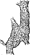
Lymphatic vessels. The roots of lymphatic vessels are lymphatic spaces. These spaces exist in all organs between the walls of blood capillaries and surrounding tissues and between individual tissue elements-cells and fibers. They usually do not have an endothelial lining. In dense connective tissue (cornea, diaphragm), by the silvering method, a system of juice spaces and canals can be discovered in the form of light spaces (Recklinghausen), communicating with lymphatic vessels. Some authors (Hueck) deny the existence of preformed lymphatic spaces in organs (e.g., around liver cords), designated by German authors as Saftrohren-system (Virchow-Köhler), Spaltraume (Ludwig).--From the tissue spaces, lymph enters the capillary vessels, already constructed on the type of blood capillaries: their thinnest wall is formed by endothelial plates lining the tissue spaces. Lymphatic capillaries form anastomosing networks of channels, gradually collecting into lymphatic trunks. The latter are already provided with valves in the form of pockets, similar to venous ones. The walls of lymphatic vessels are very thin, built of connective tissue and smooth muscles and lined with a single layer of flat endothelial cells. Valves promote the progressive movement of lymph, above the valves lymphatic vessels have flask-like expansions. Lymphatic trunks usually accompany vascular-nerve bundles, e.g., on the limbs, on the neck; they differ from veins in the thinness of their walls and the comparative poverty of their smooth muscle fibers. Lymphatic trunks, having very thin, almost transparent walls and containing colorless lymph, are not noticeable among the surrounding tissue; they become visible on corpses after filling (injection) them with colored liquid. Injection of lymphatic vessels is performed by puncturing the organ with a syringe needle. Through the needle, the injection fluid first penetrates into the intercellular spaces, from which it then fills the lymphatic capillaries and trunks. For injection, the Herot mass is commonly used, consisting of oil paint-'Prussian blue', diluted with ether, and Chinese ink, diluted with water. The latter is used for injection in living animals. Besides the mentioned puncture method for injecting lymphatic vessels, at present the method of passive movements is widely used, which consists of the following: on corpses, the Herot fluid is introduced into the serous cavity or joint by puncture, and then natural passive movements are performed. Thus, by introducing Herot fluid into the joint cavity and performing passive movements in it for 1/4-1/2 hour, the researcher obtains an injection of the efferent lymphatic vessels of this joint and the main pathways up to their entry into the lymphatic glands. By introducing Herot fluid into the abdominal cavity and performing rhythmic compression of the abdominal walls, one can obtain an injection of the lymphatic vessels of the diaphragm. From the pleural cavity, by performing rhythmic passive breathing, an injection of pleural vessels is obtained; from the pericardial cavity, by performing rhythmic compression of the pericardial sac, the vessels of the pericardium are filled; from the synovial sheaths of tendons, by performing light massage with a piece of cotton-lymphatic vessels of the limbs; and finally, from the subarachnoid spaces on children's corpses, by rhythmically pressing on the fontanelle with a finger-the lymphatic vessels of the dura mater.
Lymph capillary networks consist of tubes with uneven diameters and often with bulges on the walls in the form of blind offshoots and sacs. According to Kulchitsky, the structure of the wall of a lymph capillary essentially does not differ from the structure of a blood capillary. The wall of a lymph capillary is undoubtedly composed of a single layer of endothelial cells (Fig. 10), whereas the wall of a blood capillary may consist of two layers: endothelium and an outer membrane. Fig. 10. Lymph capillary from the silvery tendon center of the diaphragm. Fig. 11. Opened lymph vessel with its valves. The thicker wall of the lymph trunks includes three tunics, as in blood vessels. The inner tunic, tunica intima, consists of endothelium and an outward network of elastic fibers. The middle tunic, t. media, contains several layers of circularly arranged smooth muscle elements. Elastic fibers are found here only in small quantities. The outer tunic, t. adventitia, consists of fibrous connective tissue with a small admixture of thin and thick elastic fibers, which run mostly longitudinally along the axis of the vessel. Lymph trunks (like veins) are provided with valves, which are folds of the inner tunic in the form of semilunar flaps (Fig. 11). The semilunar folds, attaching to the inner surface of the lymph vessel at the same level, opposite each other, form pockets whose purpose is to allow lymph to move only from the periphery toward the center, i.e., to the place where the lymph empties into the veins. Valves are more frequent than in veins. In small vessels, valves follow one another at a distance of 2-3 mm, in larger ones 6-12 mm. At the points of attachment of the valves, lymph vessels form dilations, giving the trunks a club-shaped appearance. The walls of lymph vessels (like blood vessels) have their own blood supply system (vasa vasorum) and, according to the research of Kitmanov, Dogel, and others, are supplied with a large number of nerves, both motor and sensory. The thoracic duct is innervated, according to Lavrentiev's data (1926), by nerve fibers from the sympathetic trunk. The lymph vessel system, being an addition to the venous system, shows much in common with the venous system in its topographical position. In each particular organ, lymph trunks begin from a capillary network, which usually lies in the organs near the capillary network of blood vessels. The peculiarity of the capillary network of lymph vessels is that it forms wider and less regular intervals and is located in the tissues closer to the surface of the organ, for example, in the mucous or serous membranes. An essential difference between lymph vessels and veins lies in the fact that lymph vessels of the limbs, trunk, head, and neck, both deep and superficial, as well as vessels going from internal organs, are interrupted by lymph glands at certain places. A particularly large number of these glands is found in the axillary region, inguinal region, and neck, in the thoracic and abdominal cavities, and (comparatively in smaller numbers) in the region of the elbow bend and popliteal fossa (see below - lymph glands). L. gl. are usually located in groups, small ones - from 2 to 6, and large ones - from 10 to 40 or more glands. They occupy certain places and receive lymph from certain organs of particular regions, which is why they are called regional, i.e., they are centers through which lymph passes, brought by a certain number of vessels from organs located near these glands, as well as along the entire path of the lymph vessels from their peripheral beginning in the organs until they empty into the glands. The main superficial trunks collect lymph through lateral, small (secondary) trunks mainly from the capillary network located in the skin and subcutaneous tissue. The main deep trunks collect lymph from organs located under the fasciae and are situated together with the main blood vessels and nerves in one vascular-nerve bundle (Fig. 12). There is no complete separation of deep lymph vessels from superficial ones. Just as deep veins have anastomoses with superficial ones, mainly for the outflow of blood from deep veins into superficial ones, so deep lymph trunks give off anastomoses to superficial ones for the outflow of lymph from deep trunks into superficial ones. Deep lymph vessels take their origin from capillary networks: 1) muscles with their tendons, fasciae, and synovial sheaths of tendons; 2) bones with their bone marrow and periosteum; 3) joint capsules with their ligaments; 4) all internal organs, i.e., organs located in the body cavities, and 5) synovial and serous membranes lining the walls of synovial serous cavities. Lymph vessels are divided into secondary (small) and main (large). The former begin from the capillary lymph network of organs and empty into the main trunks (collectors). On their final course, the lymph vessels open by two trunks symmetrically on the right and left sides in the neck region, at the place where the common jugular and subclavian veins, merging, form an angle (angulus venosus). On the left side, the thoracic duct opens - the main trunk carrying lymph from organs constituting 3/4 of our body, and on the right side the right lymph duct opens, carrying lymph from the remaining (right) upper quarter of the body (Fig. 13). The thoracic duct collects lymph from the lower extremities, pelvic organs, abdominal cavity and left half of the thoracic cavity, walls of the abdomen and left half of the thoracic cage, and finally from the left upper extremity and left half of the head and neck. The right lymph duct collects lymph from organs of the right half of the thoracic cavity and its walls, right half of the head, neck, and upper extremity (Fig. 13). Part of the vessels, collecting
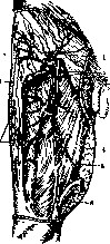
Fig. 12. Deep
lymph vessels of the thigh from the front: 1-superficial inguinal lymph gland; 2-deep inguinal lymph gland; 3 and 4-deep superior femoral lymph glands; 5-superficial femoral artery; 6-lymph nodes of the superior articular artery of the knee; 7-deep lymph vessels of the fascia; 8-superficial lymph vessels of the broad fascia. vessels, collecting 153
Related articles
Cite this page
“Vessels.” Soviet Medical Encyclopedia. English translation of Bolshaya Meditsinskaya Entsiklopediya, 1st ed. (Moscow, 1928–1936), ed. N. A. Semashko. https://sovietmedicalencyclopedia.pages.dev/article/vessels/