Kidneys
Historical document, translated for reference. It reflects medical knowledge of the 1920s–30s and is not medical advice.
Summary
This article from the 1928–1936 Soviet Great Medical Encyclopedia details the anatomy of the kidneys, including their topography, structural layers, capsules, and anatomical variations based on age, sex, and constitution.
Encyclopedia article (1928–1936)
I. Anatomy of the kidneys. The kidneys (renes) are a paired organ of characteristic bean shape (the right one is more ear-shaped, the left one bean-shaped) (Figs. 1 and 2). Both kidneys converge in such a way that their upper poles are spaced apart by approximately 7 cm, and the lower ones by 11 cm; the left kidney is located somewhat -12 12-
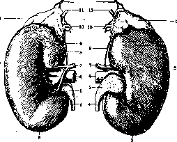
Figure 1. Right kidney from the front (a) and back (b): 1 - adrenal gland; 2 - outer border; 3 - lower pole; 4 - ureter; 5 - renal pelvis; 6 - renal vein; 7 - renal artery; 8 - inner border; 9 - inferior suprarenal artery; 10 - middle suprarenal artery; 11 - central vein of the adrenal gland; 12 - superior suprarenal artery. (After Testut.)
higher than the right, which is pushed downward by the liver. Above, the kidney does not reach the 11th rib on the right side, whereas on the left it stands somewhat higher than the 11th (Fig. 3). In relation to the spine, the kidneys occupy the space between the 12th thoracic and 2nd-3rd lumbar vertebrae, with the upper pole of the left projecting at the level of the upper edge of the 11th thoracic vertebra or slightly lower (middle of the 11th thoracic); the lower pole of this kidney lies at the level of the cartilage between the 2nd and 3rd lumbar vertebrae; the upper pole of the right kidney is located between the 11th and 12th thoracic vertebrae or slightly higher, and the lower at the height of the 3rd lumbar vertebra. The upper third of the kidney lies on the diaphragm. In general, the position of the kidney varies considerably among individuals; in women, the kidneys are located lower than in men. In newborns, the lower poles of the kidneys are at the level of the iliac crest. However, the position of the kidney changes with age (they descend), and also in connection with constitutional peculiarities of body structure (weakness of the kidney-fixing apparatus). According to Pavlenko
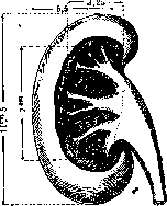
Figure 2. Dimensions of individual parts of the kidney. V59 ko, the position of the kidney can be reduced to three types: high, low, and transitional, which are related to the structural features of the lower thoracic aperture. Thus, with a wide thoracic aperture, a high position of the kidney takes place, and the angle formed by the upper ends of the longitudinal axes of both kidneys is acute; with a low standing of the kidney and a narrow thoracic aperture,
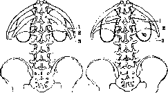
Fig. 3. Relation of the kidney to the lower border of the pleura and the 12th rib at various lengths of the latter (left - usual length of the 12th rib): 1 - lower border of the pleura; 2 - origin of the diaphragm; 3 - projection of the kidney. (After Corning.)
the indicated axes converge higher, as a result of which the renal angle becomes even more acute. The position of the right kidney is particularly variable, and the descent of the colon connected with it can serve as a cause of such kidney displacement. This displacement is also possible in the norm, for example during strong movement of the diaphragm during a deep breath. (On anomalies of the body,
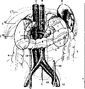
Figure 4. Topography of the kidneys: 1 - part of the kidney covered by peritoneum; 2 - part not covered by peritoneum; 3 - duodenum; 4 - adrenal gland; 5 - inferior vena cava; 6 - aorta; 7 - tail of the pancreas; 8 - spleen; 9 - colon; 10 - ureter; 11 - external iliac artery; 12 - hypogastric vein. (After Testut.)
position, and shape of the kidney, see below - pathological anatomy of the kidney.) The weight of the kidney averages 120–130 g (the left kidney is somewhat smaller than the right). The color of the kidney is usually dark brown. The upper pole of the kidney (superior pole) is broad and flat, the lower pole (inferior pole) is sharper and thicker than the upper. The anterior surface of the kidney (anterior face) is more convex, the posterior (posterior face) is flatter. In addition, the anterior surface bears variously expressed impressions - depressions of neighboring organs (Figs. 4 and 5). The consistency is elastic and rather firm. On the outside, the kidney is covered by a dense fibrous capsule (fibrous tunic), which
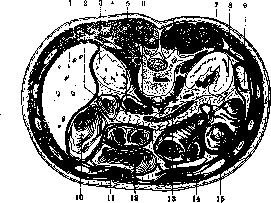
Figure 5. Horizontal section of the abdomen at the level of the first lumbar vertebra: 1 - liver; 2 - descending part of the duodenum; 3 - right kidney; 4 and 7 - pleural cavity; 5 - inferior vena cava; 6 - diaphragm and aorta; 8 - left kidney; 9 - spleen and pancreas; 10 - ascending colon; 11 and 14 - rectus abdominis muscle; 12 - transverse colon; 13 - splenic vein; 15 - left colic flexure. (After Corning.)
is weakly connected to the parenchyma of the organ and therefore in the norm is easily stripped. At the renal hilum, the capsule divides into two leaves: an inner one, which is located directly on the kidney tissue, lining its sinus (renal sinus), and an outer one, covering the structures entering and exiting...
especially in the region of
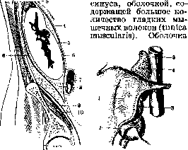
Figure 6.
Figure 7.
Figure 6. Longitudinal section through the left kidney: 1 - diaphragm; 2 - phrenic pleura; 3 - adrenal gland; 4 - peritoneum; 5 - fibrous capsule of the kidney; 6 - renal fascia; 7 - descending colon; 8 - adipose capsule of the kidney; 9 - iliac fascia; 10 - iliac muscle. (After Corning.) Figure 7. Relation of the right kidney to the parietal peritoneum: 1 - hepatic face of the kidney; 2 - position of the right colic flexure; 3 - hilum; 4 - adrenal gland; 5 - inferior vena cava. (After Corning.) this goes over the renal tissue into the hilum of the organ and lies on the papillae protruding into the renal sinus; at the base of these papillae, the muscular tunic forms a sort of muscular ring. Over the fibrous tunic there is a layer of fat (adipose capsule of the kidney), in which the kidney is immersed. It represents essentially a thickened layer of retroperitoneal fat, which is present behind the kidney in a greater amount than in front, where it is often entirely absent; this fat represents as it were a plastic case for the kidney, important as a kidney-fixing device. This fat receptacle of the kidney has characteristic sexual, age, and typological differences and plays a significant role in the pathology of the kidney. Where the layer of fat is absent on a small area of the anterior surface of the kidney, the peritoneum lies directly on the fibrous tunic (Figs. 6–8), therefore in relation to the latter, the position of the kidney must be assessed as extraperitoneal. Upon section of the kidney, two kinds of tissue of different density can already be distinguished with the naked eye: the cortical substance and the medullary substance (Fig. 9). In the fresh state,
d
, in the kidneys with difficulty one can distinguish with the unaided eye in the form of red points a very essential formation of the cortical layer - Malpighian corpuscles...
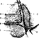
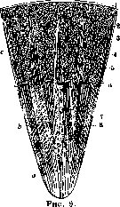
Fig. b
Figure 8. Relation of the peritoneum to the anterior surface of the left kidney: 1 - adrenal gland; 2 - splenic artery; 3 - pancreas; 4 - transverse mesocolon; 5 - colic facies; 6 - phrenicocolic ligament; 7 - splenic facies. (After Corning.) Figure 9. Diagram of the kidney structure: 1 - fibrous capsule; 2 - Malpighian corpuscles; 3 - interlobular vessels; 4 - medullary rays; 5 - labyrinth; 6 - arcuate artery; 7 - region of the urinary tubules; 8 - rays formed by vessels; a - papilla; b - medullary substance; c - cortical substance; d - base of the lobules. (After Testut.) Malpighian corpuscles). The cortex of the kidney has a yellowish-red color, the medullary substance has a partly bluish-red color; these shades depend on their varying blood supply. The cortical substance has a slightly granular appearance, easily tears in a direction perpendicular to the surface, and constitutes the main mass of the organ; the cortical part of the kidney occupies not only the superficial (cortical) layer (approx. 5-7 mm) of the organ, immediately beneath the fibrous tunic, but in places extends into its interior in the form of radial strips or partitions with transverse striation, reaching the sinus of the kidney. These strips of the cortical substance are named renal columns [columnae renales (Bertini)]. The columns are interspersed with lighter strips ascending from the base of the pyramids to the surface of the kidney in a radial direction; in the renal columns, these strips assume a horizontal position. Such processes of the medullary substance, embedded among the cortical ones, are called medullary rays or pyramidal processes [processus medullares (Ferrein)]. In the intervals between the 10-15 radial partitions, or Bertini's columns, there lies a denser, radially striated, proper medullary substance, which thus takes on the appearance of pyramids [renal pyramids (Malpighi)]; thus, each pyramid is surrounded on all sides by cortical substance. When studying the pyramids with the naked eye on a longitudinal section of the kidney, their fine radial striation (from outside inward) is visible. The base of the pyramid is convex and notched externally, while the end, shaped like a papilla facing into the sinus, is pointed (renal papilla). In the frontal plane, all the pyramids are arranged in three rows: the tallest pyramids are concentrated in the middle row, and the pyramids of the anterior and posterior rows are lower due to the smoothing of the kidney relief here. Each pyramid is about 5-8 mm high and is not sharply demarcated at its base from the cortex, but radially continues outward right up to the capsule of the organ in the form of straight strips or rays located between the cortical substance of the kidney (straight tubules). This is the striated part of the renal cortex (pars radiata). The indicated strips consist of straight tubules. The sections of the cortical substance inserted between Ferrein's rays and consisting of convoluted tubules (convoluted tubules) are called the convoluted part of the kidney (pars convoluta renis); the renal columns also consist largely of it. Each papilla of a pyramid is surrounded like a cuff by a tubular process, the so-called minor calyx (calices minores), which directly transitions here into the fibrous tunic of the kidney and into the mucous membrane of the minor calyx. Thus, a sort of soft calyx is obtained, into which the pyramid papilla is inserted and into which urine flows from the kidney through special terminal fine openings in the pyramid papilla (foramina papillaria) in an amount of 10-35 or more; these openings are located at the very apex of each pyramid (area cribrosa). Two or three Malpighian pyramids usually unite in a single papilla; the total number of such papillas in the kidney is 6 or more. The Malpighian pyramids are thinner in the middle of the kidney than at the poles. In their narrow terminal section, the minor calices transition into two (rarely three) wider reservoirs — the major calices, pelves (major calices), varying in shape and size and reflecting a number of constitutional moments (Ivanitsky) (Fig. 10). In adults, there are usually two (in 87.5% according to Ivanitsky) with an ampullary form of the renal pelvis and 3 with a dendritic one. The major calices merge into a single common reservoir — the renal pelvis. The renal pelvis (renal pelvis) has a highly variable funnel-shaped form and is flattened from front to back; the sharp part of the funnel faces away from the hilum downwards and soon transitions into the ureter (see). The fine structure of the major renal calices and pelves is similar to that of the minor calices. Three layers can be distinguished in it: the mucous membrane (tunica mucosa), the muscular middle layer (tunica muscularis), the outer

kidneys
connective-tissue, loose (tunica adventitia). Due to the fact that the musculature here does not form a dense, continuous layer, only two layers of the wall are well expressed: the inner one, consisting of stratified squamous epithelium, and that wall membrane which, in addition to the main connective tissue layer with an admixture of elastic fibers, also contains separate bundles of smooth muscle fibers. The muscle bundles are arranged irregularly, running both longitudinally and transversely. The musculature is especially developed in the wall of the renal minor calyces; here, at the base of the papillae, a annular muscle layer is formed (musculus sphincter papillae). The submucosal layer is absent. The covering stratified squamous epithelium passes directly from the inner wall of the calyx onto the papilla, covers its free surface, where it first becomes bilayered and then, at the apex of the papilla, single-layered and cuboidal. At the apex of the papilla it covers the edges of the papillary foramina and continues further into the lumen of the papillary ducts themselves. The renal pelvis is located relative to the skeleton such that it corresponds to the space between the transverse processes of L1 and L2 (somewhat lower on the right, somewhat higher on the left). They are distant from the median plane of the body by approximately 5 cm. The dimensions of the renal pelvis in women are relatively smaller than in men (Ivanitsky). Only the apex (angle) of the renal pelvis projects from the hilum of the kidney. In the human fetus and in the child, the kidney has a bumpy surface because it consists of separate lobules (renal lobes)—ren lobatus; with age, the unevenness of the renal surface is smoothed out due to the thickening of its cortex. Each lobule corresponds to one Malpighian papilla. Bertin's columns are located between the lobules of the kidney. On the basis of macro- and microscopic examination, the kidney is a gland of complex tubular structure. The entire parenchyma of the kidney consists of: 1) a complex system of urinary tubules, 2) a specially differentiated network of blood vessels, 3) a soft connective-tissue skeleton with vessels and nerves. All these systems are morphologically and functionally integrated into a complex organ of urine excretion. The urinary tubule begins with a blind, cup-shaped, invaginated expansion with a double wall (Bowman's capsule), continues further into the main part, steeply convoluted in the form of a glomerulus (convoluted part of the tubule or convoluted part of the first convoluted tubules—pars prima, s. convoluta tubuli contorti primi), then into a steep bend (Henle's loop) located in the medullary substance of the kidney, and finally passes into an intercalated convoluted section with a terminal connecting tubule. All these sections of the urinary tubule differ from one another in their extent, the character of their convolution, the caliber of the lumen, and the structure of the epithelium lining it. Each urinary tubule of the kidney, together with the Malpighian corpuscle connected to it, constitutes in the simplest organization of the kidney of lower animals a functional and anatomical unity. Into each double-walled Bowman's capsule is inserted the convoluted, glomerulus-like folded part of the capillary vascular system of the kidney, the so-called Malpighian corpuscle (glomerulus Malpighi). These two intimately connected, but different in development and origin formations constitute a new whole in anatomical and physiological respects—the Malpighian corpuscle (renal corpuscle, s. Malpighi), which is visible even at low magnification with a simple hand lens in the cortical substance of the kidney; the latter therefore appears speckled with dark (from blood engorgement) dots. Both in the primary kidney of lower animals (see Urogenital organs) and in the definitive kidney of higher animals, a urinary tubule departs from the cavity of Bowman's capsule of each Malpighian corpuscle. The conglomerate of individual sections of the urinary tubule together with the Malpighian corpuscle constitutes those architectural and physiological units that make up the main part of the parenchyma of the kidney (cortex and medulla). Thus, as many individual Malpighian corpuscles as there are in the kidney, so many are the convolutions and individual renal tubules, each of which, beginning blindly with Bowman's capsule in the Malpighian glomerulus, ultimately opens into the renal calyx at the apex of the pyramid's papilla through its special openings—urine-bearing pores, or papillary foramina. Before flowing into the minor pelvis, individual urinary tubules gather in groups of several into larger tubules of the first and second order. On the basis of onto-, phylogenetic, and physiological data, Braus proposes to call the structural complex from the glomerulus to the output tubules inclusive the nephron, i.e., the main functionally and anatomically indivisible organizational part of the kidney. These units of kidney structure in man are not separated from each other by any clear connective-tissue layers; this division can be outlined only by the arrangement of the blood vessels of individual nephrons. The nephron is complicated by the fact that not the entire mass of loops of the urinary tubule is enclosed in the convolution, but a part of the loops of the latter (7/8 to 15/16) leaves this association and returns to it again; thus, only that section of the nephrons' tubules that empties directly into the collecting tubule is terminal. The length of the urinary tubule from Bowman's capsule to the end is 55.58 mm (Peter); the length of the main part is about 14 mm. Up to its end, the urinary tubule along the way forms two kinds of loops; one of them, Henle's loop, having the appearance of a descending long loop, partly leaves the convolution, partly remains in it, constituting the first and second convoluted parts of Henle's loop—the ascending (pars contorta I) and descending limbs (pars contorta II); these loops can be either long or short, depending on how close or far from the outer surface of the cortex their initial Malpighian glomeruli are located, and moreover in inverse relations, i.e., those Henle's loops that originate from the Malpighian glomeruli closest to the medullary substance of the kidney are longer. According to Peter, there are 6–7 short loops for every long loop. Henle's loops thus represent the straight descending part of the urinary tubule inserted between the first and second convoluted sections of the urinary tubule. Therefore, the loop distinguishes a descending limb—the continuation of the first convoluted part of the urinary tubule—and another limb—the ascending one, which passes into the second convoluted part of the glomerulus; these parts are thus counter-current in the direction of the fluid flow in them, i.e., one is "papillopetal," the other "corticopetal." Henle's loop is located next to and along the length of those collecting tubules that receive urine from the corresponding Malpighian corpuscle. It is remarkable that the ascending limb of the loop always returns to its Malpighian corpuscle, lying adjacent to its afferent vessel. After the convoluted and intercalated straight sections of the urinary tubule follows the straight, terminal part of the latter, which is already properly the excretory pathway (straight renal tubules). Emusually emptying in pairs into straight fine tubules (Bellini's tubules), all collecting ducts of larger caliber approach their Malpighian papilla; the number of these ducts gradually decreases, while the caliber increases. On the papilla they finally open by means of even larger ducts (papillary ducts) in the form of small openings (papillary foramina, or urine-bearing pores). The basic principle of the structure of the Malpighian corpuscle in the adult is preserved the same as in the primary kidney. In this corpuscle, the capillary vascular glomerulus (glomerulus) and its double-walled epithelial capsule (capsule of the glomerulus of Bowman, s. Mulleri) are distinguished. The glomerulus generally has a round or close to it shape; it is located in the lower section of the convolution of its urinary tubule. In it, urinary and vascular poles are distinguished, which are not always completely polar. At the urinary pole of the glomerulus, the newly formed (provisional) urine leaves Bowman's capsule through the initial part of the urinary tubule (the latter is often narrowed, forming the neck). In the region of the vascular pole, the glomerulus is in close connection with Bowman's capsule by means of the stalk of blood vessels entering and leaving the Malpighian corpuscle—this is, so to speak, the hilum of the glomerulus; however, these vessels nowhere anatomically communicate with the cavity of Bowman's capsule. There is only a close epithelial contact (transition) from the capsule wall to the vascular glomerulus. The capillary vessels entering the Malpighian corpuscle and leaving it are arteries of the afferent and efferent type (afferent vessel and efferent vessel). The afferent artery divides into many capillary branches, which (according to Maksimov) do not anastomose with each other, but lie in lobules in the form of a series of convoluted loops, then pass into a common efferent vessel, thus forming a "miraculous net" (rete mirabile). The entire surface of the glomerulus is covered like a serous membrane of internal organs by the inner (visceral layer) membrane of Bowman's capsule, which invaginates at the hilum of the glomerulus, leaving outside a free outer surface, or parietal membrane. Between both layers of Bowman's capsule remains a capillary space (intercapsular space).
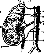
Fig. 11. Arteries of the kidney, adrenal gland, and capsule: 1 - adrenal gland; 2 - capsula adiposa renis; 3 - branches to the adipose capsule ("arcade"); 4 - branches of the internal spermatic artery to the ureter; 5 - internal spermatic artery; 6 - inferior suprarenal artery; 7 - renal artery; 8 - middle suprarenal artery; 9 - celiac artery; 10 - inferior phrenic artery; 11 - superior suprarenal artery. (After Corning.)
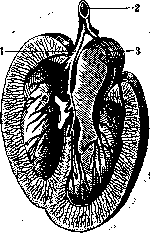
Figure 12. Distribution of vessels inside the kidney (cross section): 1 - anterior branch of the renal artery (2); 3 - posterior branch. (After Corning.)
Arteries of the kidney. The arteries of the kidney present peculiar features, inasmuch as they pass first through the rete mirabile of Malpighian corpuscles and only then connect with the capillaries of the venous bed. Blood enters the kidney through one, sometimes two or more arteries (renal artery) branching off directly from the abdominal aorta, immediately below the origin of the superior mesenteric artery (Fig. 11). The caliber of the renal artery is relatively very large and almost equals the caliber of the hepatic artery, since this artery serves not so much for the nutrition of the kidney itself as a conduit for waste products from the entire blood bed. Ventrally and next to the artery runs the vein of the same name, through which venous blood flows away from the kidney. Occasionally the vein lies behind the artery. Usually the ureter is located behind the artery. In the region of the renal sinus, the arteries and veins divide into two main branches, which are located anteriorly and posteriorly to the renal pelvis (dorsally and ventrally), and then each of them divides into 4--5 branches (Figure 12). The renal artery, single at first, branches differently: either immediately before entering the sinus (extraorgan branching type) or inside the sinus and the parenchyma of the kidney (intraorgan branching type). Very often (in 20% or more) there is also an accessory renal artery. This anomaly is usually associated with other malformations of the kidney. Having entered the parenchyma of the kidney, the artery divides into branches corresponding to the lobes of the kidney (interlobar arteries); each of these branches enters between the papillae of the pyramids into the columns of Bertin (Fig. 13). These arteries usually do not give off branches at first and de- ____
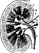
scribe incomplete arches around the pyramids Figure 13. Distribution of the (arteriae arciformes, s. ar- artery inside the kidney: cus arteriosi), which are 1 - capsule; 2 - cortical substance; located on the boundary of 3 - interpyramidal artery; 4 - the cortical and medullary Malpighian pyramid; 5 - renal substance, parallel to the sur- artery and its branches (6); 7 - face of the kidney. From the pelvis.
interlobar arteries where they form the arciform arteries, smaller-order interlobular arteries branch off (interlobular arteries, s. radiate arteries, since they go parallel to the medullary rays), which approach perpendicularly the boundaries between the cortical lobules; these arteries are tortuous, anastomose with each other, and pass directly into the so-called "admirable net" of Malpighian corpuscles.
567
While the largest number of thin branches of the interlobar arteries is distributed in the medullary substance of the kidney, the branches of the interlobular artery depart at the boundary between the medullary and cortical substance. The individual interlobular arteries divide mostly dichotomously into radial branches. Each branch gives off numerous short afferent vessels, of which the lower ones go posteriorly and the middle ones transversely to the Malpighian corpuscles. Having formed a sort of "admirable net," numerous branches and loops in the Malpighian corpuscle, the arteries reconnect into a vessel of smaller caliber than the afferent one and exit from Bowman's capsule under the name of efferent vessel. The efferent vessels immediately pass again into a thin capillary network that surrounds all straight tubules and convoluted tubules. The medullary substance of the kidney receives blood through special branches (straight arterioles) departing downward partly from the efferent vessels, partly directly from the interlobar arteries and arciform arteries. Straight arterioles are located between the urinary tubules of the medullary substance, repeatedly anastomose with each other, and surround the renal tubules with dense capillary networks up to
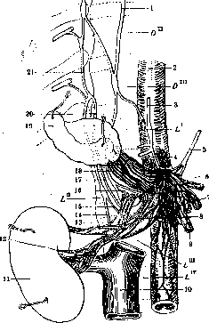
Figure 14. Distribution of splanchnic nerves: 1 - greater splanchnic nerve; 2 - aorta; 3 - phrenic nerve; 4 and 18 - branches of solar plexus; 5 - vagus nerve; 6 - celiac artery; 7 - superior mesenteric plexus; 8 and 12 - nerves to the kidney; 9 - superior mesenteric artery; 10 - inferior vena cava; 11 - right kidney; 13 - anastomoses; 14 and 21 - branch of sympathetic nerve; 15 - branch of splanchnic nerve; 16 - branchlet to the adrenal gland; 17 and 20 - lesser splanchnic nerve; 18 and 19 - adrenal gland. (After Testut.)
up to the papillae and form vascular wreaths here. The venous vessels departing from this system empty into the arciform veins. Many authors believe that the system of branches of the interlobar artery consists of "terminal" arteries, since the branches of neighboring arteries do not anastomose with each other at all. In reality, such anastomoses do exist, but in a relatively small number and of a small caliber: that is why upon ligation, embolism, or thrombosis of the renal artery, atrophy of the corresponding kidney ensues despite the clear presence of vascular connections between the system of the renal artery and several capsular arteries originating from other sources. Besides the kidney parenchyma, the renal artery supplies its branches to the renal pelvis and capsule (capsular artery). From the indicated vascular capillary networks, the blood, having passed the Malpighian corpuscles, is carried off via the venous (capillary) system through the straight venules, then via the interlobular veins and interlobar veins into the renal vein. Exactly in the same way there is also a special capsular vein, which has the peculiarity that on the surface of the kidney it forms characteristic fine radiate branches [stellate veins (Verheyen)]. Thus, in the kidney there are two capillary systems—one of them directly passes into veins and serves for the nutrition of the kidney itself, the other constitutes the system of glomeruli and participates in the process of urine secretion. - Lymph initially flows away along numerous interstitial connective tissue spaces of the renal parenchyma, after which it collects into deep lymphatic vessels following the blood vessels, and then enters the superficial ones lying near the capsule. The main outflow pathways together with the blood vessels exit into the hilum of the organ, where they empty into the nearest lumbar aortic lymph nodes. Another group of vessels passes through the diaphragm together with the splanchnic nerves and empties into the thoracic duct. The kidney is very richly supplied with nerves, which enter the organ through the hilum and reach individual Malpighian corpuscles (Fig. 14). Their endings were observed in large numbers both in the renal vessels and in the cells of the renal tubules. Their fibers are mostly unmyelinated; they originate from the celiac plexus, from ganglia embedded in the hilum of the kidney itself, and from the X-XII intercostal nerves. To these are also admixed fibers from the vagus nerve. G. Ivanov. II. Histology of the kidneys. The Malpighian corpuscle of the kidney is a spherical formation and consists of a vascular glomerulus and Bowman's capsule. The latter represents as it were a ball invaginated by the glomerulus and therefore consists of 2 layers—the inner one, closely covering the glomerulus, and the outer one. Between the layers lies a cleft—the capsule cavity, into which the fluid filtering from the vascular glomerulus penetrates. The size of glomeruli in humans ranges from 164 to 229 µ in diameter (Peter). The change in the size of glomeruli by age is seen from the following table: Table 1. Age Diameter in µ Age Diameter in µ Newborn . . 1 year . . . 4 years ..... 84.77 87.76 101.70 148.63 18 years .... 32 years ... 40 years .... 189.88 213.49 193.11 The given table (according to Peter) has only relative significance, since the variations in the size of the glomeruli are significant and since the size of the glomeruli is also conditioned by a number of functional factors, among which the diet has special significance. The number of glomeruli in humans is estimated on average at 1,700,000 for both kidneys. The total area of all glomeruli in humans averages 49.5 m2. The vessels approaching 69 the glomerulus have a somewhat different structure from other vessels. The endothelium presents no differences. The internal elastic membrane disappears mostly upon entering the capsule. The muscular coat is thin and uneven. In humans, at a short distance from the glomerulus, the muscular coat of the artery acquires cushion-like thickenings resulting from an increase in the layers of muscle cells. Special modifications of the muscular coat, the smooth muscle cells of which acquire an "epithelioid character," were discovered in rodents by Ruyter. The elastic membrane disappears, the entire region is richly supplied with nerve endings. Similar relationships were seen by Eberling in humans as well. Interestingly, sections of arteries forming the so-called arterio-venous anastomoses (nail, tip of the nose) have the same character. The functional significance of the described sections of the afferent arteriole was revealed by the works of Peterfi and Okkels, who applied micromanipulation to the living kidney of amphibians. The touch of a microneedle causes a strong and rapid contraction of the afferent vessel almost to the complete obliteration of the lumen. The contraction lasts 1-2 minutes and is observed both on the whole organ and on isolated glomeruli. Touching the glomerulus itself and the efferent vessel with a microneedle causes no contraction whatsoever. Even earlier, Richards and Schmidt, observing the living kidney of amphibians, showed that as a rule the kidney does not function entirely as a whole. While a number of glomeruli and other parts of the nephron connected with them function, other nephrons remain inactive at the same time. The number of functioning glomeruli can be greatly increased by the administration of urea, caffeine, glucose, pituitrin, and by the transection of sympathetic nerve fibers. Adrenaline and stimulation of the sympathetic nerve lead to a decrease in the number of functioning glomeruli. Thus, the periodicity of work of individual nephrons is explained by the contraction of the corresponding afferent arterioles. Upon entering Bowman's capsule, the arteriole, having lost its smooth muscle elements, breaks down into capillaries forming the glomerulus with their loops. Connective tissue, penetrating right up to the beginning of the branching of the capillaries, divides the entire glomerulus into indistinctly distinguishable lobules (numbering 4-8). Each capillary forms a strongly winding loop and empties directly into the efferent vessel. Thus, the efferent vessel receives as many capillaries as were formed during the branching of the afferent arteriole (Fig. 15). The capillaries of human glomeruli do not anastomose with each other (Wintrup, Möllendorff). The wall of the capillaries consists of endothelium and a thin connective tissue membrane lying on the outside. The boundaries between individual endothelial cells can usually not be detected either by silver or by stains. On this basis, the majority of authors deny the existence of separate cells in this section of the vascular system, considering this endothelium to be a plasmodium. However, Nussbaum, Policard, and others describe distinct boundaries between the endothelial cells of the glomerular capillaries. - The connective tissue membrane or basement membrane consists of the finest film of connective tissue. Outwardly from it between the capillary loops lie connective tissue cells, the number of which is relatively small. The nature of these cells is not sufficiently elucidated (fibrocytes or histiocytes). A small number of collagen fibers accompanies the convolutions of capillaries. The entire glomerulus is clad on the outside by the inner layer of Bowman's capsule. The long-established view that this layer represents a continuous layer of very thin epithelium must now be abandoned. The cells of the inner layer of Bowman's capsule represent strongly branched flat
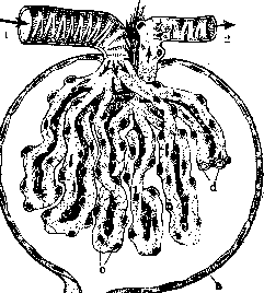
corpuscle hygiene: 1 and jj Jf^Y i i - afferent and efferent arteries; a - epithelium of Bowman's capsule; b - neck; c - covering cells; d - sectioned capillaries. (According to Möllendorff.) stellate cells which, covering the outer surface of the glomerulus, close the basal membrane of capillaries not solidly, but in such a way that significant gaps remain in which the capillary wall directly looks into the capsule lumen. These stellate cells were named "covering cells" (Deckzellen). Interestingly, similar relationships have been found recently in pulmonary alveoli. The covering cells are capable of accumulating a certain amount of vitally introduced dyes. The outer layer of Bowman's capsule in humans consists of a single-layered flat epithelium. The cellular plate is so thin that the cell nuclei protrude into the capsule lumen. In rodents (mouse, rat), the distal half of the capsule adjacent to the neck is covered by a taller epithelium which, approaching the neck, becomes cubic. The outer surface of the epithelial cells of the outer layer bears a series of depressions in the form of fine grooves into which the fibrils of the capsular basement membrane are pushed. The latter consists of a homogeneous connective tissue film penetrated by reticulin (so-called "lattice") fibers. Nephron tubules. Histologically, the nephron tubules are divided into the following sections (Fig. 16a): I. Neck. II. Main segment (synonyms: portio principalis, convoluted tubules of the first order, Hauptstück). It distinguishes - 71
KIDNEYS
67) consists of a) the convoluted part and b) the straight part (syn. pars convoluta, pars recta). III. Transfer segment (syn. descending segment of Henle's loop, thin segment). IV. Middle segment (syn. Mittelstück, incoming segment of Henle's loop, and second-order convoluted tubules). It is divided into a) the straight part (ascending limb of the loop) and b) the convoluted part (second-order tubules); the latter in turn consists of the intermediate part (Zwischenstück) and the intercalated part. V. Connecting segment, passing into VI. The system of collecting ducts (Fig. 16, 6). I. The neck in humans
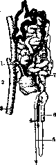
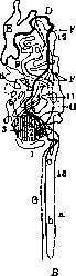
Figure 16. Nephron from the kidney of a 26-year-old human. A - wax reconstruction. Malpighian corpuscle and main segment with small dots; Henle's loop is cut; intermediate and intercalated parts of the middle segment are black. Collecting tubules are white (from Braus): 1 - vas afferens; 2 - Malpighian corpuscle; 3 - a. interlobularis; 4 - ascending part of the loop; 5 - collecting tubule; 6 - straight part of the main segment; 7 - vas efferens. B - Diagram: c-13 - bends of the main segment; a - descending part of Henle's loop; b - ascending part of Henle's loop - straight part of the middle segment; cc - intermediate part of the middle segment; D-E - intercalated part of the middle segment; F - collecting tubule; G - straight tubule. (After Peter.)
is mostly indistinctly expressed, its epithelium rapidly transitions from low to cubic, characteristic of the main segment. - II. Main segment. The lumen of the main segment, especially in its convoluted part, varies depending on the functional state of the organ. During increased diuresis, it is relatively wide (Fig. 17); during normal function, it is narrow; during anuria, it is slit-like. On tangential sections through the tubules of the second segment, it is seen that the boundaries between cells have a zigzag character, because the lateral surfaces of the cells present a series of wedge-shaped protrusions tightly inserted into the corresponding indentations Figure 17. Cross- section of a tubule of the main segment in a rabbit after increased diuresis. of neighboring cells (Figure 18). Therefore, with standard staining methods, the cell boundaries of the convoluted part of the second segment are not detected. The inner surface of the cells of the second segment, facing the lumen, bears the so-called "brush border" characteristic only of this segment. The latter

represents a specially differentiated protoplasm and strongly resembles the border of the intestinal epithelium. Under high microscope magnification, it appears to consist of a series of glued rods (or tubules according to Möllendorff). With increased activity of the organ,
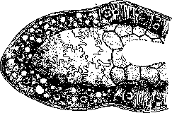
Figure 18. Straight part of the main segment. Sudden change in the character of the epithelium during the transition from the convoluted part to the straight part. The zigzag cell boundaries of the convoluted part (left) become even in the straight part (right). (After Zimmermann.)
the striation of the border becomes clearer. Its functional significance has not been sufficiently elucidated. The cell nuclei are round and lie closer to the basal part. Chromatin is scattered in uniform small clumps. The nucleolus is indistinct. The nuclei of the straight part stain more intensely than the nuclei of the convoluted part. The chromatin here lies tightly against the membrane, and the nucleolus is clearly visible. The protoplasm of the cells of the entire second segment possesses a characteristic striation. This "rod-like" striation occupies the greater part of the cell, extending to the entire basal part and the nuclear zone, leaving only an insignificant unstriated part directly beneath the brush border (Fig. 19). Since the chondriome in this epithelium is represented by long rod-like chondrioconts occupying the same area of the cell as the striation
lity, most researchers believe that the striation can be explained by the arrangement of chondrioconts. M. Heidenhain and Mislawsky assume, in addition to the chondriome, a fibrillar structure of the protoplasm. The striation is a vital formation; upon cell death, as well as in a number of pathological states, it disappears. Its functional significance has not been elucidated. Similar morphological structures are observed in the excretory ducts of the salivary glands (salivary tubes). The cells of the second part possess an extraordinarily strongly developed chondriome. Long, rod-shaped chondrioconts lie closely appressed to one another. Upon the administration of inadequate stimuli (foreign protein, cantharidin), as well as in a number of pathological processes, the chondrioconts disintegrate into a large number of granules. Thus, the state of the chondriome can serve as a measure of the normal state of the tubules and signal very early pathological changes in the organ. The Golgi apparatus is discovered in all cells of the main part. It has the appearance of a network of crossbars of varying thickness, located either between the nucleus and the brush border or as a ring around the nucleus. In some cases, a basal location of the apparatus has been described. Depending on the functional state of the cell, the Golgi apparatus changes its location, moving from the inner zone toward the outer, which coincides with the picture of dye resorption by the tubular epithelium (Yasvoyn). When granules revealed by vital staining appear in the epithelium, their location as a rule corresponds to the location of the apparatus (Yasvoyn, Nasonov). Between the brush border and the nucleus lies the centrosome, usually containing two centrioles. Inclusions in the form of protein granules of various sizes may be found in the cells of the entire second part. Their physiological significance in mammals is unclear. Fat in the form of droplets of neutral fat and lipoids is encountered in the cells of the second part in the majority of mammals. The tubules of the cat contain the most fat. In the adult human, the second part, according to the opinion of most researchers, normally contains no fat. In newborns and especially in infants, fat is found in a large percentage of cases. Fat depositions in the tubules of the second part can be provoked experimentally by the introduction of fat components into the blood of animals (soaps, fatty acids). In elderly individuals, an insignificant amount of so-called physiological pigment may be found in the cells. The straight part of the second part generally has the same cytological features as the convoluted part. The cell boundaries in the straight part become more distinct, which is connected with a change in the lateral surfaces of the cells, which become flat in the straight part. The change in the character of the cell boundaries occurs suddenly upon the transition of the convoluted part into the straight part, therefore this transition has a sharp boundary. III. The intermediate segment begins with a sharp change in the character of the epithelium, which becomes flat. At the same time, transitional forms from the cubic epithelium, characteristic of the second part, and the extremely flat epithelium of the third part do not exist. The cell boundaries here are just as uneven as in the convoluted part of the second part. The nucleus has an oval shape, elongated along the axis of the tubule. The chondriome is represented by sparse chondriosomes. The centrosome lies between the nucleus and the inner edge of the cell and is connected with a small flagellum looking into the lumen. The significance of the flagellum is unknown. It is possible that it represents a rudiment of the ciliated epithelium with which the tubules of lower vertebrates are lined. In old people, a significant amount of pigment accumulates in the epithelium of the intermediate segment. IV. The middle segment. In its tubules, the brush border is absent, which is the most characteristic distinction of this part from the second. Cell boundaries are discovered with difficulty, because, just as in the preceding parts, the lateral surfaces of the cells are uneven. An exception is formed by the terminal or intercalated part of the fourth part. The lumen of the tubules is considerably wider than the lumen of the second part. The cells are lower than in the second part. The nuclei of the cells are located closer to the lumen than to the base. The striation of the epithelium is sharply expressed. Rod-shaped chondrioconts, beginning from the base of the cell, reach almost to the lumen itself. The centrosome is connected with the flagellum looking into the lumen. The straight part of the segment passes without sharp cytological differences into the convoluted, intermediate part. The latter has taller cells and, giving a series of strong convolutions, always approaches Bowman's capsule of its own nephron. Here the tubule as a rule lies adjacent to the afferent vessel. The point of contact has a characteristic structure. The cells become narrower, as a result of which the abundance of nuclei strikes the eye. The place of contact of the intermediate part with the afferent vessel has been named macula densa. The physiological significance of this formation is unknown. The intercalated part following the intermediate is distinguished by a lesser density of the chondriome and a lighter appearance of the cells. The secretory part of the kidney tubules ends here. Here also lies the boundary between the elements that developed from the nephrogenic tissue of the mesonephros and the derivatives of the permanent kidney. The efferent system of the kidney begins with a short connecting segment (V). A characteristic distinction of the collecting system is the clear cell boundary, light protoplasm poor in the chondriome. Just as in the previous parts, the centrosome has a flagellum. The epithelium retains this character throughout the entire extent of the collecting system. As the straight tubules enlarge, the epithelium becomes taller and in the region of the papillary ducts assumes a palisade-like character. The connective tissue of the kidney. Just like Bowman's capsule, the tubules of all parts are covered by a thin connective-tissue membrane. The latter consists of a homogeneous thin film into which reticulin fibers are woven in large numbers. Thus, the connective-tissue stroma of the kidney belongs in its greater mass to the so-called lattice fibers (Gitterfasern). The fibers woven into the membrane of the tubules pass without interruption onto the blood capillaries enfolding the tubules, as well as onto the thin arteries and veins. Therefore, the connective-tissue stroma of the tubules and small vessels represents a single organic whole. The connective tissue in the region of the tubules is poor in cellular elements. The infiltration by cellular elements appearing in pathological cases must be attributed mainly to migration from the vessels. Around large vessels, especially at the border of the cortical and medullary substance, there is a significant accumulation of connective-tissue cells. In the medullary substance, as one approaches the papillae, collagen fibers appear, forming a dense network around the vessels. The capsule of the kidney (capsula fibrosa) consists of two sharply distinguishable layers. The inner layer is represented by a dense plexus of collagenous and reticulin fibers directly penetrating into the cortical substance. This layer is rich in cellular elements (fibrocytes and histiocytes). In humans, it contains smooth muscle fibers. The outer layer is represented by connective tissue poor in cellular elements, having a lamellar character. Between the lamellae is a dense network of elastic fibers. Histology of the blood vessels of the kidney (for the distribution of blood vessels, see above). Interlobar and large interlobular arteries have a well-developed elastic apparatus of the intima, which is represented by two or three elastic membranes and a dense network of elastic fibers between them. A strongly developed muscle layer borders externally on a dense layer of elastic fibers or even membranes—the external elastic membrane (membr. elastica externa). The structure of the afferent artery is described above. The veins of the cortical substance do not have a clearly expressed adventitia. The system of stellate veins (vv. stellatae) possesses a strongly expressed muscular coat. Bundles of smooth muscles run mainly in the direction parallel to the axis of the vessel and pass directly into the smooth-muscle bundles of the inner layer of the capsule (cf. trabeculae and veins of the spleen). The capillaries of the cortical layer have a well-expressed adventitia consisting of a connective-tissue membrane enfolded by lattice fibers. In the medullary substance, the lattice tissue surrounding the capillaries passes into a dense network of collagenous fibers. Morphological analysis of secretion of the kidney. Experimental morphology of the kidney is currently of outstanding interest and will obviously have a decisive role in elucidating the mechanism of urine secretion. Growth of cellular elements of the kidney in tissue culture. Upon planting pieces of the kidney in culture, the growth of both epithelium and mesenchyme is observed (Khlopin). The epithelial cells generally retain their character. Upon planting an embryonic kidney, phenomena of differentiation are noted. However, the formation of tubule systems is not reached. B. Lavrentiev. III. Comparative physiology of the kidneys. The most important physiological processes of the elimination of metabolic products from the organism, as well as the maintenance at a certain level of the osmotic pressure of internal, cavity fluids (blood), are carried out by excretory organs, the so-called emminktories.
In most animals, the main excretory organ is the kidney, which has its own micro- and macroscopic features at different stages of both phylogenetic and ontogenetic development (see Urogenital organs). The morphological features of the structure of the kidney in animals are also closely related to the features of the course of excretory processes at different stages of development and the qualitative characteristics of the kidney's excretate. However, the kidney is not the only morphological formation with which the excretory function is associated, as evidenced by the facts of comparative physiology and morphology. In a huge group of animals, there are atypical excretory organs, such as in protozoa (contractile vacuole), in coelenterates, nematodes, and finally in insects, which have so-called Malpighian tubules. Furthermore, the process of excretion and elimination of decay products, water, and salts by the corresponding organs is by no means, as commonly thought, the only form of final metabolic processes. Basically, there are two types of excretion—intracellular and extracellular. The first type is the most primitive and consists in the fact that metabolic products are deposited in the vacuoles of the cell protoplasm (e.g., in unicellular organisms) or in special cells or special tissues (such as the deposition of various crystals in the nephrocytes of the adipose tissue of insects). The deposition, isolation, and utilization of end-product decay are characteristic of a large group of animals and predominantly characteristic of invertebrates. In contrast to the continuous excretion of decay products that occurs in vertebrates, in invertebrates we encounter phenomena of deposition of these substances in the most bizarre forms. In many ascidians, next to the heart, there is a special vesicle filled with fluid in which a crystalline formation floats, increasing by accretion as the animal ages; in some of them (Synascidiae, Ciona), there are special cells near the intestinal tract into which concretions are also deposited. In echinoderms, wandering cells of the body cavity play the role of organs for accumulating metabolic products, and, joining together, they form special brown formations in the body. In many insects, especially old ones, uric acid concretions are found in the cells of the fat body. In the latter case, there is an important correlation between the intracellular deposition of decay products in the fat body and their elimination to the outside by the Malpighian tubules; in the pupal state, when the Malpighian tubules do not function, the excretory function falls entirely on the fat body. The study of these peculiar forms of excretion, found mainly in invertebrates, is especially important for understanding many phenomena occurring at higher stages, including in vertebrates. The fact is that the phenomenon of deposition of products of physiological and chemical metabolic processes of the organism, as well as the utilization of excretion products within the organism itself, has considerable biological significance and is widely encountered in the animal world. This deposition has, for example, protective significance in insect larvae. Thus, Kellin showed that secretions from the Malpighian tubules in the genera Acidia and Agromyza are of great importance for the formation of a protective cover for the pupa. Such protective formations are also the cocoons of certain silkworms (Bombyx lanestris, Bombyx quercus), the threads of which consist of uric acid-rich secretions of the Malpighian tubules. Moreover, chemical analysis of the wings, starting with the works of Hopkins, indicates [e.g., in the family of white butterflies (Pieridae)] a significant amount of uric acid in them. It is important that the wings of the same butterflies (in embryonic development) in the pupal state do not contain uric acid, which appears in them only after transformation into a butterfly, when the excretion of metabolic products by the Malpighian tubules into the gut begins. Furthermore, in the same insects, a connection between the formation of wing pigments and the products of final decay is indicated. The formation of skin pigments in other animals, including vertebrates, is also in a definite dependence on the utilization within the organism of the products of final decay (tyrosine, tryptophan, histidine, etc.). Finally, the phenomenon associated with the growth and coloration of scales in fish is extremely interesting. Here, the deposition of guanine, a probable product of purine decay, in the scales of fish is of great importance. In this same direction, the question can also be analyzed with respect to horny formations, etc. All the cited comparative physiological data on peculiar metabolic pathways in the sense of pathways for the elimination and utilization of final decay products deepen our usual understanding of these pathways, predominantly associated with the phenomena of extracellular excretion, with the activity of the kidney, and with the analysis of the products excreted by them to the outside. Meanwhile, the study of metabolic processes by end-metabolic products is unthinkable without a deep consideration of those biologically important processes of utilization of these products within the organism, which manifest themselves in varying degrees of expression at all stages of development of the animal world. This is unthinkable, of course, without studying the forms of elimination of metabolic decay products in various animal species. Strictly speaking, the processes of excretion (extracellular excretion) occur, as indicated above, with the participation of special organs of the urinary system, the central organ of which is the kidney. The basic data on the physiological characteristics of this organ were obtained based on the study of the kidneys of several laboratory animals (dogs, rabbits, frogs, and some others); meanwhile, a deep understanding of the processes occurring in the kidney is possible only through a comprehensive study of the physiology of the kidney with various morphological characteristics at different stages of animal development. As will be indicated below, in a number of controversial points of renal physiology, this particular approach has provided the most reliable ways to analyze the question. Morphological data indicate, along with differences, also features of similarity in various types of excretory organs of both vertebrates (pronephros, mesonephros, metanephros) and certain invertebrates ("metanephridium") (see Urogenital organs). The common features of the excretory organs in many animals are: the presence of cells capable of absorbing substances from the body cavity fluid and throwing them into the lumen of the channel, and the connection of these cells with the body cavity fluid on the one hand, and the presence of an excretory channel and its opening on the other. The difference is expressed further in the complication of the form and function of mainly the excretory channels and those cellular formations associated with them, as well as with the types of connection of the final excretory apparatus with the body cavity. The process of perception of decay products is associated with the accumulation of these substances by certain morphological elements and their further ejection or other (see above) utilization. In simple cases in many aquatic animals (protozoa, sponges, coelenterates), a simple form of diffusion takes place. The large surface of these animals serves for the diffusion of final decay products into the water. In these same animals, morphologically differentiated excretory organs are weakly developed or not represented at all. Already in lower animals (terrestrial, as well as aquatic forms), starting with the classical works of Kovalevsky, the presence in the organism of special "collecting" cells, the so-called athrocytes, has been established, which selectively absorb foreign substances (dyes, etc.) and decay products and have a direct relation to the processes of excretion. Usually such cells (nephrocytes) in nephridia (e.g., worm metanephridia) are equipped with cilia forming the so-called "flame bulb", with the help of which the fluid secreted by the cell into the lumen of the channel connected to it is driven further. Basically, despite the great variety of morphological structures of excretory apparatuses, there is much in common in the processes occurring in the final apparatus of these organs. This final apparatus is either open into the body cavity or blind, closed in relation to the body cavity. The first case is represented especially in lower worms, where amoeboid terminal cells of protonephridia wedge among parenchymal cells; in annelids, the funnel of the excretory organ opens directly into the body cavity by a group of special cells, solenocytes, resembling the terminal cells of protonephridia. In a number of other invertebrates (snails, cephalopods, crustaceans), the funnel of the excretory organ is also open directly into the body cavity or into corresponding formations of the body cavity. In the vertebrate series, the closed type of terminal apparatuses of the kidney is mainly encountered, where, along with the system of channels and funnels, there is a special formation, the so-called Malpighian corpuscle (see above), which has a direct, internal connection with the blood vessel system.
It goes without saying that these two types of connection between the excretory devices and the body cavity—open and closed—are related to the history of development of the circulatory system in both groups of animals: the open, lacunar type in invertebrates and the closed circulatory system in higher vertebrates. However, among lower vertebrates, there is also a form of open connection between the renal apparatus and the body cavity. We encounter this in cyclostome fishes (lampreys) and amphibians (Gymnophiona and Urodela). In particular, in the frog, as is known, the kidney communicates directly with the renal vein. Just as among many invertebrate animals (nemerteans), there is an internal connection between the terminal apparatus and the circulatory system. As for the system of tubules, comparative morphological and comparative physiological data indicate not only the peculiarity of the forms of these tubules at different stages of development, but also the peculiar physiological characteristics of its various sections, which perform certain functions thanks to corresponding cellular formations. As indicated above, the main observations on the processes occurring in the kidneys have been verified only on a limited group of laboratory animals, and to this time the comparative physiology of the kidneys and processes associated with this organ has been poorly developed. Existing theories of urine formation are based on facts obtained mainly from vertebrate animals (see Diuresis). Comparative data indicate the fact that the quality of urine depends on the morphological features of the urinary tubules. Thus, for example, the dog and the cat have very concentrated urine and along with it a very long thin part of the loop of the tubules; the same is true for rabbits, while the thin part of the loop in pigs is very short and they have more diluted urine. With a general review of urine concentration, one can see a deep dependence between the degree of concentration and the morphological features of the urinary tubules, especially the thin part of Henle's loop. From Table 2 it can be seen that in lower vertebrates (fish and amphibians, as well as reptiles) hypotonic urine is excreted, while in higher vertebrates (birds, mammals)—hypertonic. This is in direct dependence on the difference in the morphological structures of the kidneys at different stages of development. As the brilliant comparative physiological studies of Hōber showed, hypotonic urine is present in those animals in which Henle's loop is absent; thus the comparative physiological experiment gave a decisive answer regarding the processes taking place in this part of the renal tubules. In birds, as seen from the table, the urine is also hypertonic, although they also lack the thin loop of the tubules. But in these animals, as is known, the function of urine concentration is associated with other morphological formations—specifically with the epithelium of the cloaca. In addition to these clear data on the physiological characteristics of Henle's loop, it is necessary, in agreement with Ellinger, to point out that there are still no exact data on the physiological characteristics of the individual segments of the tubules. Synthesis in the kidneys. The kidneys of various animals are also the site of synthesis of various products. Thus, Bunge and Schmiedeberg (1876) demonstrated for the first time the synthesis of hippuric acid in the kidneys of the dog, which was later established in a series of works by Snapper, Grünbaum, and Neuberg in the pig, sheep, and man. Benedict demonstrated the formation of ammonia in the kidneys by comparing the ammonia figures in three blood vessels, as seen from the attached table. Table 3. Animals Amount of ammonia (in mg %) a. carotis v. cava v. renalis 0.102 0.088 0.113 0.085 0.226 0.176 The amount of ammonia in the blood in Benedict's experiments dropped sharply when the kidneys were excluded from the organism. As for invertebrates, the formation of chitin scales in the cells of their kidneys is indicated. There are indications of this regarding the nephridial cells of many worms, but these facts need verification. Finally, Pütter (1926) pointed out the formation of uric acid by the kidney cells of the apple snail. There is as yet no direct proof that the uric acid crystals actually present in the cells of the snail's kidney are products of formation (excretion) of the renal cells rather than a phenomenon of crystallization of protein-containing fluids. However, this question is very important, since Pütter connects the question of the synthetic function of the kidneys, the fact of finding uric acid in the cells of the snail's kidney, with his secretory theory of urine formation, pointing to the intrarenal formation of excretion products. Meanwhile, comparative physiological analysis shows that the formation of the main excretion products (urea, uric acid) in the animal kingdom proceeds by an extrarenal pathway; see also Diuresis.
Kh. Khachaturyants. IV. Pathological anatomy of the kidneys. Among the cadaveric changes of the kidneys, phenomena of autolysis on the part of the tubular epithelium, occurring shortly after death, deserve mention; at the same time, the protoplasm of the cells becomes granular, and subsequently the nucleus disappears. Macroscopically, this change presents a picture similar to cloudy swelling of the kidneys, differing from the latter by the uniform involvement of the entire parenchyma. Developmental anomalies of the kidneys are diverse. Complete congenital absence (agenesis) of both kidneys (aplasia) or their underdevelopment (hypoplasia) are rare and lead to the death of the fetus in utero or immediately after birth. Agenesis of one of the kidneys (more often the left) is not uncommon (twice as often in men as in women), and the ureter is usually absent as well; hypoplasia of one of the kidneys is observed less frequently. With the indicated underdevelopment of one of the kidneys, the other is always compensatorily enlarged. If the kidney primordia were located close to each other during their development, they can subsequently fuse, resulting in a single unpaired kidney (ren concretus, s. impar); such fusion most often occurs in the region of the lower ends of both kidneys, resulting in a single kidney of arcuate or horseshoe shape (ren arcuatus, syn. ren unguiformis, French ren en fer à cheval, English horse-shoe kidney), the arch of which has its convexity turned downward and is draped over the spine and aorta. Such a horseshoe kidney is encountered in approximately 0.3% of all autopsies.
It is less common for the kidneys to fuse at their upper ends and form a horseshoe kidney with an arch whose convexity is turned upward, or for a solid fusion of the kidneys to occur, thanks to which a single irregularly shaped organ is formed, lying like a cake (German Kuchenniere) in front of the spine with separate or common pelves, often located on the anterior surface of the organ. Asymmetric fused (horseshoe) kidneys can be observed. All these developmental anomalies are often accompanied by irregularities in the renal vessels, the presence of accessory vessels (Figs. 20 and 21), and anomalous development of the ureters (their doubling or, for example, in the case of a common pelvis in fused kidneys, the formation of only a single ureter). Numerous variants of the irregular shape of each of the kidneys individually are also observed. Sometimes a kidney (one or more often both) turns out to be lobated (ren lobatus), which is an expression of the persistence of embryonic clefts separating the embryonic primordia of the kidney from one another (the so-called renculi). A rarer developmental anomaly is a kidney bearing its hilum on the anterior surface; here the pelvis, often slightly stretched, appears surrounded by a ridge of renal tissue, which resembles a cheesecake or
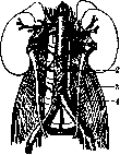
Figure 20. Accessory vessels of the kidney: 1—a. coeliaca; 2—a. mesenterica inf.; 3—ureter; 4—m. psoas. (According to Corning.) 6 81 shield and is designated as «ren scutaneus, s. scutulatus» (German Schildniere). Even rarer is a kidney in the form of a group of fused, extremely irregular lumps, which is called a lumpy, shapeless kidney (ren informis). A double kidney, i.e., bifurcated into two separate parts, usually each with a separate ureter (ren duplex), is rarely encountered. Finally, a very great rarity is the presence, alongside a normal kidney, of an accessory (usually under-
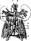
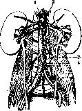
Figure 21. Accessory vessels of the kidney, a: 1—a. coeliaca; 2—a. mesenterica sup.; 3—ureter; 4—a. mesenterica inf.; 5—m. psoas. b: 1—a. coeliaca; 2—a. mesenterica sup.; 3—a. spermatica int.; 4—a. mesenterica inf. (According to Corning.)
developed) kidney. Changes in the position of the kidney see below. Of the circulatory disorders in the kidney, the most frequent is chronic congestion, which may have purely local causes in the form of, e.g., thrombosis or compression of the renal vein, or is a particular manifestation of general congestion associated with a decline in cardiac activity. The congested kidney is at first somewhat enlarged, firm, its capsule strips easily; the surface is bluish and blood-distended stellate veins are visible on it; on cross-section, the tissue is hyperemic with a bluish tint, with the medullary substance being the most cyanotic. With prolonged stasis, the kidney is somewhat reduced in size and becomes denser, remaining cyanotic (which is called cyanotic induration—induratio cyanotica renis) [see separate plate (pp. 687-688), fig. 4]. Small depressions appear on the surface with adhesion of the capsule; on cross-section, the layer of renal tissue may turn out to be thinner than normal, and the pelvis slightly dilated. Arterial hyperemia accompanies acute inflammations of the kidney, and, in addition, usually occurs in a compensatorily hypertrophied kidney; the possibility of arterial hyperemia of the kidney as a result of increased work of the left ventricle of the heart is admitted. The state of anemia of the kidney is most often observed as a partial manifestation of general anemia; anemia of a purely local character, concerning only the kidney, is possible with disorders of the patency of the renal artery of a most diverse character (trauma, inflammatory changes, arteriosclerosis, nerve spasm, etc.); anemia very soon leads to degenerative fatty change, and often to necrosis of the epithelium of the convoluted tubules. The complete closure of a branch of the renal artery, most often by means of an embolus or thrombus, results in the formation of an infa r c t, which in the kidney is usually of an ischemic character, with only a small peripheral rim appearing hemorrhagic. In small infarcts, the layer of renal tissue adjacent to the capsule often remains alive because it receives blood from the capsular vessels. The larger the caliber of the occluded artery, the greater the size of the formed infarct; upon closure of the main trunk of the renal artery, the entire kidney undergoes necrosis. Almost continuous infarction can also take place with widespread thrombosis of the intrarenal arterial branches, as happens sometimes, e.g., in eclampsia, or with spasm of the entire system of renal arteries as a result of camphor injections. Infarcts of the kidney according to general rules subsequently undergo organization, as a result of which a characteristic retracted scar is formed at the site of the infarct. Thrombosis of the renal artery can be observed with its atherosclerosis, with trauma, with inflammatory changes in the arteries, in particular with periarteriitis nodosa, and also as marantic thrombosis. Embolism in the kidney is very frequent; small emboli in the form of particles of thrombotic masses, fat droplets in fat embolism, clumps of microorganisms are retained predominantly in the capillaries of the glomeruli and, depending on the nature of the embolus, cause either necrosis of the capillary loops of the glomeruli or inflammatory changes (embolic glomerulonephritis); larger emboli occlude larger arteries, which results in the formation of an infarct. Hemorrhages into the kidney—see Hematuria. Atrophy of the kidney manifests itself in senile atrophy, expressed in a uniform decrease in the size of the kidneys while maintaining their smooth surface; often, however, on such a surface there are sunken places corresponding to areas with stronger atrophy. Microscopically, one finds atrophy of the tubules, a decrease in the volume and anemia of the glomeruli with an impoverishment of their cells, sometimes obliteration of the capillary loops of the glomeruli with transformation into hyaline lumps. In this case, there is usually some proliferation of the interstitial tissue. (Atrophy of the kidney in arteriosclerosis—see Nephrosclerosis.) An example of pressure atrophy of the kidney is hydronephrosis (see). The term granular atrophy of the kidney was formerly applied to those cases of atrophy of the renal parenchyma with proliferation of connective tissue when the depressed areas of atrophy alternate with bulging foci of preserved tissue, which gives a uniform granularity to the surface of the kidney. At the present time, this roughly morphological term is not used due to the fact that the above-mentioned picture of granular atrophy of the kidney can be observed in two completely different processes: arteriolosclerotic nephrosclerosis and the chronic period (outcome into secondary contraction) of glomerulonephritis. Necrosis in the kidney can affect not only continuous regions of renal tissue, as happens in infarcts (see above), but also selectively only the epithelium of the convoluted tubules; in the latter case, the nuclei cease to stain, the protoplasm becomes dull, granular, and subsequently the cells disintegrate. Such necrosis of the renal epithelium takes place in various poisonings (sublimate), in severe infections, sepsis, acute yellow atrophy of the liver, severe stagnant jaundice [see separate plate (pp. 687-688), fig. 2], sometimes in diabetes. Clinically, this reveals the symptom-complex of necrotic nephrosis (see). Among degenerations in the kidney, one encounters: cloudy swelling, vacuolar degeneration, hyaline-droplet degeneration (see Hyaline degeneration), fatty degeneration. All these degenerative changes concern the epithelium mainly of the convoluted tubules of the kidneys. In particular, with respect to fatty degeneration, it must be borne in mind that the presence of fat droplets in the protoplasm of epithelial cells can be assessed as a latent phenomenon only in the case of localization in the convoluted tubules, since the appearance of neutral fat in the epithelium of the intercalated parts of the tubules, loops of Henle, and in the collecting ducts usually represents a normal phenomenon, apparently related to the function of the kidney. Such physiological fatty infiltration is usually found in 1/4 of all human kidneys studied; in animals (e.g., cats) it occurs even more often. As a pathological phenomenon in the kidney, besides neutral fat, the deposition of doubly refractive lipoids can also be observed; the latter can be found in the epithelium of the convoluted tubules, as well as in the stroma, namely in the cells of the latter, which take on the appearance of xanthoma cells. This kind of lipid deposition is characteristic of lipoid nephrosis. A special kind of change is fatty infarct, manifested in the deposition of lipoids in the papillae of the medullary substance, namely into the interstitial tissue and into the membrana propria of the straight tubules; the formation of fatty infarct is usually associated with a disturbance of cholesterol metabolism, often manifesting itself simultaneously also in the deposition of cholesterol in the walls of arteries (i.e., atherosclerosis). Amyloid degeneration is expressed in the kidney by the deposition of amyloid in the capillaries of the glomeruli and under the endothelium of small arteries. The deposition of amyloid can occur in a previously unchanged kidney, as happens, e.g., in general amyloidosis accompanying chronic osteomyelitis, tuberculous caries, etc.; in these cases, they speak simply of amyloidosis of the kidney or amyloid kidney. A weak degree of change is determined only under the microscope, whereas with significant amyloidosis the kidney, while maintaining its normal size, acquires density, a grayish-white color, and a dull, lardaceous sheen, which is designated as lardaceous kidney. In other cases, amyloid degeneration of the kidney is combined with other degenerative changes of the kidney characteristic of chronic nephrosis; in such cases, designated as amyloid nephrosis or lipoid-amyloid nephrosis, the kidney presents the picture of a large white kidney, related to chronic nephrosis, but with the superimposition of a lardaceous tint peculiar to amyloid; in this case, they speak of a large lardaceous kidney or large white amyloid kidney. On the deposition of glycogen in the kidney in diabetes, see Glycogen infiltration. The kidney is characterized by various hematogenous deposits, some of which it has traditionally been customary to call by the term "infarct," which is unsuitable for this kind of change. This includes the deposition of urates in the kidney in newborns, which is called uric acid infarct. In addition, urates in the form of amorphous and crystalline masses are also deposited in the kidneys in adults with gout, being located in the form of strips and spots in the medullary substance. The deposition of blood pigments in the kidney takes place upon the destruction of erythrocytes of various origins. Thus, with hemosiderosis accompanying pernicious anemia, granular deposition of hemosiderin is found in the epithelium of the convoluted tubules; with poisoning by arsine, potassium chlorate, etc., blood poisons, hemoglobin or methemoglobin in the form of reddish-yellow or brownish drops, lumps, and continuous cylinders occupy the lumens mainly of the straight tubules of the medullary substance, which can impart a brownish tint to the pyramids (the so-called hemoglobin infarct). In jaundice of newborns, the so-called bilirubin infarct is observed (see); in jaundice of adults, yellow-green staining of the cortex takes place due to the diffuse impregnation of the epithelium of the convoluted tubules with bile pigment. The deposition of lime in the kidney can be observed in a diverse form. In various chronic changes of the kidney in their stroma and in the lumens of the tubules, small groups of small calcareous concrements can be observed.
In sublimate poisoning, alongside necrotic nephrosis (sublimate kidney), later stages of the disease may show widespread deposition of lime in the necrotic epithelial cells of the convoluted tubules, which, according to Schmidt's data, is explained by the delayed excretion of lime from the body due to the lesion of the large intestine caused by sublimate poisoning. Something similar, but to a lesser extent, occurs in severe dysentery and in naphthol poisoning. In calcareous metastases, the kidneys are a common site for the deposition of lime (see Calcareous deposits, metastases). In addition to all these types of lime deposition in the kidneys, in old age there is sometimes deposition of small calcium grains into the hyalinized interstitial tissue and into the membrana propria of the tubules of the renal papillae. This change, often combined with fatty infarction, is called calcareous infarction; in sections through the kidney in these cases, white streaks are visible in the papillae, diverging in rays from the tips of the papillae. Some call silver infarction the deposition of silver in the kidney in argyria. Regenerative manifestations in the kidneys are very frequent. With various degenerative and necrotic changes in the epithelium, the surviving cells undergo karyokinetic division, which can result in the restoration of the epithelial cover in the tubules that had perished over a considerable extent. Excessive regeneration is often observed in the form of the appearance in the tubules of voluminous protoplasmic masses with numerous nuclei (epithelial giant cells) or the formation of papillary outgrowths from the epithelium; sometimes, on the basis of regeneration, e.g., in a sclerotic kidney, the formation of true adenomatous nodules (so-called regenerative adenomas) occurs. Hypertrophy of the kidneys most commonly manifests as compensatory, vicarious hypertrophy of one kidney due to the congenital absence, surgical removal, or damage by a chronic disease process of the other. In congenital absence or underdevelopment of one kidney, the compensatory enlargement of the other kidney is based on the formation in the latter of a greater number of glomeruli and tubules, which have normal dimensions. In contrast to this, in acquired compensatory hypertrophy of the kidney, it is merely a matter of an increase in the size of the glomeruli and tubules. Inflammation of the kidneys - see Nephritis. An abscess of the kidney, i.e., the formation of a purulent cavity, and a carbuncle of the kidney—the formation of a group of abscesses—belong to purulent inflammation of the kidney (see Nephritis). Tuberculosis of the kidney manifests in various forms. Miliary tubercles in the form of pale gray or yellowish nodules, usually indistinctly contoured and located predominantly in the cortical layer, are observed in generalized miliary tuberculosis; in this case, a significant number of tubercles are usually noticed in both kidneys. In chronic tuberculosis of the lungs or other organs, individual tubercles are often discovered in the kidneys (in 1/2 of cases), appearing here as a result of hematogenous seeding. Usually they are also located predominantly in the cortical layer, while some of them develop in the medullary substance, as some think (Orth), due to the passage of tubercle bacilli through the capillaries of the glomeruli and the subsequent retention of bacilli in the tubules of the medullary layer; these tubercles, belonging to the so-called excretory tuberculosis (German Ausscheidungstuberculosis), often have an elongated shape corresponding to the course of the tubules. Sometimes the tubercles are arranged in beads along the course of the vessels. It should also be noted that in hematogenous tuberculous involvement of the walls of larger arteries in the kidneys, infarcts may develop, and in lesions of small arteries, the picture of nephrosclerosis of one degree or another (tuberculous nephrosclerosis) can be observed without signs of tuberculosis visible to the naked eye. These forms can proceed latently without any clinical manifestations. Of greater importance is another form of tuberculosis, namely chronic local tuberculosis of the kidney. A comparatively rarer type of it is the formation in the kidney of a node of the solitary tubercle type (tuberculous kidney), which has no tendency to softening and breakdown and subsequently undergoes encapsulation and petrification. More frequently observed is the caseous-ulcerative or cavernous form of chronic local tuberculosis of the kidney (renal tuberculosis, or nephrophthisis). This form can affect one kidney or both kidneys simultaneously and is expressed by the fact that a tuberculous focus (sometimes several foci) arising in the kidney undergoes caseous degeneration [see separate table (p. 687-638), Fig. 3], liquefies, and forms a cavity—a cavern; the latter may not be in connection with the pelves (closed tuberculosis) or may be in communication with the pelves (open tuberculosis). In cases of the latter kind, there is ulceration of the renal papillae and usually participation of the renal pelves (tuberculous pyelonephritis) as well as the ureter in the tuberculous process. Upon obstruction of the latter by caseous masses, the pelves appear dilated and filled with purulent or caseous contents, while the inner layer of the pelves and the surface of the renal tissue facing them consists of a crumbling caseous mass (tuberculous pyopyonephrosis). The various forms of chronic local tuberculosis of the kidney in the majority of cases have a hematogenous origin and are hematogenous metastases from a primary complex present most frequently in the lung; in the period of development of renal tuberculosis, the primary complex may already have undergone complete healing. Pyelitic forms of local tuberculosis of the kidney may, in addition, also have an ascending origin, i.e., be a consequence of tuberculous involvement of the genital organs (prostate, testicles, seminal vesicles), from which the process spreads to the bladder, ureters, pelves, and kidneys; in the ascending form, both kidneys are more often affected. In certain very protracted forms of tuberculosis of the kidney, significant development of connective tissue and wrinkling of the kidney may be observed; in such cases, an alternation of strongly depressed whitish areas with bulging areas, in which caseous nests are visible, is noticeable. Syphilitic changes in the kidneys are not uncommon. In congenital syphilis, infiltrates of lymphoid and plasma cells are very frequently found around the vessels of the cortical layer, less often of the medullary substance; sometimes a diffuse proliferation of interstitial connective tissue is noted at the same time. True gummas in the kidneys in congenital syphilis are extremely rare. Extremely peculiar findings noted by some researchers (Ribbert, Müller, etc.) in congenital syphilis in the kidneys are special large cells located in the tubules, strongly resembling Protozoa; apparently, they represent peculiarly hyperplastic and degenerated epithelial cells of the tubules. In acquired syphilis, sometimes already in the secondary period, the development of lipoid nephrosis takes place. In the later gummous period, the formation of a large number of scar-like retractions on the surface of the kidney is sometimes observed, in a word, the picture of a contracted kidney. Such a syphilitic contracted kidney may be the result of the outcome of lipoid nephrosis into contraction, while in other cases it represents nephrosclerosis developing on the basis of syphilitic changes in the renal arteries. Some, however, consider the existence of interstitial syphilitic nephritis with an outcome in contraction (fibrous multiple interstitial syphilitic nephritis) to be possible. Gummas are formed much more rarely in the kidneys. Actinomycosis of the kidney is expressed in the formation of conglomerates of small abscesses (of the carbuncle type) among dense connective tissue. The disease develops in the kidney either due to spread from neighboring parts or as a result of metastasis from other organs. Isolated actinomycosis of the kidney alone is an extreme rarity. In lymphogranulomatosis, nodules of a characteristic granuloma may be observed in the kidneys. In blood diseases, changes in the kidneys are very frequent. In anemias accompanied by the development of extramedullary myelosis, myeloid tissue often develops in the fatty tissue around the renal pelves (especially in children, so-called splenic anemia). In myeloid leukemias, infiltrates of myeloid tissue are found in the renal tissue around the vessels. In lymphatic leukemia and aleukemia, the development of multiple lymphomas in the form of whitish nodes of lymphadenoid tissue bulging under the capsule is often observed in the kidneys. In lymphosarcomatosis, both kidneys are often stuffed with whitish nodes of new growth; sometimes a predominant growth of tumor masses in the superficial layer of the cortex is noticed. Cysts of the kidneys can have various origins. Single and multiple small cysts ranging in size from a millet seed to a nut, often encountered in advanced age, are the consequence of the proliferation in the kidney of interstitial tissue compressing some of the tubules, as a result of which the overlying sections are stretched by secretion. Such cysts are observed in nephroscleroses, mainly in the cortical layer of the kidney. Their contents are either watery, colorless, or more rarely thicker, colloidal with a brownish tint. Similar single cysts of the kidney can also be met with at a young age in kidneys that are otherwise completely normal; in such cases, they are considered the results of a developmental defect of the tubular systems (absence of connection between the upper and lower sections of the tubule).
In rare cases, solitary cysts of the kidney reach very large sizes (up to the head of an adult) and in such cases can simulate hydronephrosis. In addition to this kind of cyst, the kidney is characterized by a disease in which both kidneys are permeated by a huge number of cysts of various sizes. This disease is designated as «hepar cysticum», «hydrops renis cysticus» or «cystic degeneration» of the kidney. This alteration of the kidney occurs at any age; it is also observed in newborns. Sometimes it is discovered as an accidental finding at autopsy, in other cases it is diagnosed during life. Both kidneys are usually affected to an equal degree; rarely do the kidneys retain their normal volume; usually they are greatly enlarged (up to 20–28 cm in length; weight reaches 3,000 g). From the surface of the kidney, a multitude of cysts bulges out (see separate plate, Fig. 1), and on cross-section a continuous conglomerate of cysts is visible, between which small layers of renal tissue are noticeable. The contents of the cysts are transparent, watery, sometimes yellowish, sometimes brown, rarely thick, colloid; some have occasionally observed purulent contents in part of the cysts. Microscopically, the inner surface of the cysts turns out to be lined with low cubic epithelium. Sometimes a flattened glomerulus is discovered on the inner surface of the cysts. In some cystic kidneys, islands of cartilage and bundles of muscle fibers were found in the layers between the cysts. Sometimes cystic degeneration of the kidney is combined with the presence of cysts in the liver and (more rarely) in the pancreas. Regarding the origin of the indicated alteration of the kidney, different views have been expressed. In former times, the formation of multiple cysts was looked upon as a consequence of an inflammatory process (nephritis fibrosa Arnoldi), depriving the tubules of patency. Others treated the cystic kidney as a neoplasm of the multi-chambered adenocystoma type. However, at present, the most widespread and correct view must be considered that of the cystic kidney as a consequence of a maldevelopment of the kidney, consisting in the fact that between the two systems of tubules—the upper glomerular and lower excretory, which are laid down separately—no connection occurs; in connection with this, retention cysts are formed from the upper sections of the tubular systems. This view, putting forward the delay in the normal development of the kidney as the basis for the formation of cysts, is supported by the presence of cartilaginous and muscular tissue in some of the cystic kidneys, as well as the combination of cystic kidneys with congenital cysts in other organs (see above). Tumors of the kidney can originate from the epithelium and from connective tissue. Of the epithelial tumors, adenomas are most often observed. The latter are more frequently encountered in the kidneys of elderly people in the form of one or more whitish nodules varying in size from a millet seed to a hazelnut; larger sizes of adenomas, for example the size of an adult's fist, are very rare. Most often, adenomas are located in the cortical layer beneath the capsule. Under the microscope, a picture of glandular or tubular structure (simple adenomas) or cavities with abundant papillary growths (papillary adenomas) is discovered. Very often, fat deposition in the cells of the adenoma is observed, which imparts a yellow hue to the tumor nodule; hemorrhages are the cause of the brownish color of the tumor. It is generally thought that adenomas developing during atrophic and sclerotic processes in the kidney have regenerative factors as their basis (regenerative adenomas), whereas those encountered at a young age in normal kidneys are connected with a maldevelopment of renal tissue. Cancer of the kidney usually arises in the form of a node that pushes aside and destroys the renal tissue and often reaches enormous sizes. The most frequent form of renal cancer is encephaloid cancer with the microscopic structure of adenocarcinoma, sometimes papillary. The tumor consists of soft, whitish, sarcoma-like tissue with areas of necrosis, breakdown, and the frequent formation of cysts on this basis. Much rarer are fibrous cancers of the kidney and squamous cell keratinizing cancer, originating as a rule from the epithelium of the renal pelvis. A very frequent epithelial tumor of the kidney is hypernephroma (see). Among connective tissue tumors, fibromas, fibromyomas, lipomas, fibromyolipomas are frequent, which are usually discovered in the form of small nodules, more often located in the medulla; an exception is formed by lipomas, which are usually found in the cortex directly beneath the capsule. Much rarer are hemangiomas and fibromyothelio-mas of the kidney. All these tumors in the majority of cases are connected with the maldevelopment of renal tissue and belong to hamartomas. Sarcomas of the kidney most often occur in childhood, and are also encountered in newborns; in adults they are much rarer. Sarcomas of the kidney represent rapidly growing, extremely soft tumors of a pinkish-white color, often reaching very significant sizes (up to the size of an adult's head) and weight (up to 3,000–4,000 g). Sarcomas of the kidney in adults belong to round-cell or spindle-cell forms and do not differ in any particular way in comparison with sarcomas of other organs. As for sarcomas of the kidney in childhood, they represent congenital neoplasms with a very peculiar structure, which makes it necessary to classify them as mixed tumors. Comparatively rarely do they consist only of tissue of the round-cell sarcoma type; in the majority of cases, glandular channels and alveoli lined with cubic or cylindrical epithelium are visible among such tissue, which serves as the basis for designating such tumors as adenosarcomas. In some of the glandular formations, peculiar bulgings into the lumen are sometimes observed, resembling certain stages of the embryonic development of Malpighian corpuscles. Among the interstitial round-cell tissue, regions of mucous and spindle-cell tissue, bundles of smooth and cross-striated muscle fibers (adenomyosarcoma), areas of adipose tissue, cartilage, and sometimes bone may be encountered. A tumor of such a structure grows very rapidly, destroys the renal tissue, grows into neighboring tissues, but in comparison with ordinary sarcomas rarely gives metastases (to the liver, to the lungs). Of metastatic tumors in the kidney, cancers, sarcomas, chorioepitheliomas, and other malignant neoplasms may be observed; metastases develop in the kidney hematogenously and form single or multiple nodes in them, the appearance and structure of which correspond to the tissue of the primary tumor node. Much more rarely than hematogenously, tumor metastases in the kidney occur thanks to retrograde lymphatic or venous transport of tumor cells. Of animal parasites, the single-chambered echinococcus is most often encountered in the kidney, sometimes forming a large cyst that can rupture into the renal pelvis, which results in the appearance of hooks and scoleces in the urine. Multi-chambered (alveolar) echinococcus is observed in the kidney extremely rarely (prior to 1901, Melnikov-Razvedenkov could find only one case in the literature). Cysticercus is rarely encountered in the kidneys; Schistosoma haematobium or Bilharzia frequently parasitize residents of southern countries (e.g., Egypt), settling in the mucous membrane of the renal pelvis, ureter, and bladder and causing inflammatory processes in them. Filaria sanguinis hominis, s. Bancrofti, a parasite of tropical and subtropical countries, can be found in the lymphatic pathways of the urinary tract wall, causing lymph stasis and the admixture of the latter with urine (chyluria) and hematuria. Cases of the finding of the worm Eustrongylus gigas in the human renal pelvis, frequently discovered in animals (dogs), have been described. Sometimes a calcified parasite nodule of Pentastomum denticulatum is found in the kidney. In connection with the clarification of the life cycle of ascarids and hookworms, it has become known that the larvae of these worms can occasionally be discovered in the kidney, where they cause the formation of granulation nodules with the parasite in the center.
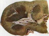
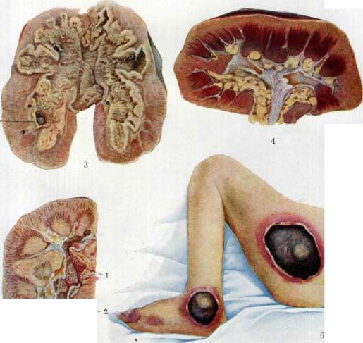
Fig. 2. Jaundiced (necrotic) nephrosis. The substance of the kidney is painted in yellowish-green. Fig. 3. Tuberculosis of the kidney, the pelvis and adjacent renal parenchyma in a state of caseous breakdown; 1 - stone. Fig. 1. Cyanotic induration of the kidney. Fig. 5. Pyelitis in nephrolithiasis: 1 - hyperemic mucosa of the pelvis; 2 - mucus-purulent masses; 3 - stone. Fig. 6. Bedsores in decubitus. 690 bundles of smooth and cross-striated muscle fibers (adenomyosarcoma), areas of adipose tissue, cartilage, sometimes bone. A tumor of such a structure grows very rapidly, destroys the renal tissue, grows into neighboring tissues, but in comparison with ordinary sarcomas rarely gives metastases (to the liver, to the lungs).>
A. Abrikosov. V. Functional Diagnostics of the Kidneys. Determining the functional capacity of the kidneys in various disorders serves: 1) to establish the form of kidney damage in each individual case: whether there is a primary disease of the kidneys or whether their disease is merely a consequence of the illness of other organs (for example, a congested kidney in heart diseases); 2) for the prognosis of the disease: whether renal function is sufficient or not; whether its restoration to normal is possible; 3) in part to establish topical diagnostics: whether there is predominant damage to the glomeruli or tubules; 4) to develop methods of treatment, or the prescription of a diet, depending on the preserved function of the kidneys. At present, the clinic possesses a large number of diverse methods for the functional diagnostics of the kidneys. All these methods can be divided into three main groups: 1) methods based on determining the nature and amount of excretion of various substances in the urine, 2) methods that ascertain changes in the blood observed during impaired renal function, and finally 3) methods where renal function is determined by observing changes simultaneously in both the blood and the urine. 1. The first group of methods for determining renal function comprises two types, one of which has the purpose of determining the nature of the excretion by the kidneys of substances characteristic of the organism and formed in it in the process of metabolism (water, salts, urea, and other nitrogenous products); the second type sets itself the task of determining the nature and degree of excretion by the kidneys of such substances that are not normally found in the organism (various coloring agents, certain salts—potassium iodide, sodium ferrocyanide, etc.). a) For determining kidney function, the simple determination of the amount of urine and its specific gravity, the establishment of quantitative and qualitative changes in diuresis: anuria, oliguria, polyuria, and the determination of the molecular concentration of urine by cryoscopic methods, the establishment of specific gravity either by the usual method or using an aeropycnometer, are of great importance. Along with these methods, the determination of the patient's weight and water balance is also important for assessing the state of renal function. Of much greater importance for determining the sufficiency, or insufficiency, of the kidneys are the water test and the concentration test. The most well-known methods in the clinic for applying this test are the tests of Strauss, Volhard, and Nordgaard (H. Strauss, Volhard, Nordgaard). Of these, the most widespread and accepted is the Volhard test. The principle of this test is as follows: since the essence of normal kidney function consists in the maximum excretion of water upon a single administration of a significant amount of it into the organism, which indicates normal function of the glomeruli, and, on the other hand, in the greatest excretion of solid substances with a small amount of excreted urine, i.e., in the ability of its maximum concentration, which gives mainly the opportunity to establish good function of the tubules, then when applying this test, the variability and degree of excretion of water and solid substances are an indicator of the state of the excretory function of the kidneys regarding the excretion of water and solid substances. Volhard's method itself consists of the following: the patient in the morning on an empty stomach, after preliminary emptying of the bladder, is weighed, drinks 1,500 cm3 of water or liquid tea within 1/2 or 3/4 of an hour, and thereafter, while remaining in bed, urinates every 1/2 hour; thus, urine is collected for 4 hours. The quantity and specific gravity of each half-hour portion are determined separately. After the expiration of 4 hours and further until the next morning, the patient receives neither soup nor other liquid, but only dry food. During this time, urine is collected every 2 hours, measured, and the specific gravity of each individual portion is determined in the same way. Thus, the first part of the experiment represents the water test, and the second part (with dry diet) represents the concentration test. In the first part of the experiment, of greater importance than the total amount of liquid excreted over 4 hours is the maximum rate of secretion, i.e., the largest half-hour portion of urine, which in a healthy kidney reaches 250–300 cm3 with a simultaneous decrease in its specific gravity to 1.000–1.002. If the drunk water is indeed excreted entirely within 4 hours, but the individual half-hour portions remain approximately equal and the diuresis curve is flat, then such a character of the water test indicates impaired kidney function. With severe damage to it, individual half-hour portions become smaller and more uniform, the total amount of urine over 4 hours decreases sharply, and the specific gravity of the urine portions decreases insignificantly. In assessing the concentration capacity of the kidneys, the primary importance is held by qualitative changes in diuresis, the absence of variability, i.e., the impossibility for the kidneys, with a low excretion of urine (or water), to excrete the same amount of solid constituents as occurs under normal conditions of excretion. The measure for determining the concentration capacity is the maximum value of the specific gravity of individual portions under a dry diet. From this point of view, a functionally sufficient kidney is characterized by the independence of the excretion of solid substances from the excretion of water, and vice versa, in renal insufficiency, this very dependence comes to the fore (Volhard). To judge what degree of renal insufficiency exists in each individual case, both tests—both for water excretion and for concentration—must be performed. The results should be evaluated as follows: 1) if the ability to rapidly excrete water and the independence of the separation of solid substances from water excretion (concentration capacity) are preserved, then renal insufficiency is excluded; 2) if both tests turn out poorly, renal insufficiency is present; the latter is the stronger, the less pronounced the extrarenal factors are—edemas (hidden and overt), cardiovascular insufficiency, elevated temperature, etc.; 3) with preserved good concentration capacity and poor water excretion, the results of the test almost always indicate a disorder of water excretion due to extrarenal factors; 4) with poor concentration and fully preserved ability to excrete water, renal insufficiency may be absent, since in this case the relative inability to concentrate can be compensated by polyuria. From this it logically follows that "the main criterion of renal insufficiency is the violation of concentration capacity, and the most important criterion for prognosis is the ability to excrete water as a mechanism for compensating renal insufficiency" (Volhard). Examples of various types of the Volhard test are given in Table 4. When evaluating the results of the Volhard test, it is necessary to take into account a number of conditions that can influence these results: the presence of edemas of nephrotic and cardiac origin, fever, diarrhea, sweats, prior restriction of fluid before the test (Volhard) or preliminary increased administration of it (Siebeck), the nature of the diet before the test, the state of the cardiovascular system, constitutional moments, disorders of the endocrine glands, anemic and cachectic states, the influences of the nervous system, individual characteristics, etc. (Munk). A correct assessment of all these conditions in each individual case, simultaneous observation of changes in blood concentration, and the use of other methods ultimately make it possible to determine with the Volhard test the state of renal function with considerable accuracy. Several modifications of the Volhard test have been proposed in order to obtain various details in determining renal function. Table 4. Volhard's test for water excretion and concentration. Unlike the Volhard test, Strauss's method determines the percentage of chlorides and the urea content in each portion of urine. On the evening before the experiment, the patient receives an omelet of 2 eggs with 1 g of salt, a roll with butter, and 200 cm3 of tea; a warning is given not to eat anything at night. The next day at 6 and 7 a.m., the patient urinates; after this, he drinks 1 liter of liquid tea and urinates every hour for 4 hours. At 11 a.m., the patient receives: 100 g of white or black bread, 100 g of cheese, and 1 g of salt; at 1 p.m.—an omelet, 1 g of salt, 1 apple; at 4 p.m., he receives the same as at 11 a.m. At the hours: 11, 1, 4, and 7, the patient urinates. Observation shows that with this test, the greatest dilution occurs in the 2nd–3rd hour after the administration of fluid, and the increased concentration after taking dry food occurs at the 4th and 7th hours. If the described test can often be limited to, sometimes with the Strauss method it is also necessary to use an "extended" experiment with concentration, in which at 10 p.m. of the experimental day, a dry supper is additionally given, and the specific gravity is determined in the urine collected after this, as well as at 6 and 7 a.m. of the following day.
If the specific gravity of the urine in the second morning urine (7 AM) does not rise to 1.025, the dry-diet test is continued throughout the following day. Above, in evaluating the results of the Volhard test, it was indicated that with its help (resp. by the method of Strauss and others), renal insufficiency is established, i.e., the insufficiency of excretion of water and those solid substances whose removal is necessary for life. This insufficiency occurs in those cases where the function of both the glomeruli and the tubules is impaired. Given the close dependence of the latter on the activity of the glomeruli, tubular insufficiency leading to renal insufficiency is closely linked with a decrease in glomerular function. Hence the conclusion: the degree of renal insufficiency depends not only on tubular insufficiency, but also on glomerular insufficiency. -- Renal insufficiency may arise 1) as a result of a decrease in P., or rather as a result of time from 1,500 of water drunk on an empty stomach at 7-8 AM, normally excreted amount specific gravity delayed amount specific gravity amount specific gravity at 8 hours 8 1/2
» 12 » Water test 120 250 320 280 200 140 120 60 40 1,011 1,015 1,003 1,015 - 1,000 1,001 1,000 1,003 - 1,001 1,006 1,002 - 1,002 1,011 - 1,003 -. - 1,003 1,015 1,014 1,012 1,010 1,010 1,011 Total 3 hours 5 », 7 » . 9 » . 12 » . Night 7 hours Total 1,530 | - Concentration test 1,017 1,021 - 1,017 1,026 1,015 - 1,019 - 1,018 1,027 1,017 1,030 1,015 1,012 1,011 1,013 1,011 1,011 1,013 1,011 quantitative change of secretory elements, kidneys; 2) due to qualitative changes in the tubules as a result of sharp degeneration followed by their atrophy, or due to pressure atrophy from urinary stasis; 3) due to primary glomerular insufficiency. According to Volhard, compensatory polyuria occurs and is sharply pronounced in the second case, in the first case it depends on the number of preserved glomeruli, and in the latter case it is absent. The most important sign of renal insufficiency is so-called hyposthenuria (Koranyi) and isosthenuria (Volhard). Koranyi named hyposthenuria a state in which the molecular concentration of urine, determined cryoscopically (see Cryoscopy), changes in such a way that "the difference between the maximum and minimum molecular concentration decreases parallel to the severity of the kidney lesion; this decrease occurs due to the simultaneous lowering of the maximum and raising of the minimum, and these values approach a certain average value of molecular concentration," and this average value is the molecular concentration of the blood. In renal insufficiency, the excretion of hyposthenuric urine is observed, the molecular concentration of which is the more constant and the closer it approaches that of the blood, the more severe the renal insufficiency. An indicator of the decreased molecular concentration of urine in clinical practice is the value of the specific gravity of urine, approaching the value of the specific gravity of blood (after removal of protein), equal to 1.010. True genuine hyposthenuria is characterized by the constancy of the specific gravity in each separate two-hour portion of urine and above all by the equally low specific gravity of the urine portions during dry diet, while polyuria often continues to exist simultaneously. Hyposthenuria is clearly pronounced both in Volhard's test and in other analogous tests. The impairment of the concentration ability of the kidneys, in which there is a coincidence of the molecular concentration of urine and blood (initially called "hyposthenuria" by Koranyi), was called "isosthenuria" by Volhard. With the current state of our knowledge, the mechanism of the origin of hypo-, resp. isosthenuria, is not yet clear. According to Volhard, this mechanism in renal insufficiency reduces to "exhaustion" and "transformation" of the cubic epithelium of the tubules towards its flattening, as a result of which it (the epithelium) takes on an endothelial character with a membrane structure. Close in principle to the method of water tests in their various modifications are the methods of so-called test renal meals. In these test meals, the adaptability of the kidneys is also mainly investigated when consuming food usually taken by a healthy person, and therefore the goal here is to observe the disruption of the normal work of the kidneys. The center of attention in these methods is concentrated on the change in the qualitative features of excretion, as expressed in the course of water excretion over time and in fluctuations in specific gravity. Of the test "renal meals," the most widespread are: 1) the test meal of Schlayer and Beckmann; 2) the meal of Hedinger and Schlayer; and 3) the meal according to Lichtwitz. The meal of Schlayer and Beckmann in its composition corresponds to the principle of sparing dietary therapy in relation to the kidneys, contains few stimulating substances, and consists of the following: first breakfast at 7 o'clock in the morning - 1 cup of milk, a roll or a slice of black bread; at 10 o'clock second breakfast - the same; at 12:30 p.m. dinner: a plate of mucous soup without salt and 500-600 g of bread; at 4 o'clock the same as at the morning breakfast, and finally in the evening at 7 o'clock 500-600 g of porridge and a cup of milk. Usually, when testing renal function using test meals, both the Hedinger and Schlayer meal as the strongest irritant and the Schlayer and Beckmann meal as a weak irritant in relation to the kidneys are used for comparison. Such use of both meals can give a number of details in determining the nature of renal function disorders. When carrying out these tests, the disorder of renal function is expressed in a change in both the nature of water excretion and the specific gravity, as occurs in the usual water test. According to the nature of urine excretion during the test meal, Schlayer distinguishes several types of the state of irritation of the kidneys: 1) State of mild irritation: continuously strong excretion of urine in the hours following food intake, as well as an increase in the total daily amount of urine without nycturia; the specific gravity, although showing significant fluctuations, has a width of these fluctuations smaller than normal. 2) State of severe kidney irritation expressed by the separation of uniform two-hour large portions of urine, with a low specific gravity remaining within 1.005-1.010; isosthenuria is observed. 3) State of severe irritation with a reduction in the parenchyma capable of secretion, when, regardless of food intake, the amount of excreted urine slowly increases during the day, reaching a maximum only at night. 4) A type bearing, according to Schlayer, the name of torpor of the renal vessels, indicates a severe lesion of the renal vessels. In this type, the specific gravity stands at high figures (1.025-1.030), a slow increase in two-hour portions of urine is observed during the day, daytime urine excretion is insufficient, and nycturia is absent. In addition to the described "test" meal, mention must also be made of the "test" renal diet of Lichtwitz (see below). The methods of determining kidney function described above had as their task the determination of the total work of the kidneys - in relation to both water excretion and the excretion of salts and nitrogenous products. Along with these methods, methods are used whose purpose is to elucidate the function of the kidneys partially: in relation to the excretion of salts by the kidneys - NaHCO3, NaCl, CaCl2, etc. - or nitrogenous products - urea, creatinine, etc. These methods are called "load tests". Regarding the NaCl loading test, the most well-known methods are: 1) The old method for determining the NaCl balance: after a preliminary three-day period of administering a specified amount of water and NaCl, the patient is given 10-15 g of NaCl in cachets, and the amount of excreted water and NaCl is determined, while the patient's weight is also determined. If, in a normal state of the kidneys, the indicated amount of NaCl should be excreted entirely within 2 days, with the maximum falling on the 1st day itself, then in renal patients the excretion of NaCl occurs: a) either in the same period, but delayed, with the maximum falling on the 2nd day, or b) delayed for a period longer than 2 days, or c) the entire amount of introduced NaCl is not excreted at all (Monakow). 2) The NaCl loading method according to Lichtwitz has the purpose of determining the effect of the addition of 10 g of NaCl on the change in water excretion and the degree of concentration. Lichtwitz's method consists in the fact that on the 1st day the patient is given a "test" meal of a defined composition; on the 2nd day, ¾ l of water is added to the test meal at 10 o'clock in the morning; on the 3rd day, the test meal is given with the addition of 10 g of NaCl. With this method, it is possible to determine not only the height of the concentration capacity, but also its duration; changes in height and duration indicate the nature of the kidneys' performance. In a healthy state of the kidneys, a certain degree of concentration occurs in a timely manner and without a decrease in the amount of urine; in kidney diseases, a change in both is observed either at the beginning of the daily diuresis curve or during its entire course. Verification of methods for determining kidney function by means of NaCl loading has shown that the results of these tests depend on the preceding period and in particular on the amount of salt that the patient received earlier. This consideration can also be applied to all methods with NaCl loading. Frequently, even in a normal state of the kidneys, NaCl loading can lead to either a decrease or an increase in diuresis, which depends on the close relations that exist between water metabolism and common salt metabolism; therefore, any transition, for example, from a regimen poor in NaCl to a regimen with a high NaCl content (during loading), can in itself affect the results of excretion, resp. the results of the test, either in the direction of increasing or in the direction of decreasing the excretion of both water and salt. This can be especially sharply observed in the presence of edema. The effect of NaCl on metabolism in general and in particular on the extraperenal mechanism in most cases makes the results of determining kidney function using the described method problematic and often inaccurate, not to mention the fact that in very many patients this method is not applicable, since it can intensify phenomena of irritation from the kidneys (hematuria). This explains why at present the NaCl loading method is rarely used in clinical practice.
To judge the partial function of NaCl excretion, it is usually sufficient to limit oneself to the fact that during the Volhard test, the determination of chloride content is performed simultaneously with the determination of specific gravity in the obtained portions, as is done in Strauss's method. Less frequently than with NaCl, loading with other inorganic salts is used in determining kidney function. Among such methods, the following should be mentioned: 1) The CaCl2 loading method according to Glaser. Urine is collected via catheter, 10 cm3 of a 10% CaCl2 solution is injected intravenously, and the Ca content is determined in portions of urine collected before and after the CaCl2 infusion according to de Waard. According to the author, with normal kidney function, the Ca content after the infusion rises to 15--25 mg %, while in renal diseases the difference does not exceed 4--9 mg %. Close to this method is the method of Hetenyi and Nogradi. 2) The NaHCO3 loading method according to Sellards: every 2 hours 5 g of NaHCO3 is given by mouth until the urine becomes alkaline in reaction to litmus. With normal kidney function, this moment usually occurs after giving 5, maximum 10 g of sodium bicarbonate; when function is impaired, a larger amount of soda is required. Close to this method is the method of Rehn and Gunzburg; in this method, 2 hours before the experiment, the patient receives on an empty stomach 20 drops of diluted hydrochloric acid in 300 cm3 of water; 50 cm3 of 4% sodium bicarbonate is injected intravenously, and at short intervals in portions of urine before and after the soda infusion, the pH is determined according to Michaelis. If with a functionally healthy kidney, an increase in pH by a minimum of 1.0 is observed already after 2--7 minutes, then in cases of renal insufficiency, the ionic concentration before and after the sodium bicarbonate infusion remains the same. In the method of Rosenberg, Hellfors, and Lebermann, the intravenous administration of sodium bicarbonate is replaced by its administration by mouth. According to these authors' method, at 8 o'clock in the morning the patient receives 10 cm3 of a 10% HCl solution in 300 cm3 of water; urine is collected every half hour for 2 hours and its pH is determined according to Michaelis; at 10 o'clock in the morning 15 g of soda in 300 cm3 of water is given, and again the pH is determined in each half-hour portion of urine. When the kidneys are damaged, a restriction in the excretion of alkalis of varying degrees is observed. The latter method is also suitable and used for determining kidney function in relation to the excretion of acidic and basic valencies by the kidneys. Proceeding from the premise that the kidney is the organ regulating acid-base balance, and that in its disease its role in this regard must be disturbed, the above-described methods with the administration of acid and alkali have been proposed. Along with these methods, Beckman's method should also be mentioned as the most practical in a clinical setting and corresponding to physiological principles. The basis of this method is monitoring the change in urine pH when prescribing an "acidic" or "basic" diet. The test is carried out for two days: on the first day, a diet with a predominant content of acidic valencies is prescribed, and on the second, predominantly alkaline valencies. These diets are as follows: Table 5. Breakfasts, lunch, afternoon snack, and dinner "Acid" diet "Basic" (alkaline) diet 1st breakfast ... 300 cm3 of coffee, roll ... 300 cm3 of oat soup, 50 g of sausage and 1/2 roll. 2nd breakfast ... 300 cm3 of oatmeal, 200 cm3 of coffee ... 300 cm3 of coffee with a roll. Lunch ... 100 g of meat, 300 cm3 of coffee with milk, 300 cm3 of milk, 300 g of vegetables, 200 g of potato puree ... 300 cm3 of coffee with milk, 300 g of vegetables, 200 g of potato puree. Dinner ... 200 g of oatmeal, 200 cm3 of coffee ... (no entry). During the experimental day, urine is collected during the day every two hours, and at night for 12 hours. In individual portions, the following are determined: quantity, specific gravity, and pH according to Michaelis; these values are plotted graphically. When analyzing the curves, it should be kept in mind that the main thing in determining kidney function by this method is to determine the variability of the reaction curve: if with normal function a significant range of curve movement is usually observed with both diets, then with significant impairment of function, constancy of the pH fluctuation curve is noted. A significant flaw of this method is that the test results are significantly influenced by the state of gastric secretion. Regarding all methods based on determining kidney function by the character of salt excretion or the character of changes in urine reaction, it is necessary to bear in mind that the research results cannot be fully attributed to the activity of the kidneys. As regards water excretion, so also regarding salts, and especially NaCl, it is necessary to take into account the significance, apart from the work of the kidneys and other factors---constitutional, nervous, endocrine, vascular, colloidal features of tissues, resp. their physicochemical state---the formation of salt depots (NaCl) of various locations and volumes in the body. Only by taking into account, correctly understanding, and properly evaluating all the indicated influences is it possible, on the basis of the described methods, to draw certain conclusions about the state of renal function in each individual case. Along with determining the character of water and salt excretion by the kidneys, elucidating the character of the excretion of various nitrogenous products by the kidneys has also taken a firm place in the diagnosis of renal function. In view of the fact that usually the content of nitrogenous products in the urine can undergo very significant fluctuations regardless of the form of kidney disease, the simple determination of nitrogen balance, as previous numerous studies have shown, has not found widespread application in clinical practice. The "loading" method is also widely used here. Among such methods, the following should be mentioned. Monakow's method: in the preparatory period for a number of days, the patient receives a low-nitrogen diet: 1,500 cm3 of milk, 150 g of bread, 3--4 eggs, 60 g of butter, 30 g of sugar, 200 g of stewed fruit, which corresponds to 2,400 calories containing 4 g of NaCl and 14 g of nitrogen. On the day of the experiment, 20 g of urea (= 9.3 N) diluted in milk is added to this food. With a healthy state of the kidneys, when consuming the same food, an excess of nitrogen is excreted equal to 7--8 g on the day of the experiment, plus 1--2 g on the following day. If excretion is insufficient or delayed, this indicates a disorder of excretion due to glomerular damage. Similar to this test is the test of Lichtwitz, McLean, and others, in which the amount of urea received by the patient ranges from 10 to 30 g. The maximum concentration of urea in the urine during such tests, reaching 2%, indicates, according to Coop and Jones, a normal state of kidney function; below 1%, its damage; at values between 1.5--2% urea concentration, there is a possibility, and at values between 1--1.5%, the probability of renal insufficiency. As a general conclusion regarding these methods, it can be said that these tests are important for establishing a prognosis and are less suitable for differential diagnosis (Lebermann). Along with the urea "loading" method, an analogous method with creatinine loading should be placed. Among the methods related hereto, O. Neubauer's method can be indicated. A special diet is not followed, but some restriction of meat and fluid in the introduced food is necessary. Observation takes place over three days: on the first day at 9 o'clock in the morning the bladder is emptied, all urine is collected for 24 hours, and the creatinine content is determined in the daily urine. The obtained content of the latter is divided by 4 to obtain the value of the average 6-hour urine excretion of creatinine. On the next (experimental) day, the subject receives 1.5 g of creatinine dissolved in 100 cm3 of sugar water; it can also be administered intramuscularly (gluteal region). The excreted urine is collected and mixed for each 6-hour period separately (at 3 o'clock in the afternoon, 9 o'clock in the evening, 3 o'clock in the night, and 9 o'clock in the morning) and the creatinine content is determined in each portion. The obtained results are compared with those obtained during the previous day. For control, observation of creatinine excretion is carried out during the day following the day of the experiment, just as is done on the first day. With a normal state of renal function, a sharp increase in creatinine excretion is observed already in the first 6-hour period, reaching 60--90% of the introduced creatinine; in the next 6-hour period, its excretion is also increased, so that over two 6-hour periods the amount of excreted creatinine reaches 70--100% of the introduced amount. In the third period, the excretion reaches a value equal to the average 6-hour value of the pre-experimental day. In disorders of renal function, both a slowdown in creatinine excretion and its retention are observed. A more valuable method for determining kidney function by creatinine excretion was proposed by Holten and Rehberg; it consists in the fact that after the oral administration of 3 g of creatinine, its content is determined every hour simultaneously in the blood and in the urine.
By this method, according to various authors, one can determine the amount of glomerular filtrate produced per minute, which for a healthy kidney ranges from 113 to 186 cm3; in renal lesions, these values change sharply. However, the clinical significance of this method is still insufficiently studied. The determination of uric acid excretion to assess kidney function was proposed by Lücke. The method consists of determining the amount of uric acid in urine portions collected during the execution of the Volhard test. In a normal state of the kidneys, on a purine-free diet, 0.3-0.4 g of uric acid is excreted in 24 hours; the fluctuations of uric acid in individual portions range from 2-5 mg % to 80-120 mg %, with 80 mg % being the lowest limiting concentration value of uric acid in healthy individuals. When the kidneys are damaged, both the total amount and the degree of concentration drop, and therefore all uric acid concentration values below 80 mg % already indicate impairment of kidney function. Any decrease in concentration must be regarded as the earliest sign of the onset of renal insufficiency, a sign of reduced adaptability of the kidneys (Lücke). It is necessary to dwell on three methods in which the functional state of the kidneys is determined by the pattern of excretion of substances that do not normally occur in the organism. All these methods can be divided into three groups: a) the tests of Schlayer, Schlayer and Takayasu, and Nyiri with potassium iodide, milk sugar, and sodium thiosulfate; b) tests with various dyes; and c) phlorhizin tests. a) The Schlayer test was proposed for the purpose of topical diagnosis of renal diseases, on the assumption that the change in the excretion of potassium iodide is associated with damage to the tubules, while the change in the excretion of milk sugar is associated with damage to the renal vessels. The test consists of the oral administration of 0.5 g of Kalii jodati and the subsequent determination of iodine content in the urine either by Monakow's method (5 cm3 urine + 1 cm3 H2SO4 + starch paste + 1-2 drops of 1% NaNO2), or by Müller's method (urine + HCl + starch paste + CaCl2 solution), or by Sandow's method (25 cm3 urine + 2 cm3 of 10% H2SO4 + 1 cm3 of 0.2% NaNO2). Linder modified the Schlayer test such that 2.0 g of KI is administered orally and the iodine content in the urine is quantitatively determined (urine + nitrous acid, extraction with chloroform followed by titration with n/100 thiosulfate). Normal function is considered to be when iodine is excreted within 44 hours; if excretion lasts 60 hours or longer, this indicates kidney damage. The use of the KI test with its intravenous administration represents a variant of it. In this case, 1.0 g of KI is administered intravenously, and observation of iodine excretion is carried out by determining iodine in hourly urine portions over an 8-hour period. In normal kidneys, 100-130 mg should be excreted back within just 3 hours. The milk sugar test according to Schlayer and Takayasu consists in the intravenous administration of 20 cm3 of a 10% milk sugar solution (the solution is pasteurized for three days at a temperature of 75-80°). After the injection, urine is collected in half-hour portions, and sugar is determined qualitatively and quantitatively in each portion (by polarimeter). In a normal state of the kidneys, 90% of the administered sugar is excreted within 4-5 hours. Both of these Schlayer tests are currently used little and rarely, and Volhard attaches no practical significance to these tests, while drawing attention to the risk of their application, expressed by an increase in hematuria in acute nephritis when using, for example, milk sugar. Recently, the sodium thiosulfate test has gained significant popularity. After emptying the bladder, 10 cm3 of a sterile 10% solution of Natr. hyposulfurosi puriss. (Na2S2O3+5H2O = <<Thioren>>) is administered intravenously. Urine is collected hourly for 3 hours and measured. To 20 cm3 of urine from each portion, about 0.5 g of purified animal charcoal is added; all of this is filtered. To 1/10 cm3 of the transparent filtrate, starch paste is added, and the mixture is titrated with a n/10 solution of iodine until a blue color appears. By multiplying the number of cm3 of the n/10 iodine solution by 15.8, the amount of thiosulfate excreted in the urine is obtained in milligrams. In a healthy state of the kidneys, the amount of excreted Natr. hyposulf. reaches 23-24% of the administered amount, never dropping below 20% (Nyiri). When function is impaired, the amount of excreted Natr. hyposulf. ranges from 0 to 23%. When conducting this test, one should bear in mind that the results of the test are influenced by: the reaction of the urine (an acidic urine reaction is necessary), the presence of leukocytes, bacteria (Bact. coli com.), and metabolic disorders (Basedow's disease). b) To determine kidney function, a whole series of various dyes has been proposed: phenolsulfophthalein, methylene blue, uranin, Natrium ferrocyanatum pur., indigo carmine, carbinol coloring substances Fuchsin S, Wasserblau, Lichtgrün, trypan blue. Of the specified tests, the most common are the following: the phenolsulfophthalein test, the indigo carmine test, and the uranin (fluorescein) test. The phenolsulfophthalein method consists of the following: 0.6 g of phenolsulfophthalein + 0.84 cm3 of a 2n NaOH solution is diluted in 100 cm3 of 0.75% NaCl, after which another 0.15 cm3 of a 2n NaOH solution is added. 1 cm3 of such a solution is injected either into a vein or intramuscularly. For the first half hour, the subject urinates every 10 minutes, and thereafter every hour. Urine is collected in a vessel containing 10 cm3 of 25% NaOH. To determine kidney function, either they limit themselves to establishing the period of time during which the dye is excreted, or its quantitative determination is performed. In the latter case, the urine portion with the highest degree of coloration, after preliminary alkalinization, is diluted with distilled water to 1 liter, and such a solution is compared with a standard solution (6 mg of dye per 1 liter) in an Autenrieth colorimeter; in this way, the degree of concentration of the dye in the urine portion is determined. According to various authors, in a healthy kidney, the dye appears in the urine after only 5-11 minutes; in the first hour, it is excreted in an amount from 43% to 70%, in the second hour, excretion reaches 55-90% of the total administered amount; after 3-4 hours, the excretion of the dye is completed; in impaired kidney function, both a slowing of dye excretion and a decrease in its concentration degree, respectively, a decrease in its percentage content in urine portions, are observed. The results of the test can be influenced by the state of blood circulation (edema of cardiac origin), the state of diuresis (oliguria), the state of the sympathetic and parasympathetic nervous system, and finally, the chemical purity of the preparation. The indigo carmine method is applied as follows: 20 cm3 of a 0.4% solution of the dye in a 0.6% NaCl solution is injected intramuscularly into the gluteal region. Urine obtained by ureteral catheterization already contains the dye in healthy individuals after 3-5 minutes, after 30-45 minutes the concentration of the dye reaches a maximum, and after 1½ hours its excretion ceases entirely. In Strauss's method with fluorescein (respectively, uranin), 1 g of uranin is given in a cup of tea; after 10 minutes, fluorescein appears in the urine in healthy individuals and persists for 40 hours; in impaired kidney function, the excretion of uranin is delayed and its concentration is reduced. The administration of uranin can be combined with the Volhard test. As a general conclusion regarding all colorimetric methods for investigating kidney function, it must be said that they do not present any significant advantages compared to those methods where the excretion of metabolic waste products is studied. The influence of extrarenal factors, various absorption conditions in the event that the dye is given orally, the possibility of chemical alteration of the substance itself during its passage through tissues, the excretion of these substances not only through the kidneys but also through other excretory organs (stomach, liver, intestines, etc.), which naturally affects the quantitative relations of the excretion of these coloring substances by the kidneys—all this makes the results of the test inaccurate and the conclusions based on them often problematic. c) Standing somewhat apart from the methods of determining kidney function described above is the phlorhizin test, proposed by Achard and Delamare based on Mering's observations that the appearance of sugar in the urine is observed under the influence of phlorhizin. Since the view that glycosuria in phlorhizin administration is of renal origin is generally accepted, the use of phlorhizin to determine renal function was proposed on this basis. In bilateral kidney lesions, the phlorhizin test according to Hetenyi can be used; the patient receives 0.02 g of phlorhizin in an alcoholic solution 3 times a day with intervals of 4-5 hours, and the sugar content is determined polarimetrically in the urine excreted during the day.
With the commonly used method of the phlorhizin test (primarily in unilateral kidney diseases), phlorhizin is administered subcutaneously in an amount of 0.01. In healthy individuals, sugar appears in the urine after 30 minutes, and its excretion ends after 4 hours. In patients with diseased kidneys, there is a delay in the onset of excretion, a prolongation of the excretion period, a decrease in the excreted sugar (the norm per 24 hours is 0.5-2.5 of sugar), or its complete absence (e.g., in kidney sclerosis). In view of the very mixed results of the test, both in unilateral and bilateral kidney diseases obtained by different authors, the test does not enjoy general recognition and, having no significant advantages over other methods of functional diagnostics, is of no great clinical value. The role that the kidney plays as an excretory organ in the mechanism of maintaining the chemical equilibrium of the organism as a whole, as well as its role in metabolism, makes it understandable that when kidney function is impaired, changes must occur both in the tissues and, in particular, in the blood. If changes in the tissues are usually inaccessible to clinical analysis, then changes in the blood, occurring in it during kidney diseases, have long since become an indicator of the degree and form of impairment of renal function. As an illustration of this general proposition, a table by Myers is given, which presents comparative values of blood composition fluctuations in normal conditions and in kidney lesions. The data of the table, despite all their conventionality and relativity of significance, nevertheless clearly show that in kidney lesions, diverse changes in the chemical composition of the blood are observed. Hence the importance of studying these changes in determining impairments of kidney function. When analyzing these data on blood changes in renal lesions, it is necessary to take into account a number of conditions that, apart from the kidneys, can also exert an influence on blood composition. Among such conditions, one can point out: the nature of the distribution of retained substances between the blood and tissues, the dietary regimen, and finally the onset of chemical changes in the blood only in severe forms of kidney lesions and often their absence in cases of mild and moderate severity. All this understandably limits the value of blood testing in determining kidney function. But along with this, with a correct evaluation, careful verification, and application on a par with other methods, blood testing in kidney diseases is a necessity and renders valuable services in determining the form, degree, and nature of the functional impairment in kidney patients. The existing methods of blood testing in renal diseases can be divided mainly into 4 groups. The first group includes methods for determining the degree of retention in the blood of various nitrogenous products; the second includes methods for determining changes in the salt composition of the blood; the third aims to elucidate changes in the acid-base balance of the blood due to impaired renal function; and finally, the last sets the goal of determining changes in the water content of the blood. The most valuable in its significance for determining impairments of kidney function is the study of various nitrogenous products in the blood. Clinical experience shows that only those renal patients die in whom both poor excretion of nitrogenous products in the urine and their accumulation in the blood are observed. On this basis, the degree of nitrogen retention in the blood can be considered as a measure of the degree of renal insufficiency. Hence the important diagnostic significance of determining in the blood: residual nitrogen, urea, uric acid, creatinine, indican, etc. Of particularly great importance, since the studies of Schottin, Strauss, and others, the determination of residual nitrogen in the blood has acquired in the diagnostics of renal function (see Blood, extractive substances of blood). Under normal conditions, residual N is excreted primarily by the kidneys, and to a lesser extent by the skin and intestines. When the excretory ability of the kidneys with respect to nitrogenous products is impaired, residual nitrogen is retained in the blood, and azotemia develops (Widal). Under normal conditions, the content of residual N in the blood and tissues is a relatively constant value and fluctuates on average within the limits of Table 6, from 25 to 50 mg%. When kidney function is impaired, this value of residual N in blood serum sharply increases, as can be seen from Table 7 (in mg%). In renal insufficiency and especially in true uremia, as a rule, an increased content of residual nitrogen in the blood is observed. The degree of increase in residual N can vary within wide limits depending on various factors: on the degree and rate of development of the kidney lesion, on the degree of preservation of other excretory functions of the kidneys (water-compensatory polyuria, chloride excretion, etc.). In cases of retention of nitrogenous products in renal insufficiency, often clinical observation alone makes it possible to determine the presence of such retention—a specific uremic odor from the mouth and the development of the clinical picture of azotemic uremia. But understandably, exact data on the degree of retention of nitrogenous products in the blood can be given only by a chemical study of the blood for its residual N content (see Blood, extractive substances of blood). When evaluating the results of determining residual N in the blood, it must be borne in mind that a number of conditions can influence the value of residual N. Among these conditions, one must first of all point to the nature of the food consumed by the patient before the study (a significant protein content in it), the presence of cardiac decompensation, fever, severe anemia, liver diseases, malignant tumors, etc. A critical evaluation of all these influences makes it possible in each individual case to establish to what extent the change in residual N of the blood should be attributed to impaired kidney function; finally, it is also necessary to take into account the magnitude of diuresis, since in the presence of polyuria there may be no increase in residual N due to sufficient excretion of nitrogenous products in the urine, or there may be a transient sharp increase in residual N during temporarily observed oliguria, which often may occur in cases of acute nephritis, or in cases of exacerbation of chronic nephritis in stage II. In these latter cases, despite high figures of blood residual N, there may be no renal insufficiency. On this basis, to clarify the degree of nitrogen retention in the blood, it is necessary to determine residual N repeatedly over a long period of time. Establishing a prolonged increase in blood residual N then makes it possible to establish the presence of its overt retention, resp. renal insufficiency. In cases of azotemic nephritis without overt retention or with minor kidney function impairment, to clarify such hidden or initial forms of azotemia, one can determine the so-called urea excretion coefficient. Lebermann's method for determining residual nitrogen consists of the following: after a preliminary determination of blood residual N on an empty stomach, the patient receives 20-40 g of peptone and 200-400 cm3 of water mixed with berry syrup, after which blood residual N is determined again at intervals of 30 minutes, 1, 2, and 3 hours. In a healthy state of the kidneys, an increase in blood residual N is observed after just 30 minutes, and after 1 hour its content reaches its highest rise, increasing by 100 percent or more compared to the initial value on an empty stomach. When kidney function is impaired, a more delayed, protracted rise in blood residual N is observed, and the difference between its values on an empty stomach and during the test, after 2-3 hours, is much smaller than it is in healthy individuals. According to the author, data from this kind of study are a much more subtle method for detecting renal insufficiency than any other methods of determining it by the "load" method, e.g., urea load (15 g of urea per 100 cm3 of water), aimed at establishing the ratio of the magnitude of urea retention in the blood and its excretion in the urine. Among the methods related hereto, one should point out: Ambard's method (see Ambard's constant) in its various modifications (McLean, Addis and Adolph, Peters, Van Slyke) and Addis's method, which have currently gained significant distribution. The latter method aims to establish the ratio between blood urea and the amount of urea excreted in the urine, and thus to determine the amount of functioning renal tissue. The study is performed as follows: at 6 a.m., the patient drinks 1 liter of water with 30 g of urea. At 7 a.m. and thereafter, every 2 hours, he drinks 2 glasses of water; urine is collected every hour, with particular attention paid to samples from 9 a.m. to 12 noon.
Blood is taken to determine urea, and the ratio is established: mg of urea in 100 cm3 of blood / urea for 1 hour; normally this ratio is 50.4 with fluctuations of ±6.61. Along with determining the residual nitrogen of the blood in assessing kidney function, the determination of the content of other nitrogen-containing substances in the blood is also used. In most cases, the values of urea and residual nitrogen increase parallel to each other, and essentially for most cases one method is sufficient—either determining urea or determining residual nitrogen. In a number of cases, to clarify the degree of renal failure, the rate of its development, and to establish a prognosis, the determination of other nitrogenous products retained in the blood when renal function is impaired is of much greater importance. Based on the studies of a number of authors (Myers, Fine a. Lough), the conclusion should be drawn that during the development of renal failure, a definite sequence is observed in the retention of various nitrogen-containing substances in the blood, namely: the concentration of uric acid in the blood increases first, followed by urea, and finally creatinine. This phenomenon depends on the varying ability of the kidneys to concentrate the indicated substances: the ratio of the degrees of their concentration by the kidneys is expressed by the values—uric acid : urea : creatinine = 20 : 80 : 100. Consequently, when kidney function is impaired, uric acid will be retained in the blood earlier as a substance with a lesser capacity for concentration by the kidneys, and creatinine will be retained last, as it is better and more intensively concentrated by the kidneys. On this basis, the determination of uric acid in the blood can be useful for establishing the early stage of nitrogen retention, resp. renal failure. Blood testing for uric acid content is performed after a preparatory period of a purine-free diet according to the Folin-Wu method; if under normal conditions the amount of blood uric acid is 3.5 mg %, then in renal failure this amount reaches 10 mg %. Determining creatinine in the blood is of even greater importance for assessing the state of kidney function. The normal blood creatinine content ranges from 1 mg % to 2 mg %; an increase in blood creatinine content to 5 mg % indicates severe impairment of renal function. It is appropriate here to mention the observations of Feinblatt, who out of 1,500 observations found a creatinine content greater than 2.5 mg % in 43 cases in the presence of other signs of kidney damage. Of these 43 individuals, in 14 the creatinine content reached 10 mg %, and all of them died within three weeks; out of 15 individuals in whom blood creatinine was determined between 5–10 mg %, 11 died within seventeen days, and three others within a year. Therefore, a number of authors attach very significant importance to the determination of blood creatinine as one of the exact methods for the diagnosis and prognosis of renal failure. (For the determination of blood creatinine, see Blood, extractive substances of blood). Finally, to determine kidney function, the determination of blood indican content and the so-called xanthoproteic reaction are also used. As for indican, its significance in determining impaired renal function is controversial. For the qualitative determination of blood indican, Machold's method can be used, which consists in adding 1 cm3 of concentrated H2SO4 to 1 cm3 of blood filtrate after its precipitation with trichloroacetic acid; in a positive reaction, a red-violet ring forms at the site of contact; the reaction occurs within 2 minutes. For quantitative determination, see Indican. Normally, the blood contains 0.026–0.107 mg % of indican (Rosenberg); in renal failure, its content rises to 0.14 mg %, and in azotemic coma up to 2.7 mg % (Haas). The xanthoproteic reaction has recently become widespread for determining renal failure. The xanthoproteic reaction, according to Becher, is performed as follows: blood (resp. plasma, serum) for protein precipitation is mixed in a 1:1 ratio with 20% trichloroacetic acid; 0.5 cm3 of concentrated pure nitric acid is added to 2 cm3 of the filtrate, and the mixture is boiled for 30 seconds, cooled, and after cooling, 1.5 cm3 of a 33% NaOH solution is added. A liquid volume of 4 cm3 is placed in a measuring cylinder and examined colorimetrically in an Autenrieth colorimeter; a bichromate solution (0.03874%) serves for comparison. The determination is best performed in daylight. Along with the xanthoproteic reaction, Millon's test is also performed, but this latter test does not detect tyrosine and tryptophan derivatives, and therefore its results are narrower than those of the xanthoproteic reaction. According to Becher and a number of other authors, this reaction is the most important indicator of renal failure and appears earlier than the accumulation in the blood of higher products of intermediary metabolism: urea, residual nitrogen, etc. It is especially often observed in far-advanced cases of kidney damage (stage III of glomerulonephritis—secondary contracted kidney), but it may not be obtained in an acute case of uremia in the absence of anuria or oliguria. Corresponding to the retention of aromatic substances in the blood in sharply expressed renal failure, the concentration of the indicated substances in the urine drops sharply, decreasing by 2–3 times compared to normal, whereas in cases of kidney damage without renal failure this concentration remains unchanged. Considering the value of the xanthoproteic reaction for determining kidney function, it still cannot be given absolute and universal significance in determining renal failure. Its presence in other comatose states (coma diabeticum, hepaticum), in pernicious anemia, and in brain tumors makes one treat the significance of the reaction with caution in kidney lesions. Nevertheless, the theoretical justification, clinical verification, and finally the simplicity of the technique make this reaction a very valuable aid for elucidating both the moment of the early onset of renal failure and its development, as well as for establishing a prognosis in the development of true uremia (see) resulting from kidney damage. Along with determining the retention of nitrogenous and aromatic substances in the blood to establish renal failure, the study of calcium and phosphates in the blood is also used. As for calcium, numerous studies have shown that in renal failure, a decrease in blood Ca is observed. This decrease is often observed simultaneously with an increase in blood residual nitrogen and phosphates; hypocalcemia is observed in true uremia and is absent in eclamptic uremia. According to Liebermann, with normal kidney function, a decrease in blood Ca is not observed, in contrast to states with reduced kidney function. In contrast to the decrease in Ca, in cases of renal failure an increase in blood phosphates up to 20–30 mg % is observed instead of 3 mg % normally. The retention of phosphates usually runs parallel to the retention of urea in the blood, and according to Myers, to the retention of creatinine as well. Since there is reason to attribute a toxic effect to phosphates, and to a much greater extent than to urea or creatinine, their determination in the blood can be of important significance for determining the degree of renal failure and for establishing a prognosis. To illustrate the relationship of blood Ca and P in cases of renal failure, a table according to Wesselow is given below: Table 8. Inorganic P of plasma in mg% | Serum Ca in mg% | 2–4 | 4–5 | 5–10 | 10 and above | 9.7 | 9.4 | 8.3 | 6.6. Many authors attach important significance to the study of blood phosphates for determining renal failure, considering the increase in phosphates as a sign of impending death. In conclusion, mention must be made of the method for determining renal failure by establishing the degree of acidosis in the blood. Since in kidney lesions, resp. renal failure, acidic products accumulate in the blood due to their poor excretion by the kidneys, acidosis develops in the blood. The presence of acidosis in nephritis can be established by: 1) determining the alkaline reserve of the blood; 2) determining the pH of the blood and urine; 3) determining the ammonia content in the urine after preliminary administration of acids; 4) determining the tension of CO2 in alveolar air; and 5) determining in the urine the ratio: free acid / total acidity. According to Anbar and Schmidt, in edematous forms of nephritis, pronounced blood acidosis is not observed; conversely, in the azotemic form, a sharp drop in the alkali reserve is observed due to poor excretion of acidic products by the kidneys. According to Lukaszczyk, a drop in the alkaline reserve below 30 indicates an impending fatal outcome; acidosis is especially frequent and sharply expressed in cases of developing or established azotemic uremia.
Accordingly, determining the degree of acidosis in kidney diseases by one of the indicated methods can be of significant assistance in determining renal failure. The preceding description of methods of functional diagnostics in kidney diseases has shown with sufficient clarity that in order to determine the impairment of renal function, it is of great importance to elucidate both the character of the excretion of various substances with urine (respectively, the study of urine) and the character of the retention of a number of substances in the blood. Hence it becomes clear that the simultaneous establishment of the relationship between the magnitude of the retention of various substances (water, salts, nitrogenous products) in the blood and the magnitude of their excretion by the kidneys must also be of no small importance for elucidating the impairment of renal function. On this principle are based methods aimed at establishing the ratio of blood urea and urine urea values. These include the Ambard and Addis tests, as well as the determination of the coefficient according to Bernhard, Jacobi, and Jenten: (Blood urea N x total urine N) / Urine urea N, or the McLean formula (transliterated as Kawahaan in text): where I is the index, q. u. is the quantity of urea. Methods for determining the constants for creatinine, uric acid, methylene blue, and phosphates are based on a principle analogous to the principle of determining renal function in the Ambard method. If the latter methods have so far received little distribution, the Ambard method has found wide distribution for determining impairments of renal function. Despite this, the significance of the Ambard constant is disputed by many. Along with researchers who attach essential diagnostic significance to the Ambard test (Rosenberg, Schlayer, Vogel and others, Lang's clinic, Zamyslova), there are not a few authors who either attach limited significance to the test or attach no significance to it at all. All the preceding presentation of the question of functional diagnostics in kidney diseases definitely shows that by this time the clinic has a number of diverse methods at its disposal for determining the state of kidney function. But at the same time, it must be pointed out that, corresponding to the multiplicity of kidney functions, there cannot be a single method alone for elucidating the state of their function. As a result of this, in clinical practice, not a single method for determining the function of diseased kidneys can have a universal character. Despite this, the presence of a significant number of diverse methods of this diagnostics makes it possible to elucidate a number of questions: a) on differential and topical diagnosis in various forms of kidney diseases, b) on establishing the stage of the disease, and c) most importantly—makes it possible to elucidate the question of whether the kidney is sufficient or insufficient in functional respect, what the degree of this insufficiency is and the rate of its development in each individual case of kidney disease. The main question of diagnosis, prognosis, and treatment—whether or not the function of the kidneys is sufficient in one case or another of kidney disease—can be resolved by various methods. Of these, essential are: with respect to the character of water excretion (apart from the presence of polyuria, and sometimes oliguria and anuria)—the results of the water test in one modification or another of it (Volhard, Strauss, and others); with respect to the impairment of the excretion of solid substances—the determination of the amount of blood residual nitrogen, uric acid, creatinine, the presence of aromatic substances (xanthoproteic reaction); the determination of the Ambard constant, Addis, and others, on the one hand, reveals the presence of azotemia and its degree, and consequently the degree of impairment of the excretory capacity of the kidneys; on the other hand, the determination of the character of the impairment of excretion with urine, respectively, retention in the blood, of certain mineral constituent parts (NaCl, Ca, and P), make it possible to answer with considerable accuracy the question of whether or not renal function is sufficient in each individual case. If there is a poor water test, impaired concentration capacity (isosthenuria), a significant degree of azotemia (increase in residual nitrogen), high creatinine figures, the presence of sharply expressed retention of aromatic substances in the blood, as well as a high content of P and low Ca of the blood and a state of acidosis, then one can speak with certainty that renal failure is present. In cases where the results of the above-mentioned studies are unclear, prolonged observation, repeated studies, and finally additional tests make it possible to establish the presence of sufficiency, respectively, insufficiency of the kidneys. The determination of renal failure makes it possible at the same time to establish the stage of the disease: the presence of persistently existing, progressing, clearly expressed renal failure in glomerulonephritis in the majority of cases makes it possible to determine stage III (secondary contracted kidney) (see Nephritis). From what has just been said, it is also evident that the application of various methods for determining the working capacity of the kidneys in the majority of cases also contributes to the differential diagnosis of various forms of kidney lesions (nephritis, nephrosis, sclerosis). The difficulty sometimes encountered in the differential diagnosis of certain forms of nephrosis from the so-called edematous forms of nephritis ("pseudonephrosis") in a number of cases requires special special research methods: determination of blood cholesterol content (Munk, Loewenthal) and birefringent substances in the urine, determination of changes in the surface tension of blood serum (Leiter). Finally, as regards the determination of the topis of the renal lesion—glomeruli or tubules—by this time the clinic does not have a single method at its disposal which would be for the indicated purpose sufficiently both theoretically substantiated and practically reliable. Taking into account the physiological connection and functional dependence between the glomeruli and tubules, as well as facts of an_at.-path. order, indicating that always a lesion of one section (glomeruli or tubules) to one degree or another is bound to have an effect in one way or another on the other section of the kidney, it should be said that with the present state of our knowledge it is permissible to doubt in general the possibility of accurate and reliable topical functional diagnostics using existing methods for determining kidney function. But if there is no possibility of an exact demarcation of isolated impairment of kidney function in their various diseases, this still does not exclude the possibility of determining in a number of cases the preferential lesion of one or another renal apparatus, even if only with some approximation. In this regard, one can point as an attempt to approach the solution of this task to Schlayer's method, on the one hand, and on the other hand, partially also to the method with the water test and concentration test according to Volhard. But it is clear that both of these methods must be given a conditional and relative significance with respect to establishing topical diagnostics. With the multiplicity and sometimes complexity of existing methods of functional renal diagnostics, for practical purposes and in everyday practice it is sufficient to use only some of these methods in order to clarify with their help the main questions of the pathology and clinic of kidney diseases. Observation and control of urine excretion and its specific gravity, determination of body weight, the Volhard test with the determination of the gross amount of NaCl excretion, determination of blood residual nitrogen, determination of urine pH according to Michaelis when prescribing alkali and acid per os—all this makes it possible to determine the most essential for elucidating the character of functional impairment in the work of the kidneys. But at the same time, it should be remembered that there is not a single method that can be considered all-encompassing and universal. In addition, it is necessary to perform functional studies repeatedly and under various conditions. Finally, for the correctness of the functional diagnosis in each case, a correct critical evaluation of the results of the functional study is necessary, taking into account a number of factors from the side of other organs, apart from the kidneys (heart, vessels, liver, etc.—the so-called extrarenal factors) and, it goes without saying, taking into account the entire complex of clinical phenomena observed in kidney diseases. Only with the observance of all these rules can and should the application of various methods for determining the work of the kidneys yield significant results for judging the character, degree, type, and rate of development and change of renal function; as well as data on their significance for the diagnosis, prognosis, and therapy of functional disorders in kidney diseases.
N. Leporsky. VI. Clinic of the kidneys. Changes in the position of the kidneys. The position of the kidneys is not stable (Fig. 22), and even in the norm they are characterized by a certain mobility depending on the respiratory movements of the diaphragm, which during deep breaths pushes the kidneys downwards; the amplitude of these movements is determined to be 2–3.5 cm. The kidneys do not have a sharply expressed ligamentous apparatus, and their position is determined by the interaction of several factors. The first of these is that, by virtue of the lumbar lordosis, the kidney is tilted backward by its upper pole and, by virtue of its gravity, strives to maintain its anat. position. According to some authors (Testut et Jacob), the kidney is fixed by its vascular pedicle, on which it is suspended like a fruit on
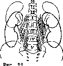
Variations in
its branch. Among the factors determining the normal position of the kidney, mention must also be made of the renal fascia, fascia renalis, which envelops the kidney like a sheath from all sides, with the exception of the inner and lower sides, where the layers of the fascia do not directly connect with each other. The renal fascia itself is fixed very firmly, because at the top both of its layers are attached to the diaphragm, the posterior layer is tightly connected with the fascia of the lumbar muscles and with the ligamentous apparatus of the spine, and the anterior layer with the peritoneum. In view of the fact that the fascial sheath is open only downwards, the kidney during its displacements slides precisely downwards, parting the plates of this fascia. The fixing role of the renal fascia in the length and position of the kidney (according to Corning) is further reinforced by the presence of the adipose capsule of the kidney and its close connection with the renal fascia. From the renal fascia to the fibrous capsule of the kidney, a multitude of thin fascial bundles stretches like a web, penetrating the layer of the adipose capsule of the kidney; a kind of soft spring, a fatty cushion on which the kidney rests, is thus created. The fixing role of this spring is proven by cases of rapid and extensive displacements of the kidney observed as a result of rapid emaciation; in these cases, the kidney is deprived of the elastic pressure of the fat and it begins to move freely in its bed. The three factors indicated above could not hold the kidneys in their normal position if not for the fourth and most important factor—intra-abdominal pressure, which, like an air bladder, presses the kidneys to their normal bed (Volkov and Delitsin). The factors listed that fix the kidneys are inconstant, subject to individual fluctuations, and can change sharply depending on the physiological and pathological states of the organism. In cases where these fluctuations go beyond the limits of anatomical and physiological norms, a persistent displacement of the kidney is obtained, causing a complex of symptoms that in a number of cases develop into a definite clinical picture. In clinical practice, displacements of the kidney are denoted by several terms: nephroptosis, displaced kidney (ren dislocatus), mobile kidney (ren mobilis), wandering kidney (ren migrans). In addition to the described category of secondarily displaced kidneys, there are cases of abnormal position of the kidney due to a disruption of the process of embryonic development of the kidney, when it does not complete its ascent upwards into the lumbar region; in contrast to acquired displacement, such a "dystopic" kidney is characterized by the firmness of its attachments and in most cases by the complete absence of mobility. In addition to downward displacement, changes in the position of the kidney can also occur as a result of rotation around its axes. When the kidney rotates around the longitudinal axis, the lateral edge turns anteriorly, and the medial edge posteriorly, and vice versa (lateroversion of the kidney according to Fedorov); when the kidney rotates around the transverse axis, the upper pole departs posteriorly, and the lower protrudes anteriorly (retroversion of the kidney), or, conversely, the upper pole tilts anteriorly, and the lower posteriorly (anteversion of the kidney). The amplitude of kidney displacement varies widely. In practical terms, the scheme of displacements proposed by Glenard is most acceptable, according to which 4 degrees of kidney displacement are distinguished: in the first, the lower pole of the kidney is palpable, but not held by the hand; in the second, the kidney can be felt and held; in the third, it is possible to feel the upper pole of the kidney and go behind it; and finally, in the fourth, the kidney is clearly palpable from all sides, even during exhalation. With severe displacements, the kidney can be found in various parts of the abdomen of the corresponding side, in the iliac fossa, and even in the small pelvis; cases are known in the literature where the kidney was found in a hernia sac in inguinal hernias. Prolapses of the kidney in the overwhelming majority of cases are observed in women: according to Legry, in women in 87%, in men in 13%; in 85–90% the right kidney is displaced (Radziyevsky). As for age, prolapse of the kidney is usually observed after 20 years and is very rarely encountered in children. Sufficient data cannot be adduced in favor of a hereditary predisposition to kidney displacement either. The fact noted above that women predominantly suffer from nephroptosis finds its explanation in the fact that, in connection with the act of pregnancy and childbirth, they experience a sharp fluctuation in intra-abdominal pressure; secondly, in the fact that, as established by the studies of Volkov and Delitsin, the renal niches in women are shallower, have a cylindrical shape, and are widely open downwards, which facilitates the easier slipping of the kidneys from their receptacle; the greater frequency of displacement of the right kidney is due to the fact that the liver lies adjacent to the upper pole of this kidney, which with its weight presses on the kidney and as it were ousts the latter from the bed it occupies; in a number of cases during operations it is established that the liver entirely occupies the renal bed. In some cases, nephroptosis is one of the manifestations of splanchnoptosis (see), although the combination of these two pathological states is not frequent: out of 113 cases of nephroptosis, Fedorov found enteroptosis 2 times, and hepatoptosis 5 times. In some cases, the direct cause of kidney displacement is trauma: acute (falling from a height, a strong jump or concussion of the body, a blow to the lumbar region, excessive and sudden muscular strain) or chronic (frequent lifting of heavy objects, jolting rides, severe cough, frequent straining [constipation], etc.). But all the listed moments are secondary in the mechanism of the origin of nephroptosis; the primary determining factor in the pathogenesis of nephroptosis is the constitution of the patient. Thus, people with a short and wide chest and a convex abdomen usually do not suffer from kidney displacement; people with a slender build, a long, elongated, and narrow chest (asthenics), on the other hand, relatively often suffer from nephroptosis. The state of primary tissue weakness also belongs to this category of causes of nephroptosis (Obraztsov, Strümpell, Tuffier). As for the state of the renal parenchyma in the displaced kidney, according to the studies of some authors, phenomena of nephrosclerosis or nephrosis develop in it (Gotlieb found phenomena of pyelitis in 21 patients out of 71 cases of nephroptosis). The upper section of the ureter usually follows the displaced kidney, undergoing curvatures, kinks, and twists, which usually disappear when the kidney is replaced in its proper position and, if movable, do not violate the patency of the ureter. If the ureter is fixed in an incorrect position, its patency is violated, pyelectasis or hydronephrosis is formed, and the kidney, having increased in weight, drops even lower; along with the vessels, the nerve plexus of the hilus is also stretched. The adrenal glands usually remain in place and do not follow the displaced kidney. In view of the close anatomical relations of the right kidney to the pylorus, duodenum, and ascending colon, displacement of the right kidney can entail the displacement of these organs as well; knowledge of this fact is very important for understanding many symptoms in the clinical picture of nephroptosis. Clinical picture. There are cases of nephroptosis, and even sharply expressed ones, that are not accompanied by any painful symptoms, and the displacement of the kidney is an accidental finding during palpation of the abdomen. These cases have no clinical significance. In a series of cases where painful symptoms are present, the French school of urologists distinguishes three varieties: 1) painful, 2) dyspeptic, and 3) nephrasthenic, corresponding to the predominance of a certain group of symptoms. The clinical symptoms of kidney displacement are of varying strength, and the intensity of these symptoms is far from proportional to the degree of kidney displacement and is often in inverse relation to it (Botkin). Most often, patients complain of dull, pulling pains in the lumbar region, which decrease and even disappear at night when lying down and resting, and worsen during the day, during physical exertions, when lying on the opposite side, during menstruation, while walking, or riding on a jolting pavement. Pains can radiate into the urinary bladder, into the leg, resembling sciatica or femoral neuralgia, and can sometimes radiate into the foot, chest, or arm. Upon palpation, the displaced kidney is always painful. The pains are of a constant character or occur in the form of severe attacks, sometimes with frequent urination. The attacks of pain give the picture of true renal colic: sharp pain in one hypochondrium, nausea, vomiting, abdominal distension; pains sometimes radiate downwards, into the region of the urinary bladder, sometimes into the epigastrium or under the scapula. Urine, despite frequent urges to urinate, is excreted in small quantities, and sometimes does not pass at all (reflex anuria). The causes of the described phenomena of "renal colic" must be considered to be entrapment of the kidney or a sudden increase in intrarenal pressure on the basis of retention of urine in the pelvis. The mechanism of kidney entrapment is understood differently: some authors believe that the kidney is entrapped in the surrounding connective tissue (in the poorly compliant fibrous capsule — Mysz) or in the subperitoneal tissue, while others believe that it is entrapped between the spine and the XII rib. A third group believes that the described pain symptom complex during entrapment in many cases depends on the kinking of the ureter and the cessation of urine outflow from the stretched pelvis; and finally, a fourth group explains it by the twisting of the renal vein and acutely occurring venous hyperemia of the kidney.
With the dyspeptic symptom complex, there is a predominance of clinical phenomena from the gastrointestinal tract: heaviness in the epigastrium, loss of appetite, eructation, heartburn, nausea, constipation, and abdominal distension; in more severe cases, strong pains in the epigastrium, vomiting, intestinal pains (phenomena of colitis) and especially frequently in the region of the cecum, increased acidity, and sometimes a disturbance of the motor function of the stomach; in some cases, jaundice is observed as a result of compression of the duodenum and the bile ducts. All of these indicated symptoms can be of reflex origin or purely mechanical. With the nervous symptom complex, general disorders of the nervous system are observed: patients are hypochondriacal, irritable, and easily upset on any occasion; they tire easily, often suffer from headaches, dizziness, palpitations; hysterical stigmas are observed: paresthesias, anesthesia of the skin and mucous membranes, hyperesthesias; in some cases it comes to psychiatric disorders. The cause of all these indicated phenomena is considered to be irritation of the nerve plexuses of the kidney itself and neighboring organs, mainly the autonomic nervous system (Radziyevsky). Regarding the urine, oliguria or polyuria is observed in nephroptosis; among pathological admixtures in the urine, protein and casts are most often found; in rare cases, hematuria is observed. Diagnosis. The accepted method for determining displacement of the kidney is the method of palpation in the patient's positions typical for kidney examination: in the position on the back, on the side with bent thighs, and in the standing position. To distinguish a mobile and strongly displaced kidney from other abdominal tumors, one can use the method of palpator albuminuria proposed by Menge: compression and massage of the displaced kidney, produced through the abdominal walls, soon after causes the appearance of protein in the urine. The most valuable and precise data on the position of the kidney are given by an X-ray picture in combination with pyelography in the lying and standing positions. This method makes it possible to reveal kidney displacement in those cases where it escapes ordinary research methods; it allows determining the amplitude of kidney displacement and its rotation around its axes, clarifying the condition of the pelvis and the upper section of the ureter. One of the auxiliary diagnostic methods is obtaining experimental colic caused by filling the pelvis with some indifferent liquid until a feeling of pain appears in the corresponding kidney. The pyelography method provides particularly valuable services in differentiating a secondarily mobile kidney from a dystopic one. In kidney dystopia, it is fixed lower, the shadow from the pelvis is constantly in one place, the ureter is shortened. In appropriate cases, to clarify the source of pain, one should resort to the method of blocking the renal segments according to Löwen (Don-Li) and determining Head's zones (Dxi-Dxn). Treatment. Kidney displacement that does not cause pain or other disorders is not a disease in itself, and therefore is not subject to treatment. In mildly expressed degrees of displacement, the patient can be helped by prolonged lying on the back combined with forced feeding. In many cases, a bandage relieves pain, and the action of the bandage should be directed only to strengthening the relaxed abdominal wall, and by no means to holding the displaced kidney in place by direct pressure on it (Fedorov). Therefore, any pads whatsoever are completely inappropriate and even harmful on the bandage, since they usually press not on the kidney at all, but on other organs (liver, stomach, colon), causing the function of the latter to suffer. Usually, an abdominal support belt is prescribed, made of dense fabric with a mesh of whalebone or steel plates embedded in it and with elastic inserts of rubber on the sides of the belt; instead of a bandage, a corset with a straight splint can be recommended. All bandages and corsets should be put on in the lying position on the back, after the possible reposition of the displaced kidney into the hypochondrium, which patients easily learn to do themselves. Along with wearing a bandage, hydrotherapy, electrification, and massage are beneficial to patients. In pronounced cases of nephroptosis, when orthopedic treatment is unsuccessful, surgical treatment is indicated, which consists in suturing the kidney—nephropexy. The general principles that should be laid down as the basis of the nephropexy operation can be formulated as follows: 1) the kidney should occupy its anatomically normal position as far as possible; 2) the kidney parenchyma must not be damaged; 3) the kidney must be stably fixed in its new position; 4) in view of the fact that kidney prolapse is not a life-threatening disease, only those surgical methods can be applied in the technique of which there are no life-threatening moments. The number of methods proposed for nephropexy is extremely large; it already approaches 150 (Gotlib). All numerous operational methods of fixing a displaced kidney can be divided into 4 main groups: 1) parenchymal methods, where the fixing material is passed through the kidney parenchyma; 2) scar methods, in which one relies on the formation of scar adhesions of the kidney with underlying tissues; 3) capsular methods, where kidney fixation is performed by its capsule; 4) fascial methods based on the suturing of the renal fascia. Numerous combinations and variants of these main moments form that diverse arsenal of surgical methods that surgeons have in reserve. The Albarran-Fedorov method with decapsulation of the kidney, suturing of the kidney by the capsule, and placing a tampon to the lower pole of the kidney is most widespread. After the operation, patients are kept in bed for 3 weeks in a position on the back or on the operated side. After the operation, it is advised to wear a bandage or corset for 4–6 months and to avoid any physical exertion for a year. None of the existing methods of nephropexy gives an absolute guarantee against the return of nephroptosis. According to recent literature data, the percentage of relapses varies among various authors over a wide range—from 50% (Klapp and Kleiber) to 8% (Gorash—on 163 operations). Regarding the question of indications for surgery in nephroptosis, there is no uniform opinion among surgeons; in general, a restrained attitude toward the nephropexy operation prevails in view of the uncertainty in the persistence of the operation results; when evaluating indications for surgery, one should take into account mainly the intensity and severity of painful symptoms depending on nephroptosis, the type and degree of kidney displacement, and finally the occupation, age, and condition of the patients (Fedorov).
V. Voznesensky. During the patient's lifetime, anomalies of the kidney. Congenital anomalies of the kidneys. Achievements of modern instrumental and X-ray diagnostics have led to the fact that the recognition of congenital anomalies of the kidneys and ureters, which was hitherto a matter of accidental autopsy or operative findings, has become possible. As can be seen from Gotlib's data, anomalies of the kidneys and ureters, diagnosed clinically, were found 54 times per 470 patients, which is 11.5%. This figure significantly exceeds previous data from autopsy material (1% per 10,000, Naumann) and surgical material (2% in Israel, 3% in Fedorov). Shape anomalies. Horseshoe kidney can exist completely asymptomatically and
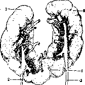
Fig. 1. Horseshoe kidney: 1—right kidney with ureter (2); 5—left kidney with ureter (3); 4—isthmus. (After Testut.)
may then not require treatment (Fig. 23 and 24). In some cases, however, it is a source of indefinite pain sensations in the abdominal cavity, depending on the abnormal mobility of one or both halves of the horseshoe kidney, on the pressure of its isthmus on the underlying nerve plexuses, or on the development in the kidney itself of one or another pathological process (uro-, respectively pyonephrosis, stone, tuberculosis, etc.). A horseshoe kidney can be the cause of stubborn constipation, an obstacle to childbirth, etc. In individual cases, it is possible to make a correct preoperative diagnosis using palpation (palpatory albuminuria). The most perfect method of recognition is pyeloureterography. Gotlib summarizes the characteristic findings in it in the following provisions: 1) both renal pelves are located on both sides lower than usual;
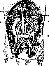
Figure 24. Topography of a horseshoe kidney: 1 - v. cava inf.; 2 - a. et v. renalis dex.; 3 - isthmus; 4 - a. et v. renalis sin.; 5 - aorta. (According to Corning.) 2) the ureters are shortened; 3) both renal pelves tend to approach each other with their lower poles, which is why the lower calyces are closer to the spine, while the upper ones are further apart than normal. The lower calyces are sometimes so close to each other that they partially cover the spine; 4) the longitudinal axes of the renal pelves intersect in the caudal direction, forming an acute angle open upward; 5) the ureter, as it approaches the pelvis, moves away from the spine; 6) the renal calyces are located medially to the renal pelvis; 7) in relation to the ureter, the renal pelves are located either medially or on both sides of it; 8) the renal pelvis may be of atypical shape, and the renal calyces may be elongated in length. - Since the horseshoe kidney frequently becomes diseased itself or causes pain, dyspeptic, and nervous disorders, it is often necessary to resort to surgical treatment, which, according to Botez, has yielded a mortality rate of only 6.6%. Of the various types of renal operations, the most indicated according to the case are: 1) suturing (nephropexy) of one or both halves of the horseshoe kidney as a means of eliminating pain in its abnormal mobility; 2) heminephrectomy for unilateral disease of the kidney (hydro-, resp. pyonephrosis, sometimes stones) and with an isthmus suitable for division; 3) the latter
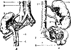
Figure 25. Two cases of renal ectopia. A: 1 - aorta; 2 - v. cava inf.; 3 - kidney lowered into the lesser pelvis; 4 - a. hypogastrica; 5 - ureter. B - unilateral location of the kidneys: 1 - aorta; 2 - right kidney with ureter (3); 4 - ureter of the left kidney; 5 - left kidney. (According to Testut.)
this condition must also be taken into account when performing an operation to dissect the isthmus in order to eliminate pressure on the aorta, resp. its nerve plexus; 4) pyelotomy, and specifically its anterior variety, for renal pelvis stones. When the horseshoe kidney is located low (pelvic and iliac dystopias), a transperitoneal approach to the kidney is preferable; in other cases, it is also possible to operate extraperitoneally through a lumbar incision. A double (fused) unilateral kidney also belongs to the conjoined forms of developmental anomalies of the kidneys. In this anomaly, both kidneys, fused by their poles, are located on one side of the spine and therefore resemble a single, but significantly elongated kidney (ren elongatus) (fig. 25B). Much less frequently, a double kidney is formed due to the fusion of the kidneys along their lateral surfaces or at an angle. Each of the kidneys has its own pelvis and ureter, which do not communicate with each other and usually open at normal sites in the urinary bladder; in this case, the ureters may cross at one height or another of their course. The pelves of both kidneys either face the same side or different sides. The complete mutual independence of the urinary system explains the possibility of an isolated disease of one of the halves of a double kidney (retention tumors, tbc, stones, tumor). The comparatively large weight of a double kidney easily causes a downward displacement of the kidney.-In the diagnosis of a double unilateral kidney, a major role is played by simple palpation of the kidney, the use of the experimental colic method, radiography with preliminary catheterization of the ureters using radiopaque catheters, and especially pyeloureterography.-The functional independence of both constituent parts of the kidney makes it possible to intervene surgically in the event of disease of one of the halves of the double kidney, up to the removal of one of them while the other is healthy. Conservative operations are all the more possible, such as: nephropexy for abnormal mobility of the kidney, pyelotomy and nephrotomy, e.g. for stones. The normal approach route (except for cases of an especially low position of the double kidney) is an extraperitoneal operation through a lumbar incision. 62 cases of kidney hypoplasia have been described, of which in 20 cases the diagnosis was made only during surgery (Gotlieb). The possibility of intravital diagnosis of kidney agenesis and hypoplasia is created by the combined use of cysto- and chromocystoscopy, and especially pyeloureterography in combination with pneumorenal imaging. Since unilateral absence of a kidney is often combined with compensatory enlargement of the second kidney, finding an unusually enlarged kidney during surgery requires keeping in mind the possibility of this anomaly to avoid the fatal removal of the sole kidney. On the other hand, as experience shows, a solitary kidney fully permits conservative-type operations.-Duplication of a kidney (ren duplicatus) on one side, in the presence of a third kidney on the other, is encountered more frequently than a true supernumerary kidney. Thus, according to Graser, only 13 such cases are known in the literature, and in two cases there were 4 kidneys. It goes without saying that in the event of disease of both a true supernumerary and a duplicated kidney, any surgical intervention is permissible provided the remaining reserve of renal tissue in the patient is healthy. The diagnosis is made using pyeloureterography. Circulatory disorders in the kidneys can either be local in nature or be caused by general circulatory disorders. The first group of disorders includes embolisms and thromboses of the renal arteries and their individual branches, leading to the development of larger or smaller infarctions. Kidney infarctions are usually a concomitant phenomenon in endocarditis (especially septic) and in thromboses of the heart and blood vessels caused by stagnant and inflammatory phenomena. Clinically, infarctions manifest as acutely onset pains in the lumbar region on the affected side and the appearance of a significant admixture of fresh blood in the urine. Characteristic in this regard is the absence of other pathological formed elements in the urine and the relatively rapidly transient (2-3 days) nature of the hematuria.-As a manifestation of a general circulatory disorder in heart defects and myodegenerations, venous congestion is usually observed in the kidneys, sometimes reaching extremely sharp degrees. The kidneys on cross-section in these cases appear sharply hyperemic and cyanotic, sometimes bluish-black. Such intense congestion corresponds to characteristic changes in the urine, expressed to varying degrees depending on the duration and intensity of the congestion. Clinically, the congested kidney is characterized mainly by a decrease in the amount of daily urine, albuminuria, a drop in sodium chloride excretion with a high specific gravity and saturated color of the urine, and the presence of urobilin in it. Albuminuria can reach 10‰ or more, but its degree is by no means parallel to the intensity of the congestion. The absence of hypertension is also characteristic.-A circulatory disorder in the kidney also causes a special constitutional disorder known as orthostatic albuminuria (see). Injuries to the kidneys, especially isolated ones, are not among the frequent ones. Their combinations with injuries to the ribs, spine, and abdominal cavity organs are frequent and significantly worsen the prognosis. Closed and open kidney injuries are distinguished. The latter, in accordance with the features of the wounding weapon, are divided into stab-incised and gunshot wounds. The kidney is accessible to injury from a wide variety of directions, which explains the possibility of simultaneous damage to not only the abdominal but also the thoracic cavity.-The mechanism of blunt kidney injury boils down to a bruise of the lumbar region, compression of the kidney between two hard surfaces (e.g., buffers, being run over by a carriage wheel), or counter-blow (e.g., sharp concussion of the kidney when falling from a height onto extended legs); a definite and important role in the rupture of the kidney as an organ containing a large amount of fluid (blood, urine) is played by hydraulic pressure. As a rarity, closed kidney injuries occur depending on energetic muscular contraction (lifting a weight with a jerk, wrestling). A pathologically changed kidney is sometimes more easily injured. According to the widespread Küster classification, a distinction is made between: 1) ruptures of the adipose capsule of the kidney without damage to the renal parenchyma; 2) ruptures of the parenchyma not penetrating to the calyces or pelves; 3) ruptures of the parenchyma penetrating into the pelvis; 4) laceration of the kidney; 5) ruptures of the constituent parts of the hilus (pelvis, ureter, artery, vein) without damage to the kidney itself. With differences in the nature and intensity of even an isolated kidney injury, the clinical symptom complex cannot be uniform. The main signs of a fresh kidney injury are hematuria, a tumor in the thoracoabdominal region, pain; in cases that are not fresh and are infected, the phenomena of putrid phlegmon (urinary infiltration) join. The picture of general phenomena includes: phenomena of shock (especially with simultaneous damage to abdominal organs) with increasing anemia, a drop in blood pressure, and an increase in pulse rate in the case of profuse blood loss.-Hematuria may be absent altogether (with superficial damage, in the case of detachment of the kidney from the ureter, with blockage of it by blood clots), be only microscopically detectable or macroscopically apparent, up to profuse bleeding. There is no definite correlation between the intensity of hematuria and the degree of kidney injury, all the more so because, if in some cases the main mass of blood flows through the ureter into the urinary bladder and further outward, in others it predominantly impregnates the nearby layers of cellular tissue (adipose capsule, retroperitoneal cellular tissue) or through a rupture of the peritoneum enters the abdominal cavity. Descending into the layer of cellular tissue, the hemorrhage can be detected in the groin, scrotum, or labium majus in a woman, on the thigh. The admixture of blood in the urine lasts for various periods, sometimes disappearing quickly, sometimes weakening only slowly and gradually. An increase in hematuria or its appearance again after the blood in the urine has already disappeared depends on an added infection. Such late, secondary, often repeated hemorrhages can be life-threatening and require the urgent removal of the damaged kidney. Hematuria can proceed painlessly or be accompanied by attacks of the most severe colic (blockage of the ureter by clots). Blood flowing abundantly into the urinary bladder can lead to the formation of massive blood clots in it with urinary retention, which serves as an indication to free the bladder from clots by washing with aspiration through an evacuator (if necessary after preliminary crushing of the clots with a lithotrite) or by high section of the urinary bladder. Where hematuria persists for some time, protein and casts (hyaline, granular, blood) can be detected in the urine. A simultaneous decrease in the amount of urine, and often, conversely, polyuria, then the appearance of edema on the extremities, face, and even the entire body indicate an added chronic nephro-nephritis, which can be classified as "traumatic" if in the patient's past nephritis can be excluded with certainty.
Such traumatic nephritis develops either shortly after the trauma or weeks and months after it. Flatulence and absence of gas passage as a manifestation of paresis of the large intestine, depending on the nutritional disorder of its wall caused by the infiltration of extravasated blood into both the cellular tissue and the intestinal wall itself, are not uncommon and, if misinterpreted, may give grounds for a laparotomy for a non-existent intestinal obstruction or suspected peritonitis. The detection of a tumor located in the region of the damaged kidney is not always possible in the first hours after the injury due to pronounced rigidity of the lumbo-abdominal musculature (defensive contracture). This tumor is formed due to hemorrhage and urinary infiltration. Superficial and shallow ruptures of the kidney either are not accompanied by any outflow of urine at all, or it stops quickly without causing further complications if the urine is aseptic. Pain is a usual phenomenon in subcutaneous injury to the kidney; it is especially severe when the ureter is blocked by a blood clot (renal colic) and with simultaneous rib fracture. The presence of these symptoms, which appeared in connection with a trauma to the lumbo-abdominal region, makes it possible to diagnose subcutaneous injury to the kidney with a fair degree of certainty. Any doubt that may sometimes arise as to whether the bleeding depends on damage to the kidney or the urinary bladder is easily resolved by cystoscopy. Diagnosis. Determining the detailed nature of a renal injury based solely on clinical examination data is not feasible. Ureteropyelography under these conditions can hardly be considered widely indicated and harmless. Practically, greater importance attaches to the assessment of the injury from the standpoint of the patient's general condition; the phenomena of shock, rapidly increasing anemia, with the simultaneous rapid formation of a tumor in the lumbar region, indicate the seriousness of the situation and require surgical exposure and inspection of the damaged kidney. The task of diagnosis also includes the requirement not to miss the presence of possible simultaneous injuries to the organs of the abdominal cavity, less often of the peritoneum itself. Treatment. Even if a very significant part of isolated kidney injuries ends safely with strictly conservative treatment (quiet lying on the back, a wide bandage tightening the abdomen and the lower chest, ice), in case of doubt, it is more correct to perform early (after the phenomena of shock have passed) exploratory exposure of the kidney with a view toward possible conservatism in operating. At the same time, under no circumstances should one depart from the basic requirements—quite reliable arrest of bleeding, preservation of a good blood supply to the kidney, and creation of favorable wound conditions. Methods for arresting bleeding from ruptures of the renal parenchyma that do not penetrate into the pelvis include: suturing the rupture, by itself or after preliminary tamponade of the rupture with pieces of fat, or tamponade with gauze. A completely detached piece (for example, a pole) of the kidney must be removed, and bleeding stopped by circumostention, tamponade, or suture after partial resection of the kidney. A shattered kidney, as well as one torn from the hilus or ureter, is subject to removal (nephrectomy). In case of vital indications for surgery, the fact that the function of the second kidney has not been examined cannot serve as a contraindication to nephrectomy. Damage to a known infected kidney, as well as simultaneous damage to the abdominal organs along with the kidney, requires immediate intervention, in the latter case by the transperitoneal or combined route. Under such conditions, Israel begins the operation with a median laparotomy and, having finished the surgical intervention in the abdominal cavity, including the mandatory closure of any possible rupture of the peritoneum in front of the kidney, then proceeds to the operation on the kidney itself via the extraperitoneal route. For isolated kidney injuries, the standard is an operation through a lumbar incision. Among open kidney injuries, which are even rarer than subcutaneous ones, stab-cut wounds and gunshot wounds are distinguished. Their external difference is the disruption of the integrity of the integuments and the presence of urine outflow outward in most of them. The latter and simultaneous hematuria are the most characteristic symptoms of an open kidney injury. The admixture of urine to the blood flowing from the wound is recognized by the presence of a urine odor, an acid reaction, and urea, the determination of which may be required only in case of doubt about the nature of the outflow. The wounding weapon (knife, bayonet, bullet, etc.) can penetrate to the kidney both from the back and from the side, from the abdomen, through the chest. It is therefore understandable that combined kidney injuries are just as common here as isolated ones. The damaged organs are the pleura, diaphragm, lung, peritoneum, liver, spleen, gastrointestinal tract, spine. Such complex combined wounds justly have a bad reputation and unquestionably serve as an indication for urgent surgery. The situation is different with regard to
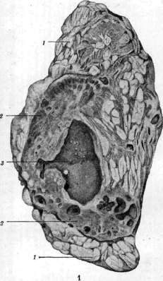
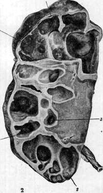
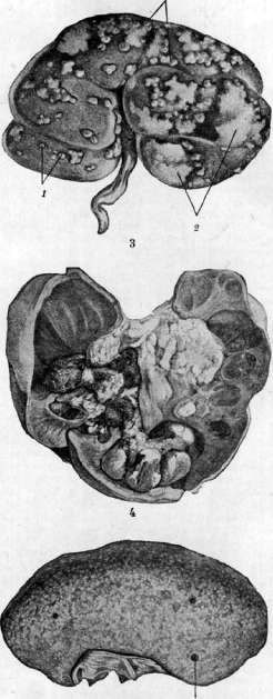
Figure 1. Urolithiasis and chronic pyelonephritis with renal sclerosis; severe paranephric obesity: 1-paranephrium; 2-small cysts and scar zones of the kidney; 3-dilated pelvis with sand and small stones. Figure 2. Hydronephrosis due to obstruction of the ureter by a stone (j): 1-sharply thinned cortical layer; 2-sharply stretched calyces. Figure 3. Suppurative embolic nephritis with the development of infarcts (2): 1-small abscesses. Figure 4. Kidney stones. Figure 5. Renal cirrhosis (secondary contraction): 1-cyst.
isolated injuries of the kidney, where, with the exception of cases of particularly severe kidney damage, recovery is quite possible even with purely conservative treatment. Nevertheless, elevating the latter to a principle would be incorrect, because it is impossible on the basis of clinical examination data alone to form a clear idea of the nature of the actual kidney damage. Therefore, the desire to expand the indications for primary exposure of the kidney in the case of undeniable, even if isolated, damage to it must be considered justified. In addition to the already mentioned combined injuries, cases with severe bleeding (both external and internal) urgently require primary intervention; cases of blind gunshot wounds from artillery shrapnel and even rifle bullets; prolapse of the kidney, especially in the case of its injury. On the other hand, such isolated kidney injuries that do not cause any immediate concerns for the patient's fate can be subjected to conservative-expectant treatment. Under such conditions, the suturing of the external wound, resp. tight tamponade of the wound canal, are undesirable (fear of urinary infiltration); on the contrary, one must ensure free outflow, leaving the wound under a well-absorbing aseptic dressing and even expanding the wound canal if necessary. The outflow of urine ceases sooner or later by itself, unless there are obstacles to the flow of urine into the bladder. Secondary operations may be needed in the event of successive hemorrhages, infection, or urinary infiltration. One cannot delay the operation here, and it must ensure a thorough stop of bleeding and the opening of urinary streaks; under these conditions, it is most correct to remove the kidney. When performing an operation in the case of a fresh kidney injury, the measures will generally be the same as in closed injuries. The non-aseptic nature of any open kidney injury, especially gunshot wounds and specifically from artillery shrapnel, expands the indications for primary nephrectomy. In isolated open kidney injuries, the lumbar approach is indicated—in some cases by expanding the existing wound of the lumbar region, in others by a separate lumbar incision. Cases of combined injury generally require a combined operation, i.e.: 1) in case of a wound of the abdominal cavity, the operation begins with a median celiotomy and only after closing the abdominal cavity (with suturing of the posterior parietal peritoneum) is the kidney approached by the lumbar route; 2) in case of a wound of the thoracic cavity, the operation begins with the latter, preferably using an apparatus with increased pressure (suturing of the lung, diaphragm, elimination of open pneumothorax), ending the operation with an intervention on the kidney by a lumbar incision. Prolapse of a viable kidney requires its preservation by reduction and tamponade; otherwise, the kidney is removed. Purulent inflammation of the kidney. Among the purulent processes in the kidney, anatomically and clinically, one distinguishes: purulent nephritis (see) (see sep. tab., fig. 3), pyelitis (see), pyelonephritis (pyelonephritis) (see Pyelitis) and pyonephrosis (see). A peculiar, infrequently occurring variety of suppuration in the kidney, described by Israel under the name of kidney carbuncle, is clinically recognized only upon inspection of the kidney exposed during the operation. Common to all named varieties of purulent processes is the infection of the kidney by microbes capable of causing suppuration. But at the same time, the clinical picture will be significantly different, depending on whether we are dealing with an intestinal infection or staphylococcal-streptococcal. In the former, despite high fever, the patient's general condition may suffer little and the course of the disease itself may be mild and even prone to spontaneous recovery; accordingly, the indications for early surgical intervention are not so urgent. The suppurative process in the kidney proceeds completely differently if it is caused by a staphylococcus or streptococcus: here general symptoms of a severe septic process come to the foreground. Common, further, is the method of introducing infection into the kidney, which has as its primary source some local process, proceeding sometimes almost imperceptibly for the patient himself (boil, pustular eczema, infected abrasion, diaper rash, suppuration of the accessory nasal sinuses, tooth caries, etc.), or some previously transferred general infectious disease (scarlet fever, smallpox, typhus, postpartum sepsis, etc.). The infection is introduced into the kidney by the hematogenous route (descending, metastatic type of infection) or ascends from the lower parts of the urinary tract (predominantly from the urinary bladder) along the column of stagnant urine or along the lymphatic pathways of the inner layers of the ureteral wall and the surrounding tissue (ascending, urogenital form of infection). Much less often, the infectious principle passes to the kidney from a nearby focus of suppuration or by its rupture into the renal pelvis. Accordingly, the hematogenous route of infection leads predominantly to the formation of abscesses in the cortical substance of the kidney (see Nephritis). With the urogenital route of infection, the renal papillae and medulla are affected first of all, and the infection spreads to the periphery of the kidney in the form of fan-shaped diverging strips of suppuration. In advanced cases, it is not possible to distinguish both varieties of pyelonephritis with complete certainty. The purulent process in the kidney involves the fibrous and adipose capsules of the kidney. The involvement of the latter is manifested either simply by edematous impregnation or by typical suppuration (see Paranephritis). Common to all varieties are changes in the urine. These latter are expressed the least in a limited number of small abscesses located exclusively in the renal cortex. As long as and while there is no communication of them with the cavity system of the kidney, there is no pyuria. The urine contains an insignificant amount of protein, isolated leukocytes, epithelial cells and red blood cells, sometimes even only in the form of shadows; casts are generally absent. In the urogenital form of pyelonephritis, pyuria and urinary casts are present. The amount of urine is reduced even with unilateral affliction, and with bilateral it can come to anuria. From a surgical point of view, it is especially important that suppuration in the kidney, especially of a hematogenous nature, is often unilateral. Common, finally, to all varieties is their more or less severe effect on the general condition of the organism, and with a unilateral purulent process, its indirect harmful effect on the other, uninfected kidney in the form of toxic nephritis. The latter passes without a trace after the liquidation of the process on the affected side. The indicated commonality of many manifestations of the purulent process in the kidney does not, however, exclude the possibility of intravital differential diagnosis. Thus, pyelitis is characterized by the presence of pyuria with an unincreased, non-palpable kidney; palpation is painless, but concussion of the kidney is painful. If with renal pyuria in urine rich in protein and pus, there are casts, if there is a picture of a severe septic affliction, if the kidney is probed enlarged, and the palpation itself is painful, if the kidney is functionally weakened, then clinically there is reason to diagnose pyelonephritis. With pyonephrosis, which is an expression of a long-existing suppuration in the kidney, proceeding under conditions of insufficiently free outflow from the kidney, there is an enlarged kidney, usually immobile due to inflammatory adhesions. A sharp violation of renal function is a consequence of the massive destruction of the renal parenchyma. Reliable recognition of a kidney carbuncle before surgery is hardly possible. The presence of a gateway for infection, pain on pressure in the angle between the 12th rib and the edge of the sacrospinal muscle, a palpable and painful kidney, accompanied by a high temperature—all this gives reason to speak of a purulent process in the kidney; small changes in urine support this assumption. A feature of the kidney carbuncle is its tumor-like character and tendency to spread over the surface and deep down, with a relatively small tendency to purulent melting. This disease is not often encountered. Smirnov collected only 21 cases, of which 5 cases are from Russian literature. The absence of other localizations of metastatic suppuration in the patient's body indicates nephrectomy. The removal of the kidney performed under such conditions does not guarantee against the possibility of subsequent introduction of infection into the previously healthy second kidney. Therefore, the successfully implemented proposals to operate by the type of excision of the carbuncle, its simple incision and even resection when the focus of the disease is located in one of the poles of the kidney are understandable. The question of the treatment of purulent pyelonephritis stands in the same way. Here, when choosing a surgical aid, along with the severity of general septic phenomena, they are guided by the degree of spread of the purulent process in the affected kidney and the state of health and function of the second kidney. Thus, the method of operation is established during the operation itself after examining the diseased kidney.
The following are employed: 1) decapsulation with the opening of all visible superficial foci of suppuration; 2) nephrotomy of the type of a Pirogov section incision followed by tamponade of the incision; 3) resection of the affected pole of the kidney; 4) nephrectomy, both primary and sequential upon the failure of one of the previous methods. In the event of simultaneous disease of both kidneys, one can only speak of nephrotomy. Neoplasms and cysts of the kidneys. From a patho-histological point of view, the variety of kidney tumors is great. This circumstance does not have any special practical significance, because it is impossible to recognize the histological structure of a tumor on the basis of clinical examination data alone. Therefore, whenever the presence of a dense kidney neoplasm is established, even in the absence of hematuria, cachexia, metastases, and pain, one should suspect a malignant kidney neoplasm. Benign kidney tumors (fibromas, lipomas, angiomas, adenomas, etc.), although not uncommon, are so small, asymptomatic, and harmless to their host that they are usually only incidental findings at autopsies. A benign kidney tumor that, as an exception, has reached such a significant size that by its pressure on surrounding organs it begins to trouble the patient, is an exceptionally rare phenomenon and requires surgical intervention. Malignant kidney neoplasms are encountered either in early childhood (up to 5 years of age) or at the age of 40-60 years. The latter age is affected particularly often, preferably the male sex, with a prevalence of disease of the right kidney. In early childhood, kidney neoplasms of a sarcomatous character predominate. In adults, we have a greater variety: hypernephromas (Grawitz's tumors), carcinomas (nodular and diffuse), and sarcomas (round- and spindle-celled, of the endo- and perithelioma type). Kidney hypernephromas are encountered most frequently (see). As for cystic tumors of the kidneys, such as: solitary kidney cysts, multiple cystic degeneration of the kidneys, and kidney echinococcus, these do not belong to the number of frequent kidney diseases. If polycystic kidney degeneration as an affliction, deeply destroying the renal parenchyma and moreover usually bilateral, inevitably leads to renal function insufficiency up to uremia, then solitary kidney cysts and echinococci become burdensome to patients only under the condition of significant size or secondary infection. Solitary kidney cysts have a smooth surface and give a sensation of dense elasticity, sometimes fluctuation. They are distinguished from hydronephrosis by the constancy of the tumor's size, which is prone only to further gradual increase, unchanged kidney function, and the picture of a normal pyelogram; from an ovarian cyst, by the absence of a connection with the genital sphere and the growth of the tumor from the upper parts of the abdomen; from hydrops of the gallbladder, by the retroperitoneal character of the tumor and the absence of a history of biliary colic. Treatment consists in total extirpation of the cyst; more often the operation boils down to marsupialization of the cyst with possible trimming of the excess cyst wall, which is then sutured into the surgical incision; finally, nephrectomy may also be required. The bulk of kidney cysts, however, does not reach significant size and has no clinical significance. Polycystic kidney degeneration as a congenital disease can be encountered already in early childhood. Unilateral affliction is encountered rarely, specifically in 3-4% (Lubarsch); as a rule, the affliction is bilateral. The symptom complex of the affliction is far from homogeneous. Coming to the fore are phenomena resembling chronic nephritis: low specific gravity of urine with a simultaneous increase in its daily quantity; a small amount of protein, usually in the absence of casts; lowered functional activity of the kidneys; often elevated blood pressure; hypertrophy of the left ventricle (up to uremic phenomena, which may be the first and sudden manifestation of far-advanced renal affliction). In some cases, the picture is complicated by pain, hematuria (even of tumor type), infection (pyuria). All these manifestations of the disease become understandable if one takes into account the progressive replacement of the renal parenchyma by multiple cysts that compress and disrupt the vital activity of the still-preserved renal tissue. Recognition is possible if, amidst phenomena of chronic nephritis, the presence of bilateral nodular enlargement of the kidneys is determined. The nodular character of the tumor and its unchanging size speak against hydronephrosis. Still, in a considerable number of cases, the final recognition can be made only after exploratory exposure of the kidney. Usually, the bilateral character of the affliction, even if variously expressed on both sides, rules out the possibility of radical operation, i.e., removal of the kidney. Palliative operations aimed at eliminating pain sensations turn out to be quite expedient. The type of such an intervention is the Payr operation, consisting in the emptying of cysts by puncturing them with an electro- or thermocautery, which significantly reduces the volume and weight of the diseased kidney and facilitates (by reducing intrarenal pressure) the function of the still-preserved renal parenchyma. The subsequent fixation of the kidney treated in this manner to the edges of the surgical incision (atypical nephropexy) eliminates the mobility of the kidney and the pain sensations associated with it. Proceeding in this manner, Fedorov obtained favorable results in 15 cases operated on by him. An even greater rarity are perirenal cysts; their relation to the kidney is not always ascertained even during the operation, which consists either in the removal of the cyst alone or of the cyst together with the kidney. In case of special technical difficulties, one has to be limited to the opening and drainage of the cystic tumor. Tuberculosis of the kidneys. For pathological anatomy and pathogenesis, see above. Tuberculosis of the kidney at the outset in a large number of cases is a unilateral affliction; for a significant period it is limited only to the kidney, proceeding slowly, chronically. A moderate secondary affliction of the urinary bladder often accompanying it is capable of regressing to a complete, at least subjective cure, under the condition of the timely removal of the primary source of tuberculosis in the urinary tract, i.e., the affected kidney. Infection of the second kidney occurs either by the ascending route from the tuberculous-affected urinary bladder or by the hematogenous seeding of the infection. The symptom complex of renal tuberculosis is far from homogeneous. First of all, it is very important not to lose sight of the fact that in a number of cases the patient's complaints do not relate to the kidney and not even to the lumbar region in general, but to disorders originating from the urinary bladder—see Urinary bladder, tuberculosis of the urinary bladder. Cystoscopy data are unquestionably conclusive for recognizing the tuberculous affliction of the urinary sphere and in particular the kidney, if they reveal: 1) on the part of the urinary bladder, the presence in the mucous membrane of tuberculous nodules or ulcerations secondarily developed from them; if, as happens in initial and not far-advanced cases, these formations specific for tuberculosis are localized near the orifice of one of the ureters, or if the affliction of the urinary bladder is present only on one side of it, then this usually also indicates the side of the affliction, especially when excreting normal urine from the ureter of the opposite side. In the case of far-advanced affliction, especially with a secondarily joined banal pyogenic infection, the cystoscopic picture loses its character specific for tuberculosis. 2) On the part of the bladder orifice of the ureter, the gaping of a funnel-shaped retracted bladder orifice of the ureter with infiltrated or eroded edges, working sluggishly and discharging turbid urine; a certain diagnostic significance is attributed by Casper to the vesicular edema in the vicinity of the ureteral orifice. A cystoscopically healthy state of the urinary bladder despite the presence of pronounced symptoms of cystitis can be observed in tuberculous affliction of the kidney (reno-vesical reflex). Observation in the cystoscope of the character of the urine discharged from the ureters (pyuria on one side), the delay in the excretion of methylene blue on the side of the pyuria, and finally separate collection of urine (ureteral catheterization) will make it possible to recognize the side of the disease here as well. The tuberculous character of it is definitively established not always by the easy finding of Koch's bacillus through bacterioscopic examination of the urinary sediment treated with antiformin, or by inoculation of urine into a guinea pig. The presence of palpable changes in the kidney (its enlargement in cases of caseous-cavernous kidney and tuberculous pyonephrosis), the cystic section of the ureter (established in women by bimanual examination), and simultaneous tuberculous affliction of the prostate gland, seminal vesicles, or epididymes facilitates recognition. Since kidney tuberculosis, at least initially and for some time, is a unilateral and descending process, then, taking into account the usual fruitlessness of conservative treatment, the question must be raised about the earliest possible removal of the diseased kidney. The latter is conceivable only with sufficient function of the other kidney.
Therefore, functional diagnostics, if only in the form of simple chromocystoscopy, is mandatory. Unfortunately, cystoscopy in a shrunken bladder that does not tolerate stretching may be technically unfeasible. In these cases, in the absence of sufficient data to answer the question of whether both kidneys or only one and which one is affected, the diagnosis is aided by: 1) exploratory exposure of the kidney, supplemented by a functional dye test, with urine collected through a small incision in the upper part of the ureter on both sides or only on the side of the healthy, respectively better externally preserved kidney; upon completion of the study, the ureter incision is closed with a catgut suture, while the diseased kidney, in the absence of contraindications, is removed; 2) exploratory switching off of the kidney by means of temporary interruption of the patency of the latter with a ligature or a soft clamp after lumbar exposure of the kidney and the upper part of the ureter; the urine necessary for functional diagnostics is collected through a catheter inserted into the previously washed bladder. In both cases, the described study takes place during the operation itself. Radiographs and pyelograms play only a subsidiary role in the recognition of renal tuberculosis. The former allows the detection of more significant lime deposits both in caseously degenerated areas of the kidney and in the detritus of a cavity; the latter occasionally reveals erosion of the contours of the renal calyces or the presence of cavities (caverns) communicating by narrow passages with the renal pelvis. As for hematuria, in the picture of renal tuberculosis, at least as macroscopic hematuria, it does not play a special role. Stronger bleedings are occasionally observed either at the beginning of the disease, especially with ulceration of the renal papillae, or in far-advanced cases of ulcerative lesions of the urinary bladder. After all that has been said, it becomes clear how difficult the conditions are for recognizing renal tuberculosis where there are no subjective complaints from patients regarding the urinary sphere, where the presence of the disease is externally expressed by symptoms of a general character, such as general weakness, weight loss, persistent subfebrile temperature without an obvious reason for it. Careful examination of such patients must take into account even insignificant manifestations of tuberculosis of another localization in the patient's body and, especially, not leave without attention even small changes in the urine in the form of persistent albuminuria, especially if it is unilateral and the urine simultaneously contains even a small number of leukocytes. Thorough searches for tubercle bacilli in the urine, diagnostic tuberculin reactions not evaluated equally by all, and Wildbolz's reaction with the patient's own urine can help the cause of early recognition of renal tuberculosis. The standard treatment for recognized unilateral renal tuberculosis is considered to be the removal of the diseased kidney, of course provided there is functional sufficiency of the other kidney. To obtain the best results, in particular to protect the bladder and the second kidney from subsequent infection, the operation should be as early as possible. The presence of a bladder lesion in itself does not serve as a contraindication to nephrectomy; on the contrary, without removing the diseased kidney, there are no chances for a cure or even alleviation of bladder suffering. For the technique of kidney removal, see below. Here we can note only the features of nephrectomy for tuberculous kidneys: 1) It is necessary in every possible way to protect the removed kidney from breaking its integrity, especially in its cavernous and pyonephrotic varieties, to avoid specific infection of the wound cavity. 2) To avoid generalization of the process, when isolating the kidney, one must manipulate it so as not to mash or squeeze it; wide access to the kidney (large incision, rib resection) greatly facilitates the isolation of the kidney. 3) Great attention must be paid to the ureter of the removed kidney as a possible source of wound infection and the formation of non-healing fistulas. With a clearly tubercularly changed ureter, instead of its total excision, cauterization of the entire mucosa of its lumen with concentrated carbolic acid after blunt isolation of it is currently used, followed by suturing of the ureter stump into a separate incision of the abdominal wall. With a partial lesion of the ureter, it is advisable to truncate it within the limits of a macroscopically healthy section and in any case below the stricture closest to the urinary bladder (Fedorov). Even in cases of a visibly unchanged ureter, 10-12 cm of its length must be removed simultaneously with the kidney. When transecting the ureter, measures must be taken against the ingress of its contents into the wound cavity (transection between two clamps, on a laid gauze compress, with immediate lubrication of both lumens with concentrated carbolic acid or iodine tincture). 4) If one cannot deny the rationality of the requirement put forward by some authors of simultaneous removal of the fatty capsule of the kidney with the kidney, as it may contain tuberculosis nodules, then, on the other hand, the systematic implementation of this operating principle would deprive one of the opportunity to use subcapsular nephrectomy, which is convenient under the given conditions and has proven itself in a large number of cases, which are moreover technically difficult. Therefore, the removal of the fatty capsule cannot be considered mandatory under the given conditions.—The question remains about the possibility of surgical assistance in bilateral renal tuberculosis. Surgical assistance here is an exception, permitted only in particularly severe conditions: with very severe pain, high temperature (acute pyonephrosis), severe and persistent bleeding. Nephrectomy is also permissible here only with tolerable functioning of the less affected kidney; in general, we are talking predominantly about nephrostomy. The results of nephrectomy for renal tuberculosis both in terms of operative mortality (2.4%) and long-term treatment results (up to 60%—Epstein) are unquestionably good, which justifies the currently widespread attitude toward the earliest possible surgical treatment of renal tuberculosis. VII. General operative surgery of the kidneys. Operations on the kidneys for their diseases bear a pronounced character of radicalism, boiling down to the removal of the diseased kidney in the presence of a healthy or functionally sufficient other kidney. Operations of a conservative type, very tempting in idea (e.g., plastic operations on the renal pelvis and ureter), proposed in large numbers to eliminate secondary uro-, respectively hydronephrosis (see Hydronephrosis), have generally not justified themselves and do not enjoy any significant application. Resection of the kidney as a method of operating on a kidney partially affected by a malignant neoplasm or tuberculosis is rightly considered contraindicated. Nevertheless, recently voices have been raised again in favor of expanding the indications for conservative surgery of certain renal diseases in view of the importance for the organism of functionally suitable remnants of the renal parenchyma of the diseased organ, especially in cases of inferiority of the other kidney or out of fear of its possible disease, respectively deterioration of the lesion already existing in it, in direct dependence on the suddenly increased functional load. Since in the event of failure of conservative intervention on the kidney, the state of affairs can be remedied only by its subsequent removal and since such secondary nephrectomy is always an operation more difficult for the patient and technically more difficult, it goes without saying that the selection of cases of renal diseases suitable for conservative surgery must be especially careful. The issues of anesthesia in kidney operations are extremely simplified by the circumstance that such widespread ether anesthesia with preliminary injection of morphine, respectively atropin-morphine (morphine or pantopon 0.01–0.02 and atropine 0.001 1¼ hours before sleep), is perfectly tolerated by this kind of patient and can be considered the standard method of anesthesia. Those operated on for so-called surgical nephritis (painful and hematuric nephritis) are no exception, whereas those suffering from diffuse bilateral nephritis poorly tolerate general anesthesia, especially chloroform anesthesia. There is no doubt that kidney operation in a number of cases can be performed with full or relative success under spinal and even local anesthesia. Also deserving of attention is the combination of ether anesthesia with hedonal, especially in nervous and highly agitated patients: 1–1¼ hours before the operation, 3.0–4.0 of hedonal is administered per rectum or repeatedly given per os 1.0 per dose with an interval of ½ hour, total 2.0 of hedonal.—The operation on the kidney is extremely facilitated by the position of the patient on the healthy side on a hard roll placed here (about 30 cm in diameter) or even better on a specially adapted operating table. This position is generally accepted for conventional surgery through a lumbar incision. Transperitoneal nephrectomy requires a horizontal position of the patient on the back. Operation on the lower section of the ureter and with pelvic dystopia of the kidney is significantly facilitated by the Trendelenburg position. The kidney can be exposed by extraperitoneal, transperitoneal, paraperitoneal, and mixed routes.
The most common of these, in view of the extraperitoneal position of the kidney and the possibility of performing the operation extraperitoneally, is the extraperitoneal approach through a lumbar incision. But it must still be acknowledged that the transperitoneal approach to the kidney deserves more attention than it usually receives.-The extraperitoneal, resp. lumbar, method of exposing the kidney. Among the numerous lumbar incisions, the Bergmann-Israel incision is particularly good and universally applicable. Having a slightly S-shaped curve and beginning in the angle formed by the XII rib and the edge of the sacrospinal muscle, it passes in front of the anterior superior iliac spine by 11/2-2 fingers. After incision of the skin with subcutaneous tissue and the external oblique abdominal muscle, by repeated short cuts of the knife through the muscle layer in the middle part of the incision, the easily recognizable by eye transversasa fascia (characteristic white-yellow color and vertical striation) is exposed and a finger-sized hole is cut in it. Then, scissors are taken and, bluntly separating the peritoneum and using the finger to protect the displaced peritoneum and large intestine from accidental injury, the muscle layers (internal oblique and transversus muscles) are cut downwards and upwards, for the entire length of the skin incision. Having pulled the anterior edge of the incision forward and slightly separated the kidney with a finger from the posterior abdominal wall, the retrorenal fascia is torn using two toothed forceps, more posteriorly, again in the direction of the initial incision. Thus, the adipose tissue surrounding the kidney (adipose capsule of the kidney) is exposed, from which the kidney must be isolated. Both the isolation of the kidney and the protection of the peritoneal sac from accidental injury are facilitated by grasping the anterior edge of the opened retrorenal fascia and underlying fat in a pair of Luer fenestrated forceps, pulling the grasped tissue forward. During further isolation of the kidney, the operator's hand is kept all the time in contact with the surface of the kidney, avoiding careless pressure on the kidney (danger of rupture in pyonephrosis, tuberculous cavities, fracture of branched stones, bruising and crushing of the renal parenchyma). The kidney, isolated from all sides, can then be brought out to the extent of the length and mobility of the renal pedicle. The necessary facilitation of access to the kidney in difficult cases can be achieved by: 1) lengthening the lumbar incision downwards; 2) subperiosteal resection of the XII, and sometimes XI ribs; 3) transection of Henle's costovertebral ligament. When resecting a rib, one must remember the possibility of accidental opening of the pleura and the necessity of immediate tight closure of the opening in such a case—first of all with a hermetic suture (pleura, muscles), rather than a tampon.-On the kidney exposed in the indicated way and surrounded by tampons (if there is a danger of contamination of the wound cavity), the operation required in the given case is performed, after which the surgical incision is closed, which is sutured in layers: 2 rows of catgut sutures (deep and superficial) are placed on the incision of the muscle layer, avoiding catching the trunk of the iliohypogastric nerve adjacent to the posterior lip of the surgical incision in the loops of the suture (subsequent neuralgias!); a single-row interrupted thread suture is placed on the skin with subcutaneous tissue. Closing tightly is permissible only as an exception, with certainty in the absence of subsequent accumulation of wound discharge and asepsis of the extensive surgical field. As a rule, drainage (gauze, rubber) is needed, brought out through the middle part of the incision. Transperitoneal (transabdominal) method of exposing the kidney. Patient in the supine position. Conventional laparotomy, usually by a pararectal or oblique incision of the abdominal wall. Upon opening the abdominal cavity, the presenting intestinal loops and omentum are retracted to the opposite side of the abdomen, the posterior parietal peritoneum is incised longitudinally, stepping somewhat outward from the ascending, resp. descending, colon and parallel to it, and is bluntly displaced from the surface of the renal tumor along with the colon toward the midline. Upon removal of the diseased kidney, with complete dryness and presumed asepsis of the surgical field, especially in cases that proceeded smoothly and easily technically, everything can be closed tightly, without drainage devices: 1) tight suture of the incision of the posterior parietal peritoneum and 2) layered suture of the anterior abdominal wall. If drainage is necessary, the bed of the removed kidney is best drained posteriorly, through a specially made opening toward the lumbar region, after which one proceeds as just stated, or else, by suturing the like edges of the incisions of the anterior and posterior parietal peritoneum and reducing the size of the surgical incision by suturing both its ends, a corridor running from front to back is obtained in the abdominal cavity, isolated from the general abdominal cavity, which is then drained anteriorly. The first method—with drainage toward the lumbar region—deserves preference. Decapsulation of the kidney (decapsulation of the kidney). If the kidney is freed from the surrounding tissues for therapeutic purposes along with the fibrous capsule covering it (Rovsing's operation), this is called nephrolysis. The removal of the fibrous capsule from the surface of the kidney, which is easily achieved after incision of the fibrous capsule on a grooved probe along the convex edge of the kidney, is called decapsulation of the kidneys (see). Decapsulation of the kidney is a prerequisite for the operation of fixing a mobile kidney (nephropexy) where one operates according to Albarran's method or its modifications. - Fixation of the kidney according to Albarran (nephropexy). Conventional, somewhat shortened, oblique lumbar incision. The dislocated outward kidney is decapsulated. Each of the halves of the fibrous capsule pulled from the kidney is divided by a transverse incision into two parts. Using the four flaps thus obtained, firmly grasped with catgut, the kidney is fixed to the posterolateral abdominal wall in such a way that catgut threads from the two upper flaps of the fibrous capsule are passed from inside outward through the XI intercostal space and the kidney is suspended from the XII rib; the lower pair of catgut sutures fixes the kidney to the anterior and posterior edges of the muscle incision; a gauze tampon is placed at the lower pole of the kidney, under which the adipose capsule can also be gathered. Otherwise, the usual suture of the abdominal wall incision. Patients should be kept in the supine position for 3 weeks after the operation. Conditions for a positive result of nephropexy: 1) healthy state of the kidney and ureter (absence of infection, retention, etc.), 2) correct position of the fixed kidney (avoiding forceful stuffing of the kidney as high as possible under the ribs, whence it gets an incorrect installation in the position of anteversion) and 3) free outflow of urine from the pelvis. It goes without saying that the dependence of the patient's complaints precisely on the abnormal mobility of the kidney must not be subject to doubt. Removal of the kidney (nephrectomy) is permissible only with a functionally sufficient paired organ. If the exposure and delivery of the kidney outward does not encounter difficulties, then removal of the kidney is an easy matter and requires only careful ligation of the ureter and the vascular part of the renal pedicle separately. Nephrectomy for a dense renal tumor of large sizes can be facilitated by the above-mentioned methods of expanding the surgical field. Puncture of voluminous hydronephrosis acts in the same direction. Obstacles depending on dense inflammatory adhesion of the diseased kidney to surrounding tissues (e.g., in hydronephrosis) can be bypassed by the so-called subcapsular nephrectomy, i.e., such a method when the kidney is removed from its fibrous capsule, which constitutes with the surrounding tissues a continuous and immovably sitting calloused mass. This operation, which provides an excellent way out of the most difficult situations, was proposed by Ollier and described in detail by Albarran; with us it is associated with the name of Fedorov. The first moment—the capsule of the kidney is incised along the entire convex edge of the kidney, after which enucleation of the kidney is begun. The second moment—stepping about 1 cm from the hilum of the kidney, a semilunar incision is made only through the fibrous capsule from the front. The third moment—a semilunar incision through the fibrous capsule at the hilum from behind. With a finger inserted into these incisions surrounding the hilum like a ring, the pelvis and ureter are bluntly and carefully isolated as much as possible. The fourth moment—the vessels of the hilum of the kidney and the ureter are isolated from the adipose tissue with a finger and, having ligated them separately, the kidney is severed. If the ureter is not found through the semilunar incision, the capsule is incised downward and, having found the ureter in the periureteral tissue, it is transected. If it is impossible to apply clamps by eye, this can be done by touch, avoiding the application of the clamp close to the spine, especially on the right (danger of entrapment of the duodenum and subsequent intestinal fistula). If, out of necessity (short and infiltrated renal pedicle), the clamp is left in situ, it must be removed 2-3 days later.-Closure of a large cavity wound, if during the operation there was no contamination of it with virulent contents of the infected kidney, is performed by the already indicated method.
Under opposite conditions, the wound cavity is thoroughly cleansed (by washing with solutions of silver nitrate, hydrogen peroxide, and the like, or by wiping with gauze moistened in alcohol) and thoroughly drained (e.g., tamponade according to Mikulicz). The disinfected (by smearing with tincture of iodine) and carefully ligated stump of the ureter is either simply lowered into the wound cavity or tightly enclosed by a fold of the peritoneum. Where the ureter requires treatment, it can, in the interest of easy accessibility, be sewn into the surgical incision or separately. In nephrectomy for renal tuberculosis, it is not without reason recommended to also remove the adipose capsule of the kidney, as it may contain tubercular nodules. Incision of the kidney (nephrotomia) is performed in some cases for diagnostic purposes, in others for therapeutic purposes, e.g., for removing a stone (nephrolithotomia), or for draining the kidney (nephrostomia). The danger of hemorrhage during the operation is prevented by temporarily clamping the renal pedicle (with an elastic clamp, fingers); definitive hemostasis is ensured, in the absence of contraindications, by careful suturing, for the strength of which the presence of the fibrous capsule of the kidney is important. Kümmell's suture of the renal incision consists of: 1) mattress sutures placed through the thickness of the kidney above the renal pelvis along its incision and tied on the anterior surface of the kidney; 2) interrupted sutures connecting both halves of the kidney in the usual way, and 3) an additional suture on the fibrous capsule of the kidney. All sutures are preferably catgut. In many cases, one can dispense with mattress sutures, which are not indifferent to the blood supply of the kidney. A good hemostatic agent is the perirenal fat, on a pedicle or free, placed between the surfaces of the renal incision, followed by its suturing. The site of a typical renal incision is its convex margin (section line) and specifically its middle part. Experience shows that moving the incision line 0.5-1 cm posterior to the section line, as recommended by Zondek, is not mandatory (Kümmell). Successive hemorrhages after nephrotomy may require urgent nephrectomy for their cessation.
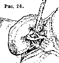
Among operations on the renal pelvis, pyelotomy (pyelotomia) is performed more often than others for the purpose of removing a pelvic, and partly also a renal, stone (pyelolithotomia, pyelonephrolithotomia) (fig. 26-28). Posterior pyelotomy (i.e., the incision is made on the posterior surface of the pelvis) is considered the norm; as an exception, anterior pyelotomy may also prove more convenient. Fedorov proposed pyelotomy (posterior) in situ, i.e., on a kidney that is not mobilized, but with the lower pole of the kidney and part of its posterior surface freed from the adipose capsule. The incision of the pelvic wall, sufficient for extracting the stone whole and moreover without bruising and crushing the edges of the incision, is closed with a careful, where possible 2-layer, catgut suture. Reinforcement of the suture line, especially of a single-row or clearly non-herniated one, by sliding peripelvic fat (as well as by suturing a free flap of fat) is highly desirable. A pedicled flap from the fibrous capsule of the kidney according to Payr is also suitable for the same purpose.
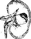
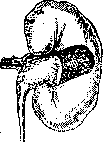
Figure 27.
Figure 28. Provided the ureter is freely patent (figure), the pelvic incision heals without the formation of a fistula even with moderately infected urine. The ever-existing possibility of even temporary leakage of urine also contraindicates closing the surgical wound tightly here.
c. Musc. VIII. Syphilis of the Kidneys. Diseases of the kidneys of syphilitic origin were first described by French authors. Rayer in 1840 linked the cases of late kidney lesions observed by him in syphilis with syphilitic infection—amyloid degeneration and the contracted kidney. Earlier authors attributed albuminuria and other signs of kidney involvement in syphilis solely to manifestations of mercurialism, i.e., poisoning of patients during treatment with mercury. Syphilitic kidney lesions of the secondary period were described by Perroud (1867) under the name of syphilitic albuminuria. This author already pointed out the involvement of the tubular epithelium. Dieulafoy described "early syphilitic nephritis" with edema, massive albuminuria, etc., in particular detail, emphasizing the anatomical features of this syphilitic large white kidney as a purely epithelial lesion, without the participation of connective tissue or vascular elements of the kidney. Thus, this description sufficiently reveals the characteristic features of kidney disease of the secondary period, corresponding to the concept of "nephrosis" in modern classification. Subsequently, the doctrine of syphilitic nephrosis underwent special development in the works of F. Munk, who pointed out in 1913 the characteristic disorders of lipid metabolism and, in particular, lipoiduria in edematous nephroses, especially in syphilitic nephrosis. Later authors, including the founder of the modern classification of diffuse kidney lesions, Volhard, describe lipoid nephrosis as the most characteristic kidney lesion of syphilitic etiology. However, in the secondary period of syphilis, aside from purely nephrotic forms, French authors have also described inflammatory glomerulonephritides with bloody urine and cardiac hypertrophy, starting as early as 1883 (Brault). Among late syphilitic kidney lesions, besides amyloid and the contracted kidney, specific changes in the form of gummas and sclerogummatous nephritis, as well as specific changes in the renal vessels (syphilitic endarteritis), which have lesser clinical significance, were described as early as the mid-19th century. A separate category should be assigned to congenital syphilis of the kidneys described by Hutinel and other authors. The indicated diversity of manifestations of syphilitic infection in the kidneys in various stages of the syphilitic process complicates and hinders a rational classification of syphilitic kidney lesions. The basis of existing classifications is the differentiation of individual forms either according to the periods of evolution of the syphilitic infection (predominantly French authors) or according to individual anatomical forms (German authors). Widal, Lemierre, and Pasteur Vallery-Radot provide the following classification: A. Nephritides of the primary period. B. Nephritides of the secondary period: 1) of the type of massive involvement of the tubular epithelium, 2) of the vascular-tubular type, and 3) with a predominance of connective tissue and vascular lesions. C. Tertiary syphilis of the kidneys: 1) specific lesions—a) gummas, b) sclerogummatous nephritis; 2) non-specific lesions—a) amyloid, b) subacute, and c) chronic nephritis. D. Congenital syphilis of the kidneys: 1) of nursing age, 2) of second childhood, 3) renal aplasia.—Thompson distinguishes: A. Early forms: a) transient albuminuria, b) acute and subacute nephritis; B. Late forms: a) chronic interstitial and parenchymatous nephritis, b) amyloid kidney, c) gummas. It should be noted that in the given classifications, "acute nephritis of the secondary period" is almost completely equivalent to "nephrosis" in the classification of Volhard and Fahr.—Among Russian works, one can cite the following classification by Yanovsky: 1) hereditary syphilis of the kidneys, 2) tertiary syphilis of the kidneys: a) gummas, b) amyloid, c) sclerosis, 3) syphilitic nephrosis. In the subsequent presentation of the clinical features of kidney syphilis, the main clinical forms are described according to the following brief scheme, close to the schemes of Thompson and Yanovsky: 1) early forms—syphilitic nephrosis (with a brief indication of benign syphilitic albuminuria and mixed forms of diffuse kidney lesions); 2) late forms: a) amyloid, b) gummas, c) chronic nephritis and syphilitic contracted kidney; 3) congenital syphilis of the kidneys—early and late. (Rarer forms of kidney syphilis of minor clinical significance are left unconsidered.) Early forms. Syphilitic nephrosis. The most characteristic clinical form of the manifestation of syphilitic infection in the kidneys, especially in the secondary period, is considered to be syphilitic lipoid nephrosis—a purely degenerative kidney lesion (see Nephrosis). It should be pointed out, however, that similarly to disseminated parenchymal lesions of the kidneys in other infectious diseases, also in early syphilis, besides pure degenerative forms, mixed and inflammatory forms may also be observed (glomerulonephritides with a lipoid-nephrotic tinge or hemorrhagic nephritis), which to a certain extent justifies the synthetic term of French authors nephrite aigue syphilitique. However, in view of the predominant significance of nephrotic disorders in the clinic, pathogenesis, and therapy of early syphilitic kidney lesions, the description of syphilitic lipoid nephrosis is taken as the basis of the further presentation, while mixed forms and nephritis are mentioned only briefly. On the other hand, transient albuminuria of the secondary period, in view of the benignity of its course, deserves special highlighting, although, according to Munk, it represents only a milder form of the same symptom complex—lipoid nephrosis.—a) Benign syphilitic albuminuria. Albuminuria is rarely observed in the primary period of syphilis; thus, Dumet out of 500 carriers of a syphilitic chancre saw albuminuria of syphilitic origin in only 2 cases. In the secondary period, albuminuria is stated more frequently (2–3% of cases). The amount of excreted protein ranges from 0.1 to 0.3‰. Talamon, however, observed the excretion of protein in the amount of 34.0 g per day just a few days after the development of the syphilitic ulcer, lasting only 10 days. The excretion of protein is not accompanied by edema or any other clinical symptoms. If the patient has general symptoms, they depend not on kidney involvement, but on the syphilitic intoxication of the body. In other cases, besides albuminuria, cylindruria, mild edema of the face or legs may be observed, disappearing in a few days or weeks. These cases represent a transition to true lipoid nephrosis. The course is favorable, which is reflected in other names for this disease form—simple, transitory, or curable syphilitic albuminuria. Treatment is specific—mercury, neoarsphenamine.—b) Syphilitic lipoid nephrosis in its clinical manifestation presents all the characteristic features of lipoid nephroses in general (see Nephrosis), moreover often expressed with particular relief. The onset of the disease is usually gradual. The first indication of lipoid nephrosis may be the detection of protein in the urine during control urine analyses performed during the course of specific treatment. Less commonly, the onset of the disease is stormy, with pain in the lower back, elevated temperature, a sensation of marked weakness, dyspeptic phenomena, rapid development of edema, as in "nephritis a frigore" or as in scarlet fever. Edema appears on the face or lower extremities, rapidly reaching enormous proportions, presenting the picture of anasarca, cavity dropsies, and visceral edema (edema of the lungs, larynx). The patient has the characteristic appearance of "Bright's disease patients" with a pale edematous face, anasarca, etc. The edematous fluid has a pseudochylous appearance, and the cholesterol content is significantly increased in the blood serum (see Nephrosis). Urine is scanty; less frequently, diuresis remains normal; complete anuria may set in. Urine is saturated in appearance, resembling cloudy broth. Specific gravity is high, sometimes reaching very figures (partly due to protein)—up to 1.060 (Karvonen). Albuminuria is of moderate degree or more sharp; its degree usually corresponds to the magnitude of the edema. In syphilitic lipoid nephrosis, the excretion in the urine of an exceptionally high amount of protein has been described, namely—55‰ (Chauffard and others), 64‰ (Widal), and even 110.0 per 800 cm3 of urine. Urine protein consists predominantly or even exclusively of albumins. Delamare described the excretion of globulins alone. In the sediment—epithelial cells, various casts, many leukocytes, isolated erythrocytes. When viewed under a polarizing microscope, the fatty-degenerated epithelium, casts, and fat droplets turn out to consist of birefringent formations, i.e., lipoiduria is present. Syphilitic treponemes have been found in the urine by individual authors. The nitrogen-excretory function of the kidneys is not impaired. However, cases with an elevated Ambard constant and with delayed excretion of methylene blue and phenolsulfonphthalein have been described. However, it is possible that in these cases with impaired excretory function of the kidneys with respect to dyes, there was not a pure nephrosis, but mixed forms (glomerulonephritis with a lipoid-nephrotic tinge). All the more should the participation of the glomerular apparatus be recognized in cases with significant azotemia [an increase in blood urea up to 150 (Widal) and even 500 mg % (Caussade) has been described].
From the cardiovascular system, pure cases of syphilitic lipoid nephrosis are characterized by normal or even lowered blood pressure and the absence of cardiac hypertrophy; the described rise in blood pressure, and even more so significant hypertrophy of the left ventricle and the early appearance of gallop rhythm, are likewise more correctly attributed to syphilitic nephritis with nephrotic overlays. Pathogenesis of syphilitic lipoid nephrosis. The occurrence of syphilitic lipoid nephrosis, as well as of syphilitic albuminuria, should be attributed to the action of pale treponema toxins on the renal parenchyma. A direct effect of the syphilis pathogen itself on the renal tissue is less likely, since the nephrotropism of the syphilitic virus is insignificant. Of great importance in the pathogenesis of lipoid nephrosis is the disturbance of protein and fat metabolism, which, even in the absence of kidney lesions, undergoes significant changes in syphilitic infection. Neumann attaches importance to the irritation of the kidneys as a result of the hemolytic action of spirochetes and their toxins. Body cooling may also act as a predisposition factor (Dieulafoy, Chauffard). Course of the disease. Syphilitic lipoid nephrosis most frequently affects men aged 20-30 years (according to Munk, however, women fall ill more often). As a rule, nephrosis sets in during the first year of syphilitic infection, according to Dieulafoy usually on the 2nd-3rd month of the disease, according to Thompson most often on the 5th month. There are indications that persistently recurring forms of secondary syphilis particularly often lead to nephrosis. Symptoms of nephrosis usually appear simultaneously with a new outbreak of secondary phenomena on the part of the skin and mucous membranes. The disease may, however, develop even before the syphilitic exanthem—the so-called proroseolous nephrosis. Mühling described the onset of the disease two weeks after the appearance of the primary affect. On the other hand, syphilitic nephrosis can also develop later, 3 years (Mauriac) and even 13 years (Vorpahl) after infection. According to Volhard, with fresh or recurring secondary syphilis with syphilitic angina and exanthem, a relatively rapid course of the disease is observed, and in later stages nephrosis begins gradually and proceeds as a chronic disease with the same clinical manifestations. At the height of its development, syphilitic nephrosis can present a severe disease with burdensome symptoms—marked dyspnea (from hydrothorax, pulmonary edema), headaches, vomiting, diarrhea, sometimes bloody. Among complications, erysipelas, lymphangitis, pneumococcal peritonitis are observed, which can lead to sepsis and death. A fatal outcome can also ensue from heart failure. Syphilitic lipoid nephrosis can last several weeks or months and end in complete recovery; more often, residual phenomena remain—insignificant albuminuria, a propensity for edema. In cases where death occurred already within a few weeks from the onset of the disease with phenomena of anuria and "major azotemia," or acute eclampsia with amaurosis developed, the kidney lesion likely had a mixed character from the very beginning (glomerulonephritis with a lipoid-nephrotic shade). Volhard did not observe uremia in his material of syphilitic lipoid nephrosis. In protracted cases with persistent edema and a cachectic appearance of the patients, lipoid-amyloid nephrosis often develops, which subsequently leads to amyloid contracted kidney and uremic intoxication. The rarest outcome of syphilitic lipoid nephrosis must be recognized as "nephrotic contracted kidney"—a disease proceeding in the same way as amyloid, with polyuria, normal blood pressure and the absence of cardiac hypertrophy, and leading to death from uremia. Most authors doubt the existence of this form in a pure state; thus, Wohlwill could not find an uncontroversial case of syphilitic nephrotic contracted kidney.
Differential diagnosis. The recognition of syphilitic lipoid nephrosis is usually easy, but in some cases it may present significant difficulties. In treatment with mercury, against the diagnosis of mercurial kidneys and in favor of syphilitic nephrosis speaks the absence of other signs of mercurialism—stomatitis, hemorrhagic colitis, the presence of edema and (according to Munk) the discovery in the urine of doubly refracting lipoid cylinders and droplets, as well as the peculiarity that the cessation of mercury treatment does not lead to the rapid disappearance of these symptoms. In salvarsan nephrosis, albuminuria also does not last long. In favor of syphilitic lipoid nephrosis, besides other symptoms of syphilis, speaks a positive Wassermann reaction, although not unconditionally, since a significant disturbance of lipoid metabolism, "nephrotic alteration of colloids" (Munk), characteristic of any lipoid nephrosis, can give a nonspecific Wassermann reaction (O'Hare). In favor of syphilitic lipoid nephrosis speaks the persistence of edema, unresponsive to conventional therapy, as well as very significant albuminuria. According to Yanovsky, frequent and significant fluctuations in the amount of excreted protein are especially characteristic of syphilitic nephrosis. Gouget emphasizes that massive albuminuria in syphilitic lipoid nephrosis may not be accompanied by oliguria and may even proceed with polyuria, as well as in amyloid kidneys. Evidence for syphilitic kidney disease is the detection of Treponema pallidum in the urine, but its differentiation from non-pathogenic spirochetes presents significant difficulties. Lipoiduria is characteristic of syphilitic lipoid nephrosis, but is not pathognomonic for this disease, as Munk was initially inclined to believe; Waldorp and Behr consider the finding of doubly refracting substances in the urine to be an unconditional indication of syphilis—acquired or congenital. The diagnosis of syphilitic lipoid nephrosis ex juvantibus can be made only with certain limitations, although Pasteur Vallery-Radot, as well as older authors, says that the therapeutic test decides the diagnosis and that syphilitic lipoid nephrosis is cured only under the influence of specific treatment. However, syphilitic nephrosis can improve or disappear even without specific treatment (Bauer, Munk, and others). In cases of fresh or recurrent secondary syphilis with syphilitic angina and exanthem, Volhard saw a relatively rapid disappearance of edema and complete recovery under the influence of diuretics only and a diet poor in water and salt. On the other hand, specific treatment can worsen, especially at first, the symptoms of kidney damage even in syphilitic lipoid nephrosis or remain without effect on the course of the disease (Fishberg), especially in cases of late "metasyphilitic" nephroses. Thus, the diagnosis of syphilitic lipoid nephrosis should be made taking into account all the indicated features of the clinical picture and the course of this disease. Syphilis should constantly be suspected as the cause of lipoid nephroses of unclear etiology. Prognosis. Kidney involvement is the most severe complication of secondary syphilis, although the prognosis of syphilitic lipoid nephrosis is recognized as better than that of lipoid nephrosis of another etiology, and this nephrosis is considered in general to be a curable kidney disease. Of 12 patients of Karvonen, only one died of uremia during a relapse of the kidney disease; according to Fournier, one out of three patients dies. The height of albuminuria does not determine the severity of the case; the persistence of edema is of greater importance. The prognosis is better in the early stages, when nephrosis coincides with exanthem. According to Volhard, specific treatment of nephrosis in fresh secondary syphilis almost always leads to a rapid cure. With a later development of lipoid nephrosis, the disease proceeds more severely and stubbornly. With the transition of nephrosis into chronic forms, the prognosis worsens significantly. Treatment and prophylaxis. For the purpose of prophylaxis of kidney damage, Renon and Dieulafoy proposed to conduct a milk regimen in all cases of syphilis from the beginning of infection to the 3rd month. The treatment of syphilitic lipoid nephrosis should be carried out first according to the general rules of treatment of edematous nephroses (see Nephrosis); as diuretics, organic mercury compounds of the salyrgan type are especially recommended, possessing at the same time a weak antisyphilitic action. Upon the disappearance of edema, or respectively in case of ineffectiveness of conventional therapy, they proceed to treatment with neoarsphenamine, the good effect of which in syphilitic lipoid nephrosis has been noted by many. According to Rosenberg, neoarsphenamine can be safely used in usual doses every fourth day, starting from 0.15–0.3–0.45. The total amount of neoarsphenamine per course should reach 4–5.0 (Lichtwitz). Pulay recommends introducing neoarsphenamine in a 5% solution of the preparation Urotropin-Chlorcalcium, which leads to an increase in diuresis and a faster excretion of neoarsphenamine. Widal, Lereboullet, and Pasteur Vallery-Radot recommend caution in the treatment with neoarsphenamine and begin with doses of 0.05, monitoring especially the urea content in the blood. Lesné cites a case of death from uremia after an infusion of 0.15 of neoarsphenamine. Mercurial preparations were widely used with good success by old clinicians (Fournier, Dieulafoy): in the case of Mosny, over 12 days of treatment, protein dropped from 72‰ to 0.5. Chauffard, Ferrand, Sireday, and others, on the contrary, note the ineffectiveness and even harm of mercurial treatment. As mentioned above, former authors attributed even all kidney lesions in syphilis to mercury. Of the various forms of administration of mercury, the inunction of gray ointment (Munk, Hoffmann) apparently produces a good effect. Most authors speak against injections of insoluble salts. Intravenous infusions of mercuric cyanide, which possesses diuretic properties similar to salyrgan, are especially recommended. Treatment with mercury is recommended by many authors after the arsphenamine course to consolidate the therapeutic effect. Bismuth preparations have also been used with good results (Rosenberg and others), although they can also worsen the course of the disease (Labbé, Widal, and others). Iodine salts were prescribed more widely before, but now find fewer supporters (Lichtwitz, Volhard, Yavein, Rosenberg) and are used mainly in combination with neoarsphenamine, etc. Treatment with iodine can even prove dangerous in renal failure: cases of death from 2.0 (Wolff) and even 1.0 of iodine salt (Rendu) have been described. c) Syphilitic nephritis. Although Volhard (1931) considers "the existence of true diffuse hypertensive syphilitic nephritis unproven," other authors describe various forms of syphilitic nephritis. Hein singles out as an independent form glomerulonephritis haemorrhagica acuta luetica praecox. French authors, as stated above, from the very beginning of the study of syphilitic kidney lesions, noted glomerular lesions. Yanovsky describes syphilitic "nephroso-nephritis." From the point of view of modern terminology, it should be recognized that in the secondary period, besides pure lipoid nephrosis, there can be observed its combination with various degrees of glomerular changes, starting from minimal and ending with the most severe forms, syphilitic glomerulonephritis with secondary lipoid nephrosis (syphilitic nephroso-nephritis) and syphilitic (hemorrhagic) glomerulonephritis. Indications of various authors for hematuria (Jaccoud), azotemia, hypertrophy of the left ventricle, early appearance of gallop rhythm, eclampsia, etc. in kidney damage of the secondary period should rightfully be attributed to the participation of the glomerular-vascular apparatus. Similarly, indications of low tolerance to iodine, partly to neoarsphenamine (in azotemia), etc., also indirectly speak against purely epithelial lesions in given cases. Late forms. 1) Amyloid is a very frequent complication of syphilis; often lipoid nephrosis in a protracted course passes into lipoid-amyloid nephrosis with the further development of amyloid contracted kidney, or amyloid degeneration is combined with chronic nephritides, gummas, nephrocirrhosis. Sometimes it is a matter of patients with persistently recurring lesions of the skin and mucous membranes, prolonged bone-periosteal suppurations, and chronic lesions of internal organs. Usually, however, amyloid develops in syphilis without prolonged purulent processes. The frequency of amyloid kidneys in syphilis is evidenced by the fact that Wagner among 63 cases of syphilis with symptoms of kidney damage found amyloid degeneration in 35. Therefore, in every case of syphilitic kidney damage, especially with protracted edema and massive albuminuria, one should reckon with the possibility of this complication (see Nephrosis), which is unfavorable in prognostic terms. For therapeutic purposes, the prescription of iodine can be tried.—2) Gummas of the kidneys in most cases do not manifest themselves during life with definite symptoms and are therefore inaccessible for recognition, being as a rule an accidental anatomical finding. Sometimes albuminuria, pyuria, and hematuria are observed; occasionally lumbar pains with nocturnal paroxysms. The general state may not be disturbed, fever is absent. The so-called pseudosurgical form of gummatous syphilis of the kidneys, proceeding under the guise of a kidney tumor, is singled out.
Diagnosis is established by exploratory exposure of the kidney and biopsy of the tumor (Fedorov). Diffuse gummous infiltration of the kidney may also lead to organ enlargement and produce the impression of a fixed, insensitive, or very painful tumor with an uneven surface. Upon rupture of a gumma into the renal pelvis, there may be a discharge of thick, dark-brown, turbid urine with an abundant sediment of erythrocytes, leukocytes, casts, and cellular detritus. The excretion of such turbid urine may be intermittent in character. Gummas can cause peri- and paranephritis with subsequent opening through the skin. The recognition of renal gummas, as well as other late syphilitic lesions of the kidney, is facilitated by the discovery of gummous tumors in other organs, especially in the liver and spleen. The prognosis even for renal gummas recognized during life should be stated with caution, since there is frequently concomitant chronic nephritis, amyloid degeneration, or gummous involvement of other organs. Treatment is usual for the gummous period, taking into account the impairment of the excretory function of the kidneys. 3) Chronic nephritis and contracted kidney [see separate plate (p. 719-720), Fig. 5]. In late stages of syphilis, renal lesions may bear a specific character (sclero-gummous nephritis, luetic endarteritis) with subsequent scar formation and frequently asymmetric atrophy of the kidneys; sometimes they present the usual features of Bright's disease of any etiology. Concomitant presence of amyloid degeneration is frequent. Clinically, all these forms manifest themselves by the usual signs of chronic nephritis with varying degrees of albuminuria, elevated blood pressure, and renal insufficiency. Nephrocirrhosis on the basis of syphilitic endarteritis may manifest in significant hypertension, albuminuria (1-2‰), hematuria, azotemia, and hemorrhagic retinitis; excretion of lipoids with the urine is not observed. A similar picture is given by nephrocirrhosis on the basis of nodular periarteritis. Nephrocirrhosis on the basis of syphilitic "chronic interstitial nephritis"—a form close to the "syphilitic contracted kidney" of Munk—proceeds with a scarcely characteristic clinical picture of albuminuria without hypertension, polyuria, and renal insufficiency; combination with endarteritis is possible. In the presence of unilateral hyperplastic para- and perinephritis, such nephrocirrhosis may proceed under the guise of a renal tumor. Syphilitic chronic nephritis proceeds with the same signs as banal forms of nephritis. The presence of syphilitic stigmata and a positive Wassermann reaction in the absence of another etiological factor point to the syphilitic nature of these nephritis cases. According to some authors, every chronic nephritis with a hypertensive syndrome in young subjects is suspicious in the sense of a syphilitic etiology. Letulle and Bergeron found a positive Wassermann reaction in 12 out of 46 brightics, furthermore predominantly in young individuals. Specific treatment of the indicated forms of late syphilis of the kidney must be carried out cautiously, taking into account urinary symptoms and the nitrogen-excreting function of the kidneys. Syphilis and essential hypertension, resp. arteriolosclerotic nephrosclerosis. In connection with late forms of syphilis of the kidney, mention should be made of the influence of syphilitic infection on arteriolar lesions of the kidney of a non-specific character. In the works of older authors, syphilis was assigned a prominent place among other etiological factors of so-called "chronic interstitial nephritis," i.e., essential hypertension and arteriolosclerotic nephrosclerosis according to modern terminology. Gallavardin points out the frequent coincidence of renal and aortic lesions ("nephroaortite syphilitique"). According to Kollert's data, out of 120 nephrosclerotics, 29 (24%) had syphilis; according to Kollert, the main significance of syphilis for the kidney is in the relatively rapid development of arteriolosclerotic changes. Fahr found indications of syphilis in 10 out of 40 cases of his "malignant sclerosis." Other authors do not attach such significance to syphilis in the etiology of hypertension and nephrosclerosis. Horine and Weiss found the same frequency of syphilis among 666 essential hypertensives and 2,000 non-hypertensives. Fishberg confirms the comparatively frequent coincidence of syphilitic aortitis and hypertension, but explains this (just like Korányi) by the fact that hypertension predisposes to the localization of syphilitic infection on the aorta. Volhard, noting syphilis in the anamnesis of hypertensives frequently, and aortitis at autopsy, is nevertheless not inclined to place hypertension in a causal relationship with syphilis. Keith, Wagener, and Kernohan found a positive Wassermann reaction in only 1 patient among 75 cases of malignant hypertension, and Fishberg in one out of 12. Volhard in malignant hypertension (out of 124 cases) finds syphilis just as frequently (in 4.8%) as in benign hypertension (out of 452 cases), which speaks against the special significance of syphilis specifically for the malignant (first) form. Specific treatment in hypertensives must be carried out with great caution. Good renal function in the presence of albuminuria does not contraindicate specific treatment in small doses (preferably with bismuth, Fishberg). Lewinson observed a certain decrease in blood pressure from specific treatment. Hyposthenuria contraindicates the use of mercury, neoarsphenamine, bismuth, and even large doses of iodine (cases of death from mercurial colitis and arsphenamine hepatitis have been described—Fishberg). Congenital syphilis of the kidney. Syphilitic lesions of the kidney in infancy do not have a clearly outlined clinical picture; they may manifest only with symptoms from the urine—albuminuria, cylindruria, oliguria sometimes reaching the degree of anuria, or subcutaneous edema, somnolence, convulsions, diarrhea, vomiting, and even uremic peritonitis join in (Guthrie). Concurrently, there are usually syphilitic lesions of the liver, spleen, skin, and mucous membranes. Cases of cure from mercurial treatment have been described. Late congenital syphilis of the kidney is similar to acquired in its symptoms. Albuminuria often reaches significant proportions, cylindruria and sometimes hematuria are also observed. Among clinical signs, edema, elevated blood pressure, cardiac complaints, headaches, and vomiting are the most constant. The disease proceeds in a chronic form and gives a poor prognosis. Diagnosis is made on the basis of anamnesis and other manifestations of syphilis.
E. Tareyev. IX. Kidneys in Pregnancy. Kidney diseases occurring during pregnancy can be of three kinds: 1) diseases caused by the pregnancy itself, 2) diseases accidentally joining it, and 3) diseases observed before its onset. Among the diseases attributed to pregnancy and classified as "toxicoses," the so-called pregnancy kidney (ren gravidarum) is the most frequent. The term "pregnancy kidney" (Schwangerschaftsniere) was first proposed by Leyden (1886), who actually gave the first detailed description of the impairment of kidney function during pregnancy from clinical and patho-anatomical points of view. Subsequently, Zangemeister (1913) assigned them the name nephropathia gravidarum, which rather quickly took root in clinical practice and became widespread. However, the concept of the pregnancy kidney is still treated extremely differently to this day, there is no reasonably satisfactory classification of kidney diseases during pregnancy, and the point of view on the causes of their occurrence is also inconsistent. There are even opinions (e.g., Ivanov, Chistovich) that there is no reason to single out the pregnancy kidney into some special pathological form. As for the very concept of the pregnancy kidney or nephropathy, it seems unquestionable that these terms at this time should be reserved only for completely definite processes, and not include (as some thought and think) all without exception diverse lesions of the kidney during pregnancy both arising primarily and developing secondarily (in other toxicoses). The term pregnancy kidney should designate only initial (non-severe) functional lesions of the kidney during pregnancy. This is necessary both logically because the very concept of the pregnancy kidney cannot cover all types of kidney diseases of a degenerative and inflammatory character, and because the clinical course, accompanying complications, and final and remote results of not only inflammatory but also various degrees of degenerative processes are far from equivalent. First of all, it is necessary to distinguish between degenerative and inflammatory processes. The first type of processes of a degenerative order is the so-called pregnancy kidney, or nephropathy. It usually appears benign with complete restitutio ad integrum at the end of pregnancy and can be considered in more pronounced cases as a preliminary stage of nephrosis (but only with hypertension). The pregnancy kidney differs from physiological albuminuria of pregnancy in that in the latter the protein excretion is not accompanied by any pathological symptoms and painful phenomena of a general order, while in the pregnancy kidney, regardless of the amount of excreted protein and the presence of formed elements, there are always present one or another pathological symptoms and general disorders, and thus in the differential diagnosis between "simple" albuminuria and the pregnancy kidney, the most characteristic is not the increase in protein or the appearance of formed elements, as some believe (Gurevich), but the onset of general disorders. And here, as in more severe degrees of kidney damage, and finally as in eclampsia, a mere increase in protein cannot serve as a criterion either for diagnosis or prognosis. Similarly, it is not the increase in protein along with the development of edema that reveals the presence of the pregnancy kidney (Zangemeister), but the appearance of characteristic symptoms and disorders of a general order (e.g., decrease in diuresis, increase in edema, hypertension, myopathy, headaches, etc.). The degree of the latter, the intensity of their manifestation, can, on the other hand, serve as a boundary for differentiating "nephropathy" from more severe deviations of the same degenerative order (nephrosis) or finally from mixed forms (nephroso-nephritis). There are other explanations of the pregnancy kidney. Thus, some (albeit isolated) authors strive to identify it with nephritis; it is quite understandable that the desire to regard degenerative processes as inflammatory must be completely rejected [for example, the old understanding by Bartels of the pregnancy kidney as acute parenchymatous nephritis (Lvov, 1899); Yakovlev (1932) even proposes a name clearly contradicting the concept of the pregnancy kidney—nephritis tubularis gravidarum (!) with an attempt to substantiate it, etc.]. It also seems irrational to propose (Ivanov) to combine all pathological processes in the kidney under the general name "albuminuria of pregnancy" (including "simple" physiological) or to reduce them only to any one form. Thus, for example, Chistovich, denying altogether the peculiar character of the pregnancy kidney, considers it a pure form of nephrosis. Seitz believes that all functional and anatomical changes in the kidneys are covered by the concept of "nephrosis." Jaschke also regards all renal processes as nephrotic (attributing even physiological changes to them) and gives them the name "nephrodystrophia gravidarum" (Jaschke distinguishes three types of this dystrophy: to the first he attributes physiological albuminuria, supposedly corresponding to the first stage of nephrosis, to the second—nephrosis, identical to the pregnancy kidney and true nephrosis, and to the third—mixed forms: a combination of nephrosis with nephritis). It is hardly correct, however, to attribute the entire diverse pathology of the kidney only to any one well-known form. Schlayer quite rightly points out that although the pregnancy kidney may externally resemble the picture of true nephrosis, this identity is only external, since in reality there are significant differences, and daily observations speak against treating the pregnancy kidney as nephrosis. Rosenberg expresses himself similarly. Admitting that the pregnancy kidney could be characterized as nephrosis with sometimes very significant hypertension, Rosenberg also believes that the pregnancy kidney must be understood as a completely peculiar disease from both a clinical and anatomical point of view, a disease that cannot be attributed either to nephrosis, nor to nephritis, nor to mixed forms. Lichtwitz (with whom Zimnitsky also agrees) likewise points out that the pregnancy kidney cannot fit into the concept of nephrosis and that the characteristic triad—hypertension, eclampsia, retinitis—gives it a peculiar character, whereas anatomically it resembles epithelial nephropathy, although clinically it can also proceed under the guise of nephritis. Opinions are also diverse regarding the causes causing the pregnancy kidney. The view that arose in the initial period of the scientific study of the pregnancy kidney and subsequently dominated for a long time—that the pregnancy kidney is caused exclusively by mechanical moments (pressure of the pregnant uterus on the veins, nodes of the sympathetic nerve, compression of the ureters, increased blood pressure in the kidney, etc.), causing circulatory disturbance with subsequent anemia of the kidney, albuminuria, dropsy, etc.—is rejected at the present time by the majority of authors, who attach more importance to toxic influences. If, of course, the role of mechanical factors cannot be completely excluded, in any case they do not play the fundamental role that they were tried to be attributed to earlier or that is assigned to them by some (e.g., Zimnitsky) even now. This is also evidenced by all the latest research, clearly showing that local factors are of secondary importance, since other more serious disorders of kidney function and general disorders on the part of other various organs and systems during pregnancy are complex diseases of the entire organism, and are not some isolated, localized processes of any single organ alone. Nor can the prevailing influence of mechanical causes be supported by the fact that the pregnancy kidney is predisposed to by such moments as, for example, polyhydramnios, multiple pregnancy, narrow pelvis, etc. Although functional suffering of the kidneys is more frequently observed with them, this greater frequency is caused not by the fact that mechanical factors are aggravated, but by the fact that in such cases one is dealing either with the inability of the organism to respond to the increased demands placed upon it or generally with its inferiority, or else with general underdevelopment with the presence of "infantilismus renalis" (Strauss) or constitutional weakness of the kidneys (Martius) with a special tendency to their damage during pregnancy.—Indications of the toxic nature of the pregnancy kidney are found even at the end of the 19th century, predominantly on the part of a number of representatives of the French school (Bouchard, Riviere, etc.). Thus, they steadily pursued the view that both deviations on the part of the kidneys and all disorders in general attributed to pregnancy (salivation, incessant vomiting, eclampsia, etc.) are the consequence of the same cause—autointoxication of the organism on the basis of impaired metabolism and retention of harmful metabolic products. Authors from other countries adhered to a similar point of view.
Later, with the development of our knowledge, the establishment of the significance of the endocrine system, the role of the placenta, the discovery of enzymes in it, the detailing of the nature of metabolism in normal and pathological pregnancy, and the ascertainment of «pregnancy acidosis», the toxic theory received substantial justification, and at the present time all disorders of the kidneys in pregnancy are regarded by most authors as occurring on the basis of unidentified toxins circulating in the blood; kidney diseases are introduced into the general group of other diverse processes bearing the common collective name of «pregnancy toxicoses» (Okinchits, Chistovich, Selitsky, Benda, Veit, Freund, Rosenberg, and others). As for the essence of the process itself and the anatomical and histological changes occurring therein, some (Heynemann) believe that lesion of the glomeruli rather than of the tubular epithelium is typical for pregnancy (i.e., there is a kind of glomerulonephrosis in which capillary circulation is disrupted—capillaropathia gravidarum), while others (e.g., Yashke) assume the opposite. A significant portion of authors also believes that vascular damage occurs initially (diffuse spasm of peripheral vessels—Rosenberg), due to which tissue nutrition is disrupted with subsequent secondary degeneration of the epithelium (Hussy, Zondek, and others). This disruption of tissue nutrition is attached special importance by some; Zondek, however, believes that pregnancy toxicosis is a general disease of the tissues («hystopathia gravidarum») which arises by virtue of the increasingly progressive acidosis during pregnancy and consists (first of all) in a change in vascular tone, in capillary spasm (with subsequent hypertension, spasm of the renal vessels, sharp albuminuria and cylindruria), and tissue thirst occurring on this basis as a result of toxic damage to peripheral tissues. To speak of pathological anatomy in the literal sense of the word with regard to the kidneys of pregnant women is impossible given the modern limitation of its concept. One has to rely on cases with more severe lesions of the renal parenchyma that ended fatally, and assume that analogous changes, but to a lesser degree, can also occur in the kidneys of pregnant women and in other kidney diseases that ended in recovery. Those authors (e.g., Hussy) are completely right in this regard who say that there is actually no histological picture of the kidneys of pregnant women, since not a single case of it ending fatally is yet known; whereas in the cases described both by Leyden and by subsequent authors, the picture of changes in the kidneys could also be caused by a more severe poisoning of the entire organism and individual organs, and finally by other concomitant pathological processes [e.g., indomitable vomiting, eclampsia, sepsis, and others, and according to some authors (Pozharisky) also chloroform anesthesia]. Leyden based his opinion on the fact that 1) the intensity of changes in the kidneys in eclampsia ending fatally is not always greater than in eclampsies ending in recovery; 2) the data of urine analysis and microscopic examination of the sediment are often identical in cases that ended in recovery as well as fatal ones; 3) despite the intensity of the kidney lesion, the process has the ability to rapidly resolve after the end of childbirth (this, in his opinion, indicates that there is only strong «fatty infiltration» and not fatty degeneration of the renal epithelium). These conclusions of Leyden have not lost their significance at the present time, since with our modern view of pregnancy toxicoses, we attribute the occurrence of all individual pathological symptoms, individual manifestations, disorders of organ function, and finally completed forms of toxicoses to unknown toxins circulating in the organism of the pregnant woman and believe that they can act extremely variously depending on a whole series of endogenous and exogenous factors; by virtue of this, it is understandable that the changes encountered in the kidneys and in other organs can be very diverse both in cases ending in recovery and in fatal ones. Confirmation of this is provided by both the clinic and autopsy data, which furthermore show that insignificant changes can be discovered in the kidneys on the dissecting table, or even that the kidneys can remain completely unaffected [in exceptional cases, e.g., in eclampsia (Nikiforov)], and that, on the other hand, in cases ending in recovery, the lesions can be very significant and remain irreparable for life. Such a point of view is shared by other authors, and inter alia Zangemeister, who has worked extensively on this issue, believes that the fresh changes encountered are observed only during pregnancy and that the picture of these changes appears typical and identical for both lighter and heavier cases; the difference lies only in different degrees of lesion of the renal parenchyma. Selitsky, when drawing a parallel between indomitable vomiting and eclampsia, also pointed out that the changes both in the kidneys and in other organs have an identical character in various diseases, that only their different degrees are observed, depending on the course of the process, the nature of the intoxication, and the constitution of the patient. The changes described by Leyden, subsequently confirmed by a number of authors, consist in a general enlargement of the kidney, its pallor, and fatty degeneration predominantly of the epithelium of the urinary tubules (both straight and convoluted) (degeneration of the glomerular epithelium is encountered significantly less frequently, while the interstitial part remains unaffected). In rarer cases, thrombi in the capillaries and small arteries (on the peripheral part of the kidney), hemorrhages into the renal parenchyma, infarcts, glomerular hyperemia, and others were discovered. In the overwhelming majority of cases, as all authors indicate, the lesions are of a degenerative order, while inflammatory changes are encountered significantly less frequently (according to Prutz—in 11.6%, according to Meyer-Witz—in 17.1%) and are all qualified in the majority of cases as accidental changes or as old ones not characteristic of pregnancy. At present, there is no need to speak of any exhaustive classification; likewise, the existing classifications of kidney diseases outside of pregnancy cannot be used (see Nephritis, Nephrosis), and all kidney diseases encountered during pregnancy can only be presented in the form of a diagram. Albuminuria (physiological) (gravidarum, sub et post partum) Kidney diseases of a mixed type
and degenerative nephrosonephritis (the predominant form), nephritis associated with pregnancy (ex graviditate), inflammatory character 1 1 nephropathy, nephrosis (pregnancy kidney) ! nephritis occurring incidentally (in graviditate) 1 nephritis previously existing (chronic). The frequency of kidney diseases in pregnancy (especially of more severe forms) is subject to considerable variation and depends on a number of factors (Tables 1, 2). Table 1. Frequency of kidney diseases in pregnancy. Authors: Number of pregnancies; Including kidney diseases; Percentage of morbidity. Selitsky-Akimova: 12,501; 966; 3.7%. Mauno-Schroderus: 9,625; 2,786; 28.9%*. Zangemeister: 8,728; ... [Table notes and continued text regarding the frequency of various forms of kidney disease, mortality, eclampsia, clinical course, symptoms, hypertension, etc., corresponding faithfully to the original Russian text].
Thus, the presence of a fairly large number of stases in the capillaries, established for the first time during pregnancy by Hinselmann, and their significant increase in kidney of pregnant women and eclampsia were subsequently discovered by a number of other authors (Schreiner, Vinogradova). These observations showed that stases are more sharply expressed in severe cases, most often in eclampsia (in pregnant women without edema in 4.5%, with dropsy in 14.3%, with nephropathy in 50%, and with eclampsia in 80%), and that hypertension is higher the more frequently stases occur. Along with the described clinical symptoms, there are changes and disturbances in metabolism. Thus, disorders in the excretion of NaCl and creatinine (Orlovius) are observed, a decrease in the relative figures of urea, a certain increase in residual nitrogen, an increase in the daily amount of ammonia, delayed excretion of water and KI, and changes in carbohydrate metabolism are noted. In diagnosis, the question may be either about differentiating various forms of diseases caused by pregnancy, or about distinguishing them from chronic nephritis or nephritis that arose during pregnancy, but independently of it. The anamnesis and clarifying the question of whether there was any previous kidney disease or any childhood infections are of exceptional importance. The main difference from chronic nephritis is also the time of onset of the disease. While diseases caused by pregnancy arise (almost as a rule) in the last months of pregnancy, the manifestation of chronic nephritis begins already at its very beginning. Along with this, pathological symptoms are also different. Thus, for example, in chronic nephritis, hypertension, if present, is weakly expressed, in the initial stages diuresis is not sharply reduced (polyuria is also often observed), edema has a completely different character, the amount of protein content is different (usually small and does not reach such figures as in kidney of pregnant women) and the data of microscopic research. In addition, chronic nephritis is more often observed in multipara, and decompensatory phenomena are more frequent in it. Hussy points out that for diagnosis, along with the anamnesis, a thorough examination of the heart and eyes is important. Cardiac hypertrophy, tense pulse, and pale appearance usually always indicate a chronic disease. A differential diagnosis with accidentally occurring acute nephritis is more difficult. Here, the anamnesis is of decisive importance, since in most cases such processes have their etiology in infection (influenza, tonsillitis, typhus) or intoxication (poisoning). When making a diagnosis, it is also necessary to take into account the possibility of pyelitis; individual forms of kidney diseases are diagnosed by clinical symptoms. In prognosis, regarding both immediate and long-term results, one must largely reckon with the nature of the past process, since, as clinical observations show, there is a significant difference in the prediction for mild and severe, acutely and chronically proceeding forms. By virtue of this, it is impossible to summarily make general conclusions, calculate a general percentage of complications and mortality for all forms. The most favorable appears to be the most frequent and typical form—kidney of pregnant women. It does not produce mortality, and if such is observed, it is as an exception and depends not on the kidney of pregnant women itself, but on associated processes (eclampsia, etc.). Due to the mild nature of the diseases, there are fewer complications from a purely obstetric side (a lower percentage of premature births, postpartum diseases). Results regarding children also appear significantly better compared to other forms. Thus, while it is considered that in general with kidney disease there is a very high percentage of primary and secondary infant mortality, a large percentage of dead and macerated children (Lvov 24.7%, Meyer 3% macerated, Gurevich 39% dead and macerated, Hofmeier 57%, Tolochinov 60%), in kidney of pregnant women it is significantly lower (on average 7-8%). In more heavier cases, complications are more frequent, the fate of the fetuses is worse (Tables 5 and 6); the opinion of Mauno Schroderus is entirely justified, that the more severely the kidneys are affected, the worse the prognosis for the fetus. In severe diseases, its intrauterine death is often observed, which, on the other hand, is considered by many to be a good prognostic sign for the mother—Wine already said that when the fetus dies, albuminuria usually passes. Long-term results also depend on the nature of the past disease: changes at the end of pregnancy remain in more severe cases. In the vast majority of cases Table 6. Difference in complications in pregnant women with and without protein in the urine (Liedeberg, 1913) (1127 pregnant women, protein in 239 - 25.6%). Complications: Phlegmasia.......... Percentage of complications in pregnant women with protein in urine: 0.36; Percentage of complications in pregnant women without protein in urine: 0.24. Adherent placenta: 15.2 / 3.3. Placenta previa: 8.0 / 1.3. Bleeding ante partum: 6.9 / 1.3; post partum: 9.0 / 2.9. Cerebral complications: 3.1 / 0.12. Premature births: 10.3 / 0.47. Postpartum diseases: 14.0 / 6.2. Dead children: 7.2 / 1.79. Secondary infant mortality: 3.1 / 0.39. even after severe or acutely proceeding diseases with eclampsia, the process is quickly resolved with the end of labor, and the transition to chronic nephritis, according to the conclusion of the majority of authors, occurs as an exception [some even dispute the possibility of such a transition; Mauno Schroderus believes that there is a permanent organic kidney disease, despite the fact that the kidneys remain healthy in the intervals; Hüssy has never seen it once, Zangemeister notes it in 7%, and Baisch even cites 10% mortality in the immediate years after the suffered disease]. Clinical data show that in approximately 20% on the 10th day after childbirth all pathological phenomena pass (Koblank in 61 cases noted the disappearance of protein after the 1st week in 19 cases, after the 2nd week in 18, even later in 20; in 2 cases the phenomena remained for another 1.5 years, while in 4 cases chronic nephritis subsequently developed). Of interest is also the possibility of recurrence of the disease upon the onset of a new pregnancy. Such a possibility is not excluded. Cases of recurrent kidney disease in several pregnancies with absolute health in the intervals between them have even been described (Bartels, Dickinson, etc.); Wine described recurrent kidney of pregnant women in 4 and 9 subsequent pregnancies, Gossmann - 8 recurrences in 13 pregnancies. According to some data (Seitz), a suffered nephrosis is especially predisposed to the recurrence of the disease. The influence of the nature of the past process is also noted. Thus, Selitsky in 112 kidneys of pregnant women observed a recurrence in 10.7%, in 25 nephroses in 20%, and in 13 nephritises in 84.6% (Zangemeister believes that recurrence occurs generally about 2%, Koblank saw it 1 time per 77 kidneys of pregnant women). The nature of the recurrent disease is not always identical with the primary process—acute forms are much rarer. The frequency is subject to large fluctuations, and it is undoubtedly lower where prevention is carried out more thoroughly and where pregnant women after the disease are placed under special consultation registration. With recurrent kidney of pregnant women, eclampsia usually does not develop. There is no reason to forbid a new pregnancy (as Chistovich thinks)—only certain intervals are necessary. With a tendency to more frequent recurrence, pregnancy must be prevented, since there are indications that multiple recurrences of the disease can lead to true arteriosclerosis, depending on the ensuing vascular damage. Prevention and therapy. That large percentage of complications observed in kidney diseases, and those severe consequences with which they are accompanied for the mother and fetus, urgently put the question of prevention in the first place. This prevention in its main features should consist in the most pedantic management of each case of normal pregnancy, paying attention to the smallest deviations and taking appropriate measures from the very beginning. The prevention of transition into more severe forms, the occurrence of eclampsia, and better antenatal protection of the infant are also based on this. This prevention, carried out in the consultation and in the ward for pregnant women, should consist not only in giving hygienic and dietary advice and monitoring their implementation, but also in protecting the pregnant woman and her infant from harmful external conditions, unhealthy living conditions, and various infections. The prevention of kidney diseases, in view of their relative frequency, should be one of the direct tasks of the obstetrician-prophylactician, because with a decrease in the number of these diseases, maternal morbidity and mortality, which still stand at high figures, will also decrease. Therapy actually begins with the prevention of kidney diseases. The main rule should be hospitalization of each case and the implementation of a regimen appropriate to the nature and degree of the arisen process. Zangemeister advises bed rest even in initial forms. Therapy in general does not differ from the treatment of kidney diseases outside of pregnancy. Along with bed rest, dietary regimen is of no less importance.
It should be predominantly vegetarian, while at the same time restricting the intake of salt, carbohydrates, and fats, as well as the amount of fluid [some (Noorden) advise restricting salt intake for prophylactic purposes in the last 3 months of pregnancy for all women—not more than 5-8 g pro die]. Zangemeister also insists on a restricted fluid intake, even dry diet and an achloridated diet. Particularly good results are produced by the milk diet recommended back in 1872 by Jaccoud and propagated in its time by the famous French obstetrician Tarnier. Laxatives are also often necessary. Various kinds of diuretics and diaphoretics that were widely used in the past (diuretica, subcutaneous injections of pilocarpine hydrochloride) are now abandoned entirely by many. One must not force and excite the weakened function of urination, which almost never achieves the goal, but rather strive to create such conditions for the entire organism under which this impaired kidney function could be restored. By virtue of this, the prescription of high-temperature baths, frequent baths in general, or wet packs is irrational, since besides being unsuccessful they can exert a harmful effect, affect the entire organism, intensify the already impaired cardiac activity, and unfavorably affect respiration. This therapy, based practically on old views (the view of renal processes as isolated, segregated lesions of only a single organ), although still recommended by individual authors, is nevertheless considerably limited at the present time, since, according to the just remark of Eckelt, the view on treatment with moist heat has now changed (on the contrary, dry heat to the kidney region—flannel, hot water bottles—has a good effect, just as a dry and warm room is a necessary condition for a female patient). Among new therapeutic agents in recent years, Fisher's solution (Oettingen), Ringer's solutions, horse N-serum (Freund, Mayer), and intramuscular injections of a 15% gum-Ringer solution [20 cm3 every 3 days, and intra partum every 3 hours (Zangemeister)] have been used in a number of cases. For persistent hypertension and headaches, bloodletting finds application (Heinemann). Thyroidin, recommended by some (Lange, Eppinger, Grossi), has not gained even relatively wide distribution, although the authors who used it noted the disappearance of albuminuria, edema, and other pathological symptoms upon its administration (see Nephrosis).-In stubborn cases not amenable to therapy, the question of terminating pregnancy must also be raised to prevent the progression of the disease, the appearance of eclampsia, and to prevent long-term outcomes. Of great importance is the very method of inducing artificial premature labor—in some cases more conservative intervention is sufficient (amniotomy, metreurysis), in others faster and more careful delivery is indicated (vaginal or abdominal cesarean section) in order to avoid increasing the already increased reflex excitability (in view of the special tendency of nephropathic women to postpartum diseases, asepsis must be carried out especially carefully during all interventions). Indications for the termination of pregnancy are the gradual or rapid increase in pathological symptoms, intensification of dropsy, hypertension, the appearance of disorders on the part of the heart and lungs, changes in the fundus oculi, a sharp decrease in diuresis, phenomena of hemorrhagic nephritis, etc. Chronic nephritis and pregnancy. The onset of pregnancy in the presence of a previously existing kidney disease can quite often lead to its exacerbation even in its very initial stages; the worsening of chronic nephritis progresses with each subsequent pregnancy, and ultimately death may ensue (coinciding sometimes with labor and the postpartum period). Often in the very first months it is necessary to resort to artificial abortion, while at later dates artificial premature labor must be induced. Spontaneous termination of pregnancy is also frequent (in 1/5 of cases—Kermauner). Indications for the termination of pregnancy are not only the progression of pathological symptoms on the part of the kidneys, but also phenomena in other organs, especially the heart, and its impending decompensation. An absolute indication is changes in the eyes, retinitis albuminurica, in which cases even permanent sterilization may be indicated. Complication by eclampsia is infrequent. The prognosis is always serious, the mortality rate is significant, and a sharp deterioration is often observed even after the termination of pregnancy [thus, Hussy notes that 1/5 die, 1/10 suffer from severe complications, and when complicated by heart disease, mortality rises to 12-40% (Zangemeister)]. The prediction is also severe for the fetus, with only 20% surviving (Fehling, Fellner, Hofmeier, and others); fetal hydrops is not uncommon, and characteristic changes are observed (in 90-100% according to Zangemeister) in the placenta (the so-called albuminuric placenta). However, there are also such cases (very rare) which are cured by the onset of pregnancy; particularly demonstrative in this regard are two cases by Gerard (1926), where in two young women (20 and 18 years old) suffering for three and five years from sharply expressed albuminuria (along with irregular menstruation), the albuminuria disappeared within the next 2 months after the onset of pregnancy and did not return either during pregnancy or in subsequent years. Gerard believes that in these cases pregnancy was a factor in the general further development of the organism and its individual organs. The course of chronic nephritis during pregnancy can vary; common symptoms are protein, formed elements, edema. Azotemia (with itching), disturbances in NaCl excretion, drowsiness, retinitis, and hypertension are also observed. In view of the frequent deterioration of chronic nephritis during pregnancy, many (Chistovich and others) recommend advising against marriage in such cases, preventing pregnancy if married, and resorting to abortion if it occurs.-Chronic nephritis is more common at an older age; in young people, it is usually the consequence of a disease suffered in childhood (infections, especially scarlet fever). The therapy of chronic nephritis during pregnancy is the same as outside of pregnancy. S. Selitsky. For other kidney diseases, see Apospematous nephritis, Bright's disease, Hypernephroma, Hydronephrosis, Nephritis, Nephrosis, Nephrosclerosis, Urolithiasis.
Related articles
Cite this page
“Kidneys.” Soviet Medical Encyclopedia. English translation of Bolshaya Meditsinskaya Entsiklopediya, 1st ed. (Moscow, 1928–1936), ed. N. A. Semashko. https://sovietmedicalencyclopedia.pages.dev/article/kidneys/