Sclera
Historical document, translated for reference. It reflects medical knowledge of the 1920s–30s and is not medical advice.
Summary
The sclera, or white of the eye, forms the outer fibrous tunic of the eyeball, making up the posterior five-sixths of this layer. It is an opaque, dense connective tissue that provides structural protection to the eye.
Encyclopedia article (1928–1936)
SKLERA (sclera, sclerotica, from Greek scleros- hard), or the white of the eye, together with the cornea forms the outer fibrous capsule (tunica fibrosa) of the eyeball. The sclera constitutes the posterior 5/6 of the surface of the tunica fibrosa, extending from the point of entry of the optic nerve to the flat circular groove at the border with the cornea (sulcus sclerae externus). Unlike the cornea, the sclera is opaque and has a lesser curvature; physically it is denser and harder, its extensibility is very slight, especially in mature age. The color of the sclera is white, which depends on its poor blood supply; sometimes it takes on a yellowish tint due to a significant content of pigment cells, in other cases, especially in young age, due to the thinness of the sclera, the choroid shines through it and gives a bluish tint. The thickness of the sclera is not uniform in different parts (see Eye-anatomy). It has the least thickness under the tendons of the eye muscles. In these places, the sclera cannot maintain its normal curvature without the assistance of intraocular pressure; with a significant decrease in the latter, these areas are flattened under the pressure of the eye muscles and the eyeball takes on a cuboid shape. Medially from the posterior pole of the eye, the sclera forms an opening for the optic nerve (foramen sclerae), and the outer layers of it, constituting about 2/3 of its thickness, continue into the outer sheath of the optic nerve: part of the inner layer, breaking up into numerous crossing strands, enters into the composition of the lamina cribrosa, through the openings of which the bundles of the optic nerve penetrate. In addition, the sclera is penetrated by a large number of channels for vessels and nerves (emissaries). Around the exit of the optic nerve there are openings for the short and long ciliary arteries and nerves, behind the equator 4-5 channels for the vorticose veins, and near the edge of the cornea openings for the anterior ciliary arteries and veins. Most of these channels pass through the sclera in an oblique direction, having a length of 3-7 mm; only the anterior ciliary arteries penetrate almost perpendicularly to the surface of the sclera. Under normal conditions, all these channels do not contain open lymphatic slits through which free communication could be maintained between the perichoroidal and Tenon's spaces (Langer). But under pathological conditions, they represent a kind of locus minoris resistentiae, and malignant neoplasms, especially sarcoma of the choroid, often grow through the sclera, using the openings for the anterior ciliary vessels or vorticose veins. With its anterior edge, the sclera abuts against the cornea, but the transition of one part of the fibrous capsule into another occurs unevenly throughout the thickness of the tissue: usually the sclera somewhat overlaps the cornea from the outside, and this overlapping is especially pronounced in the area of the upper and lower edges, as a result of which the border of the cornea has the shape of a transverse oval. Sometimes the inner layer of the sclera also extends onto the cornea, forming together with the outer a scleral ring surrounding the cornea, like the frame of a watch glass. The outer surface of the sclera in its anterior part is covered by the conjunctiva, in the middle and posterior part it is adjacent to Tenon's capsule, with which it is connected by thin strips of loose connective tissue crossing Tenon's space. The inner surface of the sclera in the posterior half of the eye is not entirely smooth due to the transition onto it of the outer plates of suprachoroidea; in front it is bounded by a shallow groove (sulcus sclerae internus), the posterior edge of which protrudes somewhat into the eye (scleral process, or scleral ridge) and serves as the place of attachment of the ciliary body. The width of the groove is about 3/4 mm, on the bottom of its posterior half lies the canal of Schlemm (see below), covered by the struts of the supporting skeleton lining the angle of the anterior chamber of the eye. Histologically, the sclera consists of undifferentiated dense connective tissue, the bundles of which, interlacing with each other, pass in various directions. Only in certain areas of the sclera is the direction of the bundles more definite, in particular around the optic nerve and in the area of the scleral ridge the bundles are arranged parallel to the equator of the eye. The shape of the bundles is flat, ribbon-like, the ratio of thickness to width is approximately 1:10, but in the scleral ridge the bundles are significantly narrower. All bundles run parallel to the surface; the perpendicular bundle described by Hannover under the name tunicae scleroticae in the area of the posterior pole of the eye apparently accompanies one of the posterior ciliary arteries during its passage through the scleral canal (Salzmann). In its central part, the bundles consist of thin collagen fibrils, on the periphery are located elastic fibers. The latter are very abundant, especially in the posterior part of the sclera, relatively thin and do not form anastomoses, there are fewer of them in the child's eye than in the adult. Flat connective tissue cells, shaped like tendon cells, lie between the bundles. From the tissue of the tendon, the scleral tissue differs only in the arrangement of the bundles and the richness in elastic fibers. In places of attachment of tendons, their parallel bundles interlace and connect with the transverse and oblique bundles of the sclera. Some peculiarities in the structure are presented by the outermost and innermost layers of the sclera. The first forms the episcleral tissue, gradually passing into the loose connective tissue of Tenon's space; the fiber bundles here are finer and more tortuous, there are significantly more vessels. The episcleral tissue is especially rich in vessels in front of the attachment of the eye muscles. The innermost layer of the sclera bears the name lamina fusca sclerae and gradually passes into the lamellar tissue of suprachoroidea. It differs in a more abundant content of elastic fibers and the presence of a significant number of pigment cells (chromatophores), especially in the posterior part of the sclera. The blood vessels penetrating the sclera give off very few small branches for nourishing the scleral tissue itself; therefore, especially in the area of the equator, it contains only a few capillaries. Around the optic nerve, anastomoses of some posterior short ciliary arteries form in the thickness of the sclera a vascular circle (circulus arteriosus Zinni or Halleri), from which numerous branches depart partly to the choroid, partly to the lamina cribrosa and to the optic nerve without medulla. Fairly abundant branches of the anterior ciliary veins are located in the anterior part of the sclera outward from the canal of Schlemm. The latter represents a circular sinus (circulus venosus sclerae) with an irregularly shaped lumen, the largest diameter of which reaches 1/4 mm; in some places the canal breaks up into 2-3 or more channels of correspondingly smaller diameter. According to Leber, the canal of Schlemm is a vein; its wall consists of endothelium, closely adjacent on the outside to the scleral tissue, and on the inside to the struts of the supporting skeleton. It has no open communication with the anterior chamber, only filtration of the anterior chamber fluid into the canal cavity is possible. On the scleral side from the canal of Schlemm, vessels depart, connecting with the anterior ciliary veins. The nerves of the sclera, according to Agababov, are quite numerous and are located mainly in the deep layers. Sometimes a long ciliary nerve, penetrating into the emissaries of the anterior ciliary artery, reaches in them to the outer surface of the sclera, making a sharp turn, returns back into the eye (intrasceral nerve loop of Axenfeld). In the anterior part near the edge of the cornea, the nerves form a circular plexus. In this same part, sensory nerve endings in the form of nerve networks, terminal plates, and tree-like terminal branchings are especially abundant. Pathology. In the pathology of the eye, diseases of the sclera play a small role. Being protected in front by the conjunctiva and subconjunctival tissue, the sclera is less than the cornea subject to the influence of external harmfulness; on the other hand, being poor in blood vessels, the sclera is little inclined to diseases of a metastatic nature. Among congenital anomalies of the sclera, color anomalies are of interest. These include first of all cases of excessive pigmentation, in which in the anterior segment of the sclera fairly large ashen-gray spots are visible, depending on the abundant development of chromatophores in the episcleral tissue. The anomaly is rare, usually on one eye, and represents a partial manifestation of the general melanosis of the eye, accompanied by a strong development of pigment in the iris, sometimes pigment spots on the optic nerve disc. In functional relation, such eyes show no deviations. Even rarer is the color anomaly of the sclera known under the name blue sclera. In the cases described, the anterior segment of the eye in the area of the sclera had a rich bluish-gray, sometimes even dark blue color. The latter depends on the abnormal transparency of the scleral tissue, through which the uveal pigment shines through. It is possible that the thickness of the sclera in this case is not less than usual (Bronson, Vogt).
Especially interesting is that in persons with blue sclera, an unusual fragility of bones and progressive deafness due to otosclerosis is observed extremely frequently. According to Freytag, out of 125 cases of blue sclera, 85 showed repeated fractures of bones or dislocations and subluxations of joints. The number of fractures can reach 10-20 or more. The nature of tissue changes has not been sufficiently clarified; anatomical studies are extremely few, and the results of X-ray examinations of bones are partly contradictory. It has been established with certainty that the entire syndrome is inherited according to a dominant type. For example, in the family described by Bronson, out of 55 members of four generations, 25 had blue sclera, 20 had bone fractures and dislocations; of 8 adults examined by Bronson, 7 were deaf. Opinions differ as to the nature of the disorder of the organism underlying the anomaly. According to Freytag, there is a congenital, hereditary weakness of the entire mesenchymal system. Other authors attribute the main importance to a defect of the internal secretion glands, in particular the epithelial bodies. - Inflammation of the sclera see Scleritis. Syphilis of the sclera. Individual cases of gummatous lesions of the sclera have been described in the literature. Patho-anatomically, isolated primary disease of the sclera with a gummatous process without prior involvement of the vascular tract has not been proven and apparently in the described cases there was a gumma of the ciliary body with its extension to the sclera. Such a secondary gumma of the sclera sometimes grows to such an extent that it prevents the eyelids from closing. This lesion responds well to specific therapy. - New growths. According to Ginsberg, the development of primary tumors in the sclera itself has not been proven. Diseases of the eye that give rise to a diagnosis of tumor of the sclera are usually revealed upon patho-anatomical examination as chronic inflammatory changes of a tuberculous, syphilitic or other nature, or as the growth of an intraocular tumor along the emissaries of the sclera. New growths of the conjunctiva and cornea also rarely extend to the sclera, infiltrating in such cases mainly the episcleral tissue. Stretches of the sclera (sclerectasias) mainly occur with increased intraocular pressure, when for some reason the resistance of the entire sclera or of its individual sections is reduced. General stretching of the entire sclera is observed in childhood glaucoma (see Bull's eye). Limited stretching of the sclera is called staphyloma (staphyloma). The latter often develops in the anterior segment of the eye on the basis of abnormal pliability of the sclera, caused by previous scleritis, degenerative changes or a penetrating wound of the sclera in connection with increased intraocular pressure. Intercalary, ciliary and equatorial staphylomas are distinguished. In intercalary staphyloma (staphyloma intercalare), the area of the sclero-corneal boundary bulges out together with the root of the iris, which in such cases has already been attached to the angle of the anterior chamber. In ciliary staphyloma (staphyloma ciliare), the area of the ciliary body behind the point of penetration of the anterior ciliary vessels into the sclera is stretched. Even further back are located equatorial staphylomas (st. aequatoriale). All of them have the appearance of slate-colored bulges occupying from 1/4 to 1/2 of the circumference of the eye. In the vast majority of cases, such staphylomas of the sclera develop in eyes that have already been blinded by secondary, less often primary, glaucoma. Since they cause disfigurement of the eye and are often accompanied by pain, enucleation or exenteration of the eye is often indicated. Another type is the stretching of the sclera in the posterior segment of the eye, often observed in high degrees of myopia (see). Injuries to the sclera. Superficial wounds of the sclera are rare and are not of serious importance, as they heal quickly under the conjunctiva without any consequences for the eye. Foreign bodies penetrate the sclera 200 times less often than the cornea, which depends partly on the more protected position of the sclera, partly on its tension and firm consistency. Having a larger radius of curvature, the sclera is more capable of oscillatory movements, and therefore it is more easily indented and thus deflects the action of a fragment. In most cases, the foreign body either bounces off the dense sclera or pierces it completely and penetrates inside the eye; only rarely does it get stuck in the sclera itself. With great force of flight, the fragment, having pierced the membranes of the eye, can penetrate the vitreous body, and then, passing through the retina and choroid on the opposite side, get stuck in the sclera. Most commonly found in the sclera are particles of gunpowder, stone, and sand after explosions of gunpowder or dynamite, fragments of iron, glass, brass shells, shot. The diagnosis of a foreign body in the sclera is not always easy, especially if it lies deep and is covered by hemorrhage; the presence of a fragment is sometimes detected only after several days or weeks after the injury, when the hemorrhage has resolved and the edematous swelling has subsided. Small foreign bodies, especially grains of gunpowder, sand, and even metals, can remain in the thickness of the sclera for a long time without causing irritation phenomena; sometimes they are spontaneously discharged. Larger fragments must be removed, grasping them with forceps and, if necessary, making an incision in the conjunctiva beforehand. Great caution is required in cases where the fragment has caused a penetrating wound and one end protrudes into the eye, as when attempting to extract it, it may go into the vitreous body. If the fragment is iron, it is necessary to use an electromagnet. Penetrating injuries of the sclera are of two kinds, depending on whether they are inflicted by blunt or sharp objects. Blunt trauma causes a rupture of the sclera (ruptura sclerae), which rarely occurs at the point of application of blunt force (direct rupture), but much more often at a site distant from this point (indirect rupture). The latter usually has a typical location in the upper part of the eyeball at a distance of 2-5 mm above the limbus. The length of the rupture is about 1 cm, and the direction is concentric to the corneo-scleral border. As patho-anatomical studies show, the rupture occurs in the area of Schlemm's canal and proceeds from inside to outside, i.e., first the integrity of the supporting frame of the angle of the anterior chamber is violated, then the sclera ruptures. The conjunctiva covering it remains intact in some cases, in others it also ruptures. The course of the injury is more favorable when the rupture remains subconjunctival, although even in these cases, the prolapse of internal parts of the eye and intraocular hemorrhage are often associated with serious consequences for vision.--A typical picture is the dislocation of the lens under the conjunctiva in subconjunctival rupture. Usually the lens comes out in the capsule and lies under the conjunctiva in such a way that the rupture is covered, with one edge reaching the limbus and the other directed toward the equator. Along with the lens, part or all of the prolapsed iris usually lies. In the future, the prolapsed lens becomes cloudy, gradually decreases in size, and sometimes almost completely resorbs. In case of rupture of the conjunctiva together with the sclera, the displaced lens prolapses outward, often with prolapse of the iris and vitreous body; in view of the possibility of infection penetration, the prognosis in open rupture of the sclera is generally very unfavorable. Rupture of the sclera can result from any contusion of the eye by a more or less large blunt object. From 1/4 to 1/3 of the described cases were caused by a blow from the horn of a cow or bull, so indirect rupture of the sclera is a fairly common occupational injury among agricultural workers. Trauma by a sharp instrument leads to a cut or punctured penetrating wound of the sclera (vulnus sclerae). The course of such injuries depends on the degree of simultaneous damage to other parts of the eye, as well as on whether infection was introduced into the eye or not. Almost always, a penetrating wound of the sclera is accompanied by perforation of the choroid and retina with prolapse of the vitreous body and hemorrhage in it. Irritation phenomena and pain are slight if the wound remains within the sclera, but if it extends to the limbus and cornea, the reaction of the eye is very pronounced. Any open penetrating injury of the sclera (both rupture and wound) opens the gates for infection, as a result of which a limited
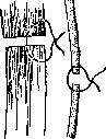
Figure 1. a-correct suture placement; b-incorrect suture placement.
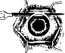
ous abscess of the vitreous body or panophthalmitis with all their fatal consequences for the eye. In other cases, traumatic iridocyclitis develops, running less severely but carrying the danger of sympathetic inflammation (see Ophthalmia) of the other eye. But even in cases of aseptic course, the consequences of penetrating injuries to the sclera are extremely serious; here not only the immediate changes caused by the trauma, such as injury and displacement of the lens, prolapse of parts of the internal membranes, but also remote consequences play a role, most often in the form of late-developing secondary detachment of the retina depending on the tension originating from the scar. Therapy for penetrating injuries to the sclera aims to prevent secondary infection and ensure rapid and firm healing of the wound. For this, the prolapsed parts of the choroid and vitreous body are cut off with scissors and sutures are applied; in small injuries, it is sufficient to include only the conjunctiva and episclera in the suture; in more extensive wounds, suturing of the sclera itself is recommended, passing the sutures not through its entire thickness (Fig. 1). In subconjunctival rupture of the lens, surgical intervention is not advisable. With the removal of the lens that has prolapsed under the conjunctiva, it is recommended to wait some time to allow the scleral rupture to heal. Operations on the sclera. An operative puncture or incision of the sclera is used in many surgical interventions on the eyeball, in particular the most common eye operations, such as cataract extraction and iridectomy, begin with an incision of the sclera at the edge of the cornea. Sclerotomy specifically refers to the operative procedure that does not limit itself to incising one sclera but also opens the internal membranes of the eye. Anterior and posterior sclerotomy are distinguished. - Technique of anterior sclerotomy. The incision is made with a narrow Graefe knife. The site of entry lies 1½-2 mm behind the cornea, 3 mm above the horizontal meridian. The knife is passed through the anterior chamber, and on the opposite side of the chamber at the same height and at the same distance from the cornea, an exit puncture is made. After the exit puncture, the field is first slowly advanced forward, and then with saw-like movements the incision is continued along the limbus, as in cataract extraction (Fig. 2). But this incision is not carried to the end, but, preserving a bridge of 3-4 mm, the knife is extracted from the second wound so that its tip returns to the anterior chamber, then by appropriate tilting of the knife handle, its tip is guided along the preserved bridge of the sclera and it is incised from the chamber side, after which the knife is extracted from the chamber. The wounds on both sides of the cornea should be 6-8 mm long (Czermak). Before the operation, eserine is necessarily introduced into the eye to pull the iris away from the angle of the anterior chamber as much as possible and prevent prolapse of the iris. After the knife is extracted from the chamber, the iris is carefully replaced. Anterior sclerotomy was proposed by de Wecker (1877) to replace iridectomy in glaucoma on the assumption that it achieves the formation of a filtering scar that maintains the outflow of fluid from the anterior chamber. Patho-anatomical studies later showed that as after iridectomy, so after sclerotomy, a filtering scar does not form. Anterior sclerotomy at present has a relatively narrow application, mainly as a repeated operation in case of insufficient effect of iridectomy and in some cases of secondary glaucoma. A number of authors recommend anterior sclerotomy in hydrophthalmos. Posterior sclerotomy is a meridional incision of the sclera with a linear Graefe knife behind the ciliary body. If the operation is performed for glaucoma, the incision is made in the outer-lower section between the outer and lower rectus muscles. The patient is made to look upward and inward, the eye is fixed in this direction with forceps or a traction suture. The knife is inserted through the conjunctiva and sclera 1 cm into the vitreous body, with the tip of the knife directed toward the center of the eyeball so as not to injure the lens. To avoid injury to the ciliary body, the entry is made at a distance of at least 6 mm behind the edge of the cornea. The incision can be continued toward the equator for 5-6 mm. Before extracting the knife, it is rotated around its longitudinal axis, which promotes the outflow of some amount of vitreous body. In glaucoma, posterior sclerotomy gives only a temporary decrease in intraocular pressure, as the opening in the sclera quickly heals with a dense scar. But it has importance as a preliminary operation for a rapid decrease in excessively high intraocular pressure in order to create more favorable conditions for another surgical intervention. Meller performs posterior sclerotomy immediately before iridectomy in glaucoma with a very narrow anterior chamber, Elschnig recommends doing posterior sclerotomy 1-3 hours before cataract extraction from a glaucomatous eye, especially in glaucoma due to swelling cataract. A meridional incision of the sclera in the middle and posterior segments of the eye is also used in the removal of subretinal cysticercus, in the extraction of parasites and foreign bodies from the vitreous body, and in the surgical treatment of retinal detachment. In these cases, the site of the incision depends on the localization of the pathological process, the technique of the operation is usually complicated by the incision of the conjunctiva and temporary transection of the eye muscle.
Figure 2.
3. Frank-Kamenetsky. SCLERADENITIS (from Greek scleros-hard and aden-gland), sclerosis of the lymph glands. Occurs mainly in chronic inflammatory diseases, sometimes in the late stages of hyperplastic and tumor processes, e.g., in lymphogranulomatosis, lymphosarcomatosis. S., accompanied by a clearly noticeable decrease in the lymph glands, is observed in old age, in exhaustion. See also Lymphatic system.
Related articles
Mentioned in
Cite this page
“Sclera.” Soviet Medical Encyclopedia. English translation of Bolshaya Meditsinskaya Entsiklopediya, 1st ed. (Moscow, 1928–1936), ed. N. A. Semashko. https://sovietmedicalencyclopedia.pages.dev/article/sclera/