Diascopy (a954)
Historical document, translated for reference. It reflects medical knowledge of the 1920s–30s and is not medical advice.
Summary
This article from the 1928–1936 Soviet medical encyclopedia explains the technique of diascopy, or transillumination, used to examine the nasal sinuses and the interior of the eye. It details the method's principles, its limitations, and the historical development of instruments for this purpose.
Encyclopedia article (1928–1936)
DIASCOPY (from Greek diaphanes- transparent and scopeo- I look). Diascopy of the nose, the transillumination of the accessory cavities of the nasal cavity by means of a light electric lamp, first proposed by Hering (1889), establishes the permeability of healthy and diseased tissue for light rays, the idea being that cavities with healthy walls containing air transmit more light than cavities filled with pathological products. In view of this, convincing results can be obtained only by comparing the diseased side with the healthy one. To illuminate the antrum and the anterior ethmoid cells, a small lamp mounted on a spatula is introduced into the patient's mouth; if these cavities are healthy, illuminated red areas are visible in the area under both eyes (in a dark room); with the eyes open, both pupils appear colored the same way, and the subject feels the light with both eyes. If there is exudate, pus, tumor, etc., in the cavity on one side, the cheek area appears darker, and the subject feels the light only on the healthy side. The accuracy of the examination is influenced by the thickness of the skull bones, the size of the cavities, the asymmetry of their location, the intensity of the light, etc. The frontal sinuses are transilluminated with the same lamp fitted with an opaque cap, which is applied to the floor of the frontal cavity at the inner angle of the eye; a light spot is marked on the front surface of the forehead and corresponds to the contours of this cavity, so its size is sometimes determined. Here, even more than in relation to the antrum, the results of the examination depend on anatomical conditions, especially since in 10% of cases the frontal sinuses are completely absent or represent only small cells. In view of the impossibility of correctly interpreting the data obtained by diascopy in all cases, this method of examination can be regarded as an auxiliary one alongside other, more perfect methods.
L. Rabotnov. Diascopy of the eye, transillumination of the walls of the eyeball to determine the presence of an intraocular tumor, first began to be applied in ophthalmology at the end of the 19th century.
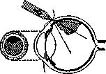
Figure 1. Anterior diascopy of the eye.
The essence of this method of examination is as follows. If a bright electric lamp enclosed in an opaque case in the form of a tube, with a strong spherical lens inserted into its opening, is applied to the sclera of the examined eye in a completely dark room, the pupil will be illuminated just as in an ophthalmoscopic examination. If a dense mass of an intraocular tumor is in the path of the rays, the light does not penetrate through it, and a shadow is obtained on a red background. When the tumor-like formation represents only a retinal detachment or consists of connective tissue formed as a result of inflammation of the eye coats, no shadowing occurs, and the pupil remains uniformly illuminated. The examination must be performed with a dilated pupil. Several types of lamps for transillumination have been proposed, but, as can be seen from the accompanying Fig. 1, they all possess one disadvantage: transillumination can be performed only through the anterior hemisphere of the eye and therefore one can see the shadow of a tumor located in front of the equator or only slightly behind it. Tumors of the posterior half cannot be determined with these lamps. To obtain a shadow from such a tumor, one must perform transillumination of the eye from behind. In 1909, Hertzell proposed a specially constructed lamp with a bright light source (about 100 candles), which was introduced through the mouth into the nasopharyngeal space and illuminated the eye from behind. But since the light had to pass through a whole series of tissues (bone, orbital fat), such transillumination did not always give satisfactory results. In 1910, Golovin proposed another, bloody, method of transilluminating the eyeball from behind. The Golovin apparatus is applied as follows: a cut is made in the conjunctiva near the limbus, and then it is dissected from the surface of the sclera and further to the optic nerve with curved scissors between two muscles (better in the upper outer part). After this, an electric lamp enclosed in a tube shaped like a curved catheter (Fig. 2) is introduced into the wound. The eye is illuminated near the posterior pole, and then, in the presence of a tumor, the shadow stands out sharply. Once the tumor is established, one can immediately proceed to enucleation of the eye. In case of a negative result, the conjunctival wound is sutured, and the patient remains under observation. It should be remembered, however, that positive results in diascopy are obtained only when the neoplasm reaches a certain size. A very small tumor may not produce shadowing. Abundant intraocular hemorrhage, however, can cause shadowing of the illuminated fundus and give the impression of a growing neoplasm. Therefore, in some cases only the combination of all the data of the examination makes it possible to make a correct diagnosis. Diascopy of the stomach, see Gastradiaphanoscopy.
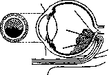
Figure 2. Posterior diascopy of the eye.
A. Merets.
Related articles
Cite this page
“Diascopy (a954).” Soviet Medical Encyclopedia. English translation of Bolshaya Meditsinskaya Entsiklopediya, 1st ed. (Moscow, 1928–1936), ed. N. A. Semashko. https://sovietmedicalencyclopedia.pages.dev/article/diascopy-2/