Nasopharyngeal Space
Historical document, translated for reference. It reflects medical knowledge of the 1920s–30s and is not medical advice.
Summary
The nasopharyngeal space, or nasopharynx, is the upper part of the pharynx located behind the nose. This anatomical section describes the varying dimensions and shapes of the nasopharynx in adults based on skull structure.
Encyclopedia article (1928–1936)
NASOPHARYNGEAL SPACE, nasopharynx, upper part of the pharynx, cavum retro-nasale or pharyngo-nasale, represents a cavity located behind the nose and directly transitioning into the middle part of the pharynx (development history—see PHARYNX). Anatomy. The size of the nasopharynx in adults varies considerably. In brachycephalic individuals, the transverse dimension is more pronounced, while in dolichocephalic individuals, the anteroposterior dimension is more pronounced. The shape and width of the nasopharynx also change depending on the greater or lesser protrusion of the main bone. Three main forms of the nasopharynx are distinguished. Most often, the nasopharynx has the form of a Gothic vault, with its vault gradually transitioning into the posterior wall at a very obtuse angle. The nasopharyngeal cavity is then located significantly higher than the soft palate. The second form (occurs in 1/3 of cases): the nasopharynx is of lesser height, and its upper wall transitions into the posterior wall in a rounded
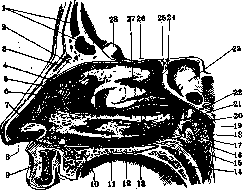
Right nasopharyngeal space with mucous membrane: 1-sinus frontalis; 2-spina frontalis; 3-os nasale; 4-carina nasi; 5-concha nasalis media; 6-atrium meatus medii; 7-limen nasi; 8-vestibulum nasi; 9-labium sup.; 10-palatum durum; 11-concha nasalis inf.; 12-meatus nasi inf.; 13-meatus nasi medius; 14-palatum molle; 15-plica salpingo-pharyngea; 16-plica salpingo-palatina; 17-elevation of m. levatoris; 18-labium post.; 19-ostium pharyngeum tubae auditivae; 20-labium ant.; 21-meatus naso-pharyngeus; 22-fornix pharyngis; 23-sinus sphenoidalis; 24-recessus spheno-ethmoidalis; 25-apertura sinus sphenoidalis; 26-concha nasalis sup.; 27-meatus nasi sup.; 28-crista galli.
at a right angle. The third form: the nasopharynx is very low, and its vault lies at the level of the hard palate. The first and second forms are found in adults, the third in children. The nasopharynx in front borders on the choanae of the nose, while above it is formed by the roof of the pharynx. On the upper-posterior wall is located the adenoid tonsil (see Adenoids). The lateral wall of the N. p. is bounded from that of the nose by the posterior nasal line (linea nasalis posterior). On the lateral wall is located the pharyngeal opening of the Eustachian tube, situated at the level of the posterior end of the inferior nasal concha (see illustration). This opening is bounded by two ridges - the anterior smaller and the posterior larger (see Eustachian tube). The posterior wall is located on the anterior surface of the bodies of Cj-n. The lower part of the nasopharynx in a state of rest is open toward the mouth. During swallowing and when pronouncing consonants, for example K, the soft palate is elevated, and the nasopharynx is separated from the middle part of the pharynx. The mucous membrane of the nasopharynx is covered with ciliated columnar epithelium, the area of distribution of which decreases with age, as it is increasingly pushed upward by the flat epithelium of the pharynx. (Topography, vessels, nerves-see Pharynx.) Physiology. The nasopharynx is part of the upper respiratory tract and a resonator for the voice. Details-see Pharynx. Examination of the nasopharynx. For examination of the nasopharynx, posterior rhinoscopy (see Pharynx), sounding, and digital examination are used. For the latter, the doctor stands to the left and behind the patient sitting on a chair. Small children are held on the arms by an assistant. The doctor places the index finger of the left hand on the patient's left cheek, pressing it between the teeth of the patient's open mouth, so that the patient cannot accidentally bite it. The index finger of the right hand is inserted into the mouth to the posterior wall of the pharynx, and, feeling the edge of the soft palate, is bent upward, thereby penetrating into the nasopharynx. In it, the choanae are first palpated, then along the posterior edge of the nasal septum the finger is directed upward to the vault of the pharynx, moves to the lateral wall, examining the opening of the Eustachian tube, and then along the posterior wall moves to the other side. The bleeding that sometimes occurs usually stops quickly (see Adenoids). Occasionally, examination of the nasopharynx with a salpingoscope, arranged on the type of a small-sized cystoscope, is used. The salpingoscope is inserted into the nasopharynx through the inferior nasal meatus. Pathology. Diseases of the nasopharynx usually extend into it from the nasal cavity itself and in turn affect the latter, and can also occur together (rhino-pharyngitis). More rarely does the disease extend from the middle and lower parts of the pharynx and pharyngeal tonsils. Inflammatory diseases of the nasopharynx are often a continuation of those of the nose (see Nose). Among acute diseases, acute inflammation of the nasopharyngeal tonsil should be noted. In adults, angina retronasalis begins with slight fever (in children, the temperature increase can reach 39.5°), complaints of general malaise, nasal obstruction, a feeling of dryness and burning in the posterior parts of the nose, moderate pain on swallowing, dry cough, sometimes tingling in the ears. On examination of the pharynx, slight redness of the posterior palatal arches and on the posterior wall of the pharynx a large amount of yellow mucus descending from the nasopharynx is noted. On posterior rhinoscopy, a swollen, highly reddened nasopharyngeal tonsil with purulent-like mucus in the folds is visible. In such cases in children, painful swelling of the cervical and occipital lymph glands is always noted, which, in the absence of sufficiently pronounced symptoms from the pharynx and nose, leads to an incorrect diagnosis- 'glandular fever'. Very often simultaneously or following angina retronasalis, angina of the pharyngeal tonsils or inflammation of the middle ear develops. In adults, a frequent consequence is a runny nose and extension of the inflammation to the mucous membrane of the pharynx, larynx, and bronchi. The disease usually passes without a trace within a few days. Sometimes, especially with enlarged adenoids and in children, the disease drags on for a longer time and can be accompanied by a number of complications from the ear and kidneys, as well as be a source of sepsis. Treatment: bed rest, a heating compress on the neck, rinsing with alkaline solutions, instillation into the nose of 2-3% protargol or 1% cocaine. With frequent recurrences-removal of adenoids, but not earlier than 2 weeks after the end of acute inflammatory phenomena. Chronic inflammation of the nasopharynx usually extends from the nasal cavity (chronic colds, diseases of the accessory nasal cavities, hypertrophy of the posterior ends of the inferior conchae, etc.) or from the side of chronically inflamed pharyngeal tonsils (chronic tonsillitis). In etiology, professional factors-inhalation of dust, smoke, irritating gases-play a large role. Catarrhal, hypertrophic, and atrophic forms of chronic inflammation of the nasopharynx are distinguished. In the hypertrophic form, redness and swelling of the mucous membrane and the discharge of mucopurulent secretion, often grayish-yellow in color, are observed. In the atrophic form, the mucous membrane is dry and covered with a large number of dried crusts. Symptoms are extremely varied depending on the form of the disease and the individual sensitivity of the patient. Patients usually complain of a feeling of pressure, scratching, tickling in the throat, discharge of mucus, as well as attacks of cough, which occur more often in the morning and sometimes reach vomiting. On examination, much yellowish-green mucus descending from the nasopharynx is visible. On posterior rhinoscopy, the same mucus or crusts are visible. Treatment consists in combating the main disease (nose, tonsils, exudative diathesis in children) and in systematic lubrication of the nasopharynx with solutions of silver nitrate in hypertrophic forms and Lugol's solution in atrophic ones. Among preventive measures, removal of the patient from the atmosphere of professional harmfulness, as well as prohibition of smoking and consumption of alcoholic beverages and general hygienic requirements in the care of the child (clean air, hardening) are important. Foreign bodies in the nasopharynx are quite rare and enter either from the nose or from the mouth during vomiting. They are rough, sharp (meat, fish bones, fragments of instruments, nails, needles, etc.) or smooth (pieces of meat, fruits, insects, leeches, etc.). The symptoms caused by foreign bodies-pain from injury to the mucous membrane of the nasopharynx and inflammatory processes in it, sometimes the formation of abscesses. Sharp foreign bodies can become embedded in the adenoid tissue of the nasopharynx. The diagnosis is made by examination of the nasopharynx (posterior rhinoscopy) and palpation with a finger. In complaints of swallowed and aspirated foreign bodies, examination of the nasopharynx should not be neglected, as the sensations of patients are often not accurate and from the side of the nasopharynx can be projected into the area of the pharynx and larynx. If the foreign body does not come out spontaneously, it is extracted with appropriately curved forceps (as for removing tumors) under the control of a mirror. Tumors of the nasopharynx. A whole series of tumors are encountered in the nasopharynx, some developing in it itself, some growing from the nose. Benign tumors of connective tissue type: nasopharyngeal choanal polyps, true fibromas on a stalk, myxomas and fibromyxomas, chondromas growing from the cartilage of the Eustachian tube and from the cartilaginous part of the primary skull, osteomas from fossa tonsillaris. Tumors of the vascular type are also encountered in the form of 1) telangiectasias, 2) cavernous angiomas, and 3) branching arterial and lymphatic angiomas. Lymphomas, fibromas, and sarcomas have also been observed. Malignant tumors of epithelial type-carcinomas and endotheliomas-are rarely encountered. Mixed tumors of the nasopharynx have also been observed: fibromyxolipomas, teratoid tumors of the nasopharynx and hairy polyps on a stalk, more often in small girls of female sex. Choanal polyps usually originate from the accessory sinuses (maxillary sinus, more rarely-others), more rarely their place of attachment is the edge of the choanae. Their removal can sometimes present great difficulties. If it is not possible to apply a loop passed through the nose, one can try to apply an appropriately curved loop through the nasopharynx. In some cases, forceps are used for removal, or one can try to remove the polyps with fingers wrapped in gauze to prevent slipping. Polyps that have formed on the basis of disease of the sinuses naturally require surgical opening of that sinus from which the polyps originate. The most common tumors in the nasopharynx are juvenile fibromas (fibroma juvenile, Basalfibroid of Germans), fibromas of the base of the skull, formerly called fibrous nasopharyngeal polyps. These tumors are usually encountered at the age of 10-25 years. Their histological structure represents a fibroma with a greater or lesser number of blood vessels (angiofibromas). From the point of view of their structure, nasopharyngeal fibromas are benign, but from the clinical point of view malignant, as they possess enormous energy of growth and recur after removal. By the age of 25, they usually undergo reverse development and resorb.
The symptoms of nasopharyngeal tumors in their early stages consist of difficulty in nasal breathing, runny nose, hearing impairment, and change in voice timbre (closed rhinolalia clausa posterior). With further growth, there may be protrusion of the soft and hard palate, swelling of the cheek, protrusion of the eye, cerebral symptoms, etc., depending on the direction of tumor growth. (For details of their origin, development, and symptomatology—see Pharynx.) The treatment of these tumors is mainly surgical and is associated with considerable difficulties, due to the impossibility of seeing the operation site, difficulty in approaching the tumor's origin, very significant bleeding, and the danger of suffocation from blood entering the respiratory tract. The operation is performed under general anesthesia in the Rose position with the head hanging over the edge of the table. The choice of operation method depends on the location of the tumor. If the latter mainly fills only the nasopharynx, removal through natural passages (mouth) is applied; with extensions into the nasal cavity, the route through the maxillary sinus (Denker's method, see Nose) is more often chosen. If it is impossible to approach the tumor in this way, various preliminary operations are used to make this approach more accessible. 1. Removal through the mouth. The soft palate is pulled back by a rubber tube passed through the nose. The tumor is separated from its attachment site with a finger, periosteal elevator, or scissors and quickly extracted with forceps (Jurasz, Denker, etc.). The very profuse bleeding that occurs stops quickly if the tumor is removed completely, by tamponade of the nasopharyngeal space (Hopmann's method). 2. Removal with preliminary operation. Route through the nose. Turning the nose downward (Ollier's method) is rarely used at present. More often, turning the nose to the side, or so-called rhinotomia superior (Mouret's method; 1902) is used. A skin incision is made on the side of the nose, starting from the inner edge of the eyebrow downward to the nasal wing. The soft parts are separated from the bone with a periosteal elevator, exposing the lower part of the frontal bone, nasal bone, nasal process of the maxillary bone up to the lacrimal bone. The nasal bone and nasal process of the maxilla are removed with a chisel and bone forceps, creating wide access to the upper part of the nasal cavity in the area of the ethmoid cells and deeper, and the tumor extensions here are easily removed. Sutures are applied to the wound. Route through the maxilla, or rhinotomia inferior (Denker's method), provides very good access to the lower part of the nose and nasopharynx through the maxillary cavity and leaves no external scars. The operation is performed under local anesthesia or general anesthesia. It is very convenient to use oral anesthesia by Kuhn or Seifert with insertion of a tube into the larynx and tamponade of the lower part of the pharynx. An incision of the mucous membrane to the bone in the vestibule of the mouth, as in the radical operation for maxillary sinusitis (see—Maxillary Sinusitis), but longer, starting from the wisdom tooth and extending 2-3 cm beyond the frenulum of the upper lip to the opposite side. The soft tissues are subperiosteally separated with a periosteal elevator and the facial surface of the maxilla, the circumference of the piriform aperture, the lower part of the nasal bone, and the nasal process of the maxilla up to the orbit are exposed. The inferior turbinate is separated from its attachment to the turbinal crest. Through the canine fossa, the maxillary cavity is widely opened, and if it is affected, all its mucous membrane is removed. Then the entire anterior wall of the maxillary cavity, the edge of the piriform aperture, and the inner wall of the cavity along with the mucous membrane are removed. Finally, the lower part of the nasal bone and the nasal process of the maxilla are removed. The profuse bleeding that occurs is stopped by tamponade. After bleeding stops, the nasal cavity, nasopharynx, and maxillary cavity are examined through the large opening obtained, and both the tumor itself and its extensions are removed. By removing the ethmoid cells with a conchotome, wide access to the main sinus can be obtained. If the mucous membrane is fused with the tumor, it is removed along with it. After removal of all pathological tissue, the wound is tamponaded, the end of the tampon is brought out through the nose, and the mucous membrane incision is sutured. On the third day, the tampons are removed, and from the 10th day, irrigation of the cavity is begun until discharge decreases. In addition to the removal of nasopharyngeal fibromas, the operations of Mouret and Denker are used for the removal of all malignant tumors of the nose and nasopharynx. For tumors located in the upper part of the nose, Mouret's method is more suitable, in the lower part—Denker's method. If the tumor extends even higher, for example toward the frontal sinus, Mouret's incision can be extended upward. If the tumor extends lower, the incision surrounding the nasal wing can be extended downward by cutting the lip in the middle and turning the cheek to the side. Thus, wide access to the tumor is created, which is then removed surgically or by surgical diathermy. The latter method (Holmgren) apparently is gaining more followers and gives better results than tumor removal with a knife. 3. Route through the mouth. Here, splitting of the soft palate in the middle (Nelaton's method) with removal of the posterior part of the hard palate and the posterior edge of the vomer is used. Still, the access obtained is not very wide. This method can be supplemented according to Krogius's proposal: to make a temporary saw cut in the middle of the lower jaw in addition to dividing the soft palate, which significantly widens access to the nasopharynx. In extreme cases, temporary resection of both maxillae according to Kocher or transverse pharyngotomy is used. Comparison of all the proposed methods for removing nasopharyngeal fibromas (there are over 55 of them) shows that external methods have not given brilliant results. In 50%, recurrence or death occurred, in 40% patients left with incompletely removed tumors. Mortality is significantly less when fibromas are removed through natural passages. Doctors' efforts should be directed toward earlier diagnosis of fibromas, as small tumors are easier to remove. At an age close to 25 years, it is better to use operations through natural passages, since the task of treatment consists in gaining time. By this age, fibromas lose their growth energy and undergo spontaneous regression. Operations with bone resection (Mouret's and Denker's methods) have to be used in cases of tumor growth into the accessory sinuses and toward the skull. Treatment. Previously used injections of various drugs into fibromas and galvanocauterization have been abandoned as giving no results. On the contrary, the use of surgical diathermy in the form of coagulation of the tumor by a series of insertions of diathermy needles has given excellent results—up to 100% recoveries. Access to the fibroma must be wide, which is achieved by using Mouret's incision so that cauterization can be performed ad oculos. The data from X-ray and radiotherapy are contradictory. Still, properly conducted X-ray treatment gives good results. Ref.: Kobyliiskii F., On the question of surgical treatment of polyps at the base of the skull, SPb, 1908; Laing G., Chirurgie de l'oreille, du nez, du pharynx et du larynx, P., 1924. See also literature for the article Otorhinolaryngology.
Related articles
Mentioned in
Cite this page
“Nasopharyngeal Space.” Soviet Medical Encyclopedia. English translation of Bolshaya Meditsinskaya Entsiklopediya, 1st ed. (Moscow, 1928–1936), ed. N. A. Semashko. https://sovietmedicalencyclopedia.pages.dev/article/nasopharyngeal-space/