Pharynx
Historical document, translated for reference. It reflects medical knowledge of the 1920s–30s and is not medical advice.
Summary
This article provides a detailed anatomical and embryological description of the pharynx as understood in the early 1930s. It covers the development of the pharyngeal structures, including the branchial arches and tonsillar development, and outlines the three anatomical divisions of the pharynx.
Encyclopedia article (1928–1936)
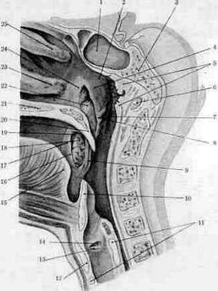
The pharynx [see separate plate (cols. 383–384), fig. 2] is a cylindrical tube, slightly compressed in the sagittal direction, 12–14 cm long, located in front of the six upper cervical vertebrae; its upper, expanded end, forming the fornix pharyngis, touches the base of the skull, while its lower, funnel-shaped narrowing end transitions into the esophagus at the level of the lower edge of the cricoid cartilage. The distance from the front teeth to the beginning of the esophagus is 15–18 cm. History of the development of the pharynx [see separate plate (cols. 395–396), figs. 1 and 2]. The pharynx develops from the cranial part of the foregut, which in a 3-week-old human embryo is still separated from the floor of the primary oral cavity by the so-called pharyngeal membrane. This membrane soon disappears, and the oral cavity (primary oral cavity) is now in broad communication with the pharyngeal gut, the so-called primary pharynx. Immediately in front of the pharyngeal membrane appears a depression of ectodermal origin, directed toward the base of the brain, the so-called Rathke's pouch, from the cerebral end of which the anterior lobe of the pituitary gland develops. In rare cases, the hypophyseal duct (canalis craniopharyngeus) remains in the body of the sphenoid bone, extending into its depth. Very often, however, remnants of this duct are observed in the mucous and submucous membranes of the pharynx [the so-called hypophysis pharyngea, or Rachendachhypophyse of the Germans (see Pituitary Gland)]. Behind the primary pharyngeal membrane lies an endodermal process, the Seessel's pouch, which is related to the bursa pharyngea. Initially, the foregut is open ventrally, but later closure occurs due to the appearance of four so-called branchial arches immediately in front of the primary oral cavity. Under each branchial arch is a so-called branchial cleft. The lower jaw develops from the first branchial arch, and the upper jaw and cheeks develop from its upward-extending process. The Eustachian tube, middle ear, and external auditory canal develop from the dorsal end of the first branchial cleft, while the ventral end undergoes regression. The arcus palatoglossus develops from the second branchial arch. The root of the tongue develops from the ventral part of the second and third branchial arches, and its anterior part, the body of the tongue, originates from an unpaired primordium between both processes of the lower jaw. The origin of the tongue from two different sources is closely connected with the development of the thyroid gland, the primordium of which grows downward toward the hyoid bone in the form of a duct (ductus thyreoglossus). The ductus thyreoglossus subsequently atrophies, and its remnant forms the foramen caecum, located at the border between the root and the body of the tongue, at the apex of the Y-shaped groove (sulcus limitans), with the apex directed posteriorly. As a result of the fusion of the medial nasal processes and both processes of the upper jaw, the primary palate is formed, separating the oral cavity from the nasal cavity. The space thus prepared (pharynx) serves both for breathing and for food, and the nasal cavities communicate with the upper part of the pharynx through the choanae. Then, behind the primary palate, two horizontal processes extend from the upper jaw, which fuse with each other and with the primary palate to form the hard palate, soft palate, and posterior palatine arches. The ventral part of the second branchial cleft disappears, and from the dorsal end remains the tonsillar sinus (sinus tonsillaris), which lies between the arcus palatoglossus and the arcus palatopharyngeus and is the site where the palatine tonsil develops. The epiglottis and thyroid cartilage develop from the fourth branchial arch, and the sinus piriformis develops from the third and fourth branchial clefts. The palatine tonsil begins to form at the beginning of the third month in the embryonic sinus tonsillaris. The epithelium covering it begins to send processes into the depth, which then split, become hollow, and form the later crypts. Subsequently, lymphoid infiltration and the formation of follicles occur. True follicles with germinal centers (reactive centers) develop only after birth. Of the original sinus tonsillaris, only the fossa supratonsillaris remains in the adult, located at the upper pole of the tonsil. However, the fossa supratonsillaris, which appears in the adult as a smooth-walled, extensive bay, is not often encountered. Its occurrence is caused by an arrest in the development of the highest part of the sinus tonsillaris, i.e., when adenoid tissue does not develop in its walls. Most often, the upper part of the sinus tonsillaris is filled with adenoid tissue, so that the tonsil continues far upward, sinking into the soft palate. This part of the tonsil is called the palatine lobule of the tonsil (pars palatina) and is permeated by channels just like the rest of the tonsil (see Figure 1). Among its channels, one is particularly noticeable, which runs anteriorly, upward, and outward (recessus palatinus superior), toward its outer corner, and often another similar one, running anteriorly and outward (recessus palatinus inferior). In addition, there is a third channel located behind the triangular fold—the anteroinferior pocket. To see the pars palatina and the channels opening there, it is necessary to pull the anterior arch outward and forward with a hook, and in suspicious cases, use probing. The depth of the channels ranges from a few millimeters to 1–1.5 cm. The development of the pharyngeal tonsil occurs through the formation of folds of the mucous membrane, which are filled with lymphoid tissue. The lingual tonsil develops due to the development of lymphoid tissue around the excretory ducts of the mucous glands of the root of the tongue. The pharyngeal lymphoid ring is still weakly developed at birth; it reaches its greatest development in early childhood, and after the period of puberty, it gradually undergoes regression. From the point of view of comparative anatomy, the pharyngeal cavity is a remnant of the original entrance to the digestive tract and acquires independent significance from the time when the palate appears, separating the oral cavity from the nose. In this form, it is found in the majority of Amniota. Among reptiles, only the crocodile has a secondary palate and a soft palate. The other reptiles possess only a primary palate, with the nasal cavity opening into the roof of the oral cavity through primary choanae. The uvula first appears in the giraffe and camel. Although lymphoid formations are found even in amphibians, the actual lymphoid organs (tonsilla palatina, tonsilla pharyngea, and tonsilla lingualis) appear only in Mammalia. The pharyngeal cavity itself is the site of the development of gills and branchial clefts. Initially, the entire gut can function as a respiratory apparatus. The greatest number of branchial clefts is found in Amphioxus. The embryonic gut in its original form is developed in those living in water and communicates with the environment through branchial clefts. In fish and amphibians in the larval stage, the branchial apparatus, constantly washed by water, functions as a respiratory organ. In reptiles, there are 5 branchial clefts; in birds and mammals, there are 4. Gradually, in animals, in connection with the development of respiratory organs, the regression of the branchial clefts occurs; therefore, although there are no gills in the embryos of higher vertebrates, the branchial clefts remain. Familiarity with the history of the development of the pharynx is important for clarifying the nature of anomalies and malformations encountered in this region. Anatomy of the pharynx. Divisions of the pharynx. The pharynx is divided into three parts: 1) epipharynx (pars nasalis pharyngis s. cavum pharyngonasale, nasopharyngeal cavity) - the upper part, located above the upper edge of the soft palate, at the place of its attachment to the hard palate in front; 2) mesopharynx (pars oralis pharyngis) - the middle part, extending downward to the upper edge of the epiglottis; and 3) hypopharynx (pars laryngea pharyngis) - down to the place of transition into the esophagus. The epipharynx represents a cavity, the size of which in an adult varies individually and on average equals the volume of a walnut. Its upper wall—the vault (fornix pharyngis)—is formed by the body of the sphenoid bone and the pars basilaris ossis occipitalis and transitions into the posterior wall at the level of the upper edge of the atlas. The anterior wall communicates with the nasal cavity through the choanae, separated in the middle by the posterior edge of the nasal septum. In the downward direction, the epipharynx communicates with the mesopharynx when the soft palate hangs freely toward the base of the tongue. However, it can be separated from the mesopharynx if the soft palate is raised and adheres to the posterior wall of the pharynx. This closure is supplemented by a roll-like protrusion of the posterior wall of the pharynx, caused by the contraction of the circular muscles of the pharynx, forming the so-called Passavant's ridge. In the vault of the nasopharynx, there is an accumulation of lymphadenoid tissue—the pharyngeal tonsil (tonsilla pharyngea). The pharyngeal tonsil often hypertrophies during the first six to seven years. After the period of puberty, it undergoes regression and in the adult takes on the character of diffuse lymphadenoid tissue. Its surface is split by five or six longitudinal grooves or clefts, penetrating deep into the lymphadenoid tissue. The middle groove, usually the deepest, runs in the sagittal direction; the lateral ones are arcuate.
The deepest furrow, the recessus pharyngis medius, is sometimes preserved even in an adult; it should be distinguished from the depression (sometimes up to 1/2 cm; Luschka), the so-called bursa pharyngea, located behind the pharyngeal tonsil. On the lateral wall of the epipharynx is the pharyngeal opening of the Eustachian tube (ostium pharyngeum tubae auditivae), located in an adult at the level of the inferior nasal concha. Anteriorly, the opening is delimited by a narrow fold of mucous membrane (plica salpingo-palatina), which merges below with the soft palate. Behind the opening, on the contrary, the so-called tubal torus (torus tubarius) protrudes sharply; its lower end continues into the lateral wall of the pharynx in the form of a noticeable fold, the plica salpingo-pharyngea. Behind the tubal torus and the plica salpingo-pharyngea, the so-called Rosenmüller's fossa (recessus pharyngeus) is formed. Here, the mucous membrane takes on the character of lymphadenoid tissue and, upon its hyperplasia, forms the so-called tonsilla tubaria. Below the tubal opening, a wide ridge is visible, running obliquely downward and medially toward the soft palate and formed by the m. levator veli palatini. The mesopharynx (see Figure 1) corresponds to the body of the II and III vertebrae and passes without any anatomical boundaries into the underlying section of the pharynx, the hypopharynx. The anterior wall of the mesopharynx is absent. The space bounded above by the lower edge of the soft palate and the uvula, below by the root of the tongue, and on the sides by the palatine arches and the tonsils located between them, is called the "fauces" (isthmus faucium) and connects the mesopharynx with the oral cavity. The lateral wall of the mesopharynx is occupied by the region of the palatine tonsil (tonsilla palatina), which is located between the anterior palatine arch (arcus palato-glossus), extending from the palatine velum near the base of the uvula downward to the edge of the root of the tongue, and the posterior palatine arch (arcus palato-pharyngeus), running from the free edge of the palatine velum vertically downward to the lateral wall of the pharynx. The m. palato-glossus is embedded in the anterior arch, and the m. palato-pharyngeus in the posterior one. From the lower end of the anterior palatine arch, a fold of mucous membrane extends, the so-called triangular fold (plica triangularis His'a), covering the lower end of the tonsil. Behind this fold, i.e., between it and the anterior surface of the tonsil, a deep pocket is often formed. The triangular space that is formed due to the divergence of the palatine arches, the so-called sinus tonsillaris, is in a normal state occupied by the palatine tonsil (tonsilla palatina), but in the upper corner, near the place where the anterior and posterior arches diverge, i.e., at the base of the uvula, a small opening remains, the so-called fossa supratonsillaris. The palatine tonsil [tonsilla palatina, see separate table (cols. 679-680), Fig. 4], located in the depression (sinus tonsillaris) between the palatine arches, represents an accumulation of lymphoid follicles embedded in lymphadenoid tissue. Its surface, facing the cavity of the pharynx, is covered with stratified squamous epithelium, lining the slit-like [spaces] of the pharynx during examination per os:
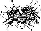
Figure 1. Mesopharynx. View 1 - fossa supratonsillaris; 2 - posterior palatine arch; 3 - probe inserted into the anteroinferior pocket; 4 - triangular fold (plica triangularis His'a); 5 - probe in recessus palatinus inf.; 6 - probe in recessus palatinus sup.; 7 - tongue; 8 - pars palatina; 9 - arcus palato-pharyngeus; 10 - arcus palato-glossus. Depressions - lacunae, or crypts, opening in the number of 12-15 on its outer surface and penetrating the tissue of the tonsil in the form of channels. Follicles approach the epithelial covering closely. The close connection between the epithelial covering and the follicles is a structural feature of the tonsils and brings them closer to the so-called lympho- (resp. lympho-ido-) epithelial organs (thymus, appendix). The outer surface of the tonsil is covered by a dense, whitish connective tissue membrane about 1 mm thick, the so-called tonsillar capsule, from which a whole series of dense connective tissue cords and plates depart, which serve as a kind of supporting framework for the tonsil. An accumulation of lymphadenoid tissue at the root of the tongue, which extends from the sulcus limitans to the epiglottis, is called the lingual tonsil (tonsilla lingualis). Together with the palatine, tubal tonsils, and pharyngeal tonsil, it participates in the formation of the so-called lympho-pharyngeal ring of Waldeyer. The latter also includes adenoid formations of an inconstant nature: the so-called lateral bands, located in the pharynx behind the posterior palatine arches, inclusions of adenoid tissue (granula) of the posterior wall of the pharynx, and in the Morgagni ventricles (tonsilla laryngea). The hypopharynx, beginning conventionally from the tip of the epiglottis and extending to the entrance to the esophagus, is located in front of the IV-V-VI cervical vertebrae and borders anteriorly with the posterior surface of the larynx. In the anterior wall there is an opening, oblong-oval in shape, connecting the pharynx with the larynx, the so-called entrance to the larynx - aditus laryngis. This opening is bounded anteriorly and superiorly by the epiglottis, and laterally by folds stretched between the epiglottis and the arytenoid cartilages, the so-called plicae ary-epiglotticae. On both sides of the entrance to the larynx, between the plica ary-epiglottica and the inner surface of the lamina of the thyroid cartilage, a depression is formed, the so-called sinus piriformis. Structure of the pharynx. The wall of the pharynx consists of a mucous membrane (tunica mucosa), a muscular layer (tunica muscularis), and a fibrous layer (tunica fibrosa). The mucous membrane in the epipharynx is covered with cylindrical ciliated epithelium, and in the mesopharynx and hypopharynx - with stratified squamous epithelium. In the subepithelial layer (lamina propria) there is a thin connective tissue plate (membrana pharyngis elastica Luschka, internal lateral aponeurosis of the pharynx of Jonnesco), which, thinning little by little downwards, continues into the cellular tissue of the esophagus, rises to the space lying between the arcus palato-glossus and arcus palato-pharyngeus, and on the sides attaches to the capsule of the tonsil. Since the superior constrictor of the pharynx does not reach the base of the skull and, consequently, the upper part of the pharynx is devoid of a muscular layer, the wall of the pharynx is formed by a thick aponeurotic plate, the so-called fascia pharyngo-basilaris (which is nothing other than a continuation of the membrana elasticae pharyngis), fused with the external aponeurosis of the pharynx. The mucous membrane of the vault of the epipharynx is closely fused with the base of the skull by dense fibrocartilaginous tissue, the so-called fibrocartilago basilaris. In the mucous membrane of the pharynx there are two kinds of glands: mucous and mixed. The former are located mainly in those parts of the pharynx which are covered with squamous epithelium. They are especially developed in the region of the Rosenmüller fossae, as well as in the plica salpingo-pharyngea and the soft palate. Glands of the mixed type are found in that part of the pharynx which has ciliated epithelium and is devoid of submucosa, as for example in the vault. The excretory ducts of these glands open into the depressions of the pharyngeal tonsil. In the posterior wall of the epipharynx, the submucosa is absent, and the glands, and partly their excretory ducts, are located in the muscular layer. In the mesopharynx, there are fewer mucous glands. The anterior wall of the hypopharynx is especially rich in mucous glands. - The muscular layer (tunica muscularis pharyngis), located outside the mucous membrane, consists mainly of three constrictors of the pharynx - mm. constrictores pharyngis (m. constrictor pharyngis superior, medius, and inferior). The fibers of these muscles take their origin from the medial plate of the processus pterygoideus, the hamulus, from the raphe pterygomandibulare, from the lower jaw, and furthermore originate from the transverse fibers of the root of the tongue and finally from the hyoid bone, the larynx, and the ligaments between the hyoid bone and the larynx. The fibers of these muscles, running in a transverse direction, cover the lateral walls of the pharynx like a sheath and meet each other on the midline of the posterior wall, forming the so-called raphe pharyngis. Since the upper part of the constrictor of the pharynx starts from the pterygoid process, it must rise posteriorly and superiorly to reach the base of the skull, as a result of which the upper end of the palatine tonsil is devoid of this muscle. Upon their contraction, the constrictors of the pharynx narrow the lumen of the pharynx. Elevators of the pharynx - m. stylo-pharyngeus and m. pharyngo-palatinus. M. stylo-pharyngeus begins from the styloid process and, descending downwards, attaches to the lateral wall of the pharynx at the upper edge of the superior constrictor. Upon contraction, this muscle widens the lumen of the pharynx and raises the pharynx and larynx. M. pharyngo-palatinus departs from the lower edge of the tubal cartilage, the medial plate of the pterygoid process, and the palatine aponeurosis and, passing in the lateral wall of the pharynx in the thickness of the arcus palato-pharyngeus, ends at the posterior edge of the thyroid cartilage. Upon contraction of this muscle, the posterior palatine arches approach each other, and the lower part of the pharynx and larynx are raised. The constrictors of the pharynx are covered on their outer surface by a dense fibrous fascia, the so-called external aponeurosis of the pharynx. The upper part of the tonsil is not covered from the outside by the superior constrictor of the pharynx, and therefore the internal aponeurosis in this section is in contact with the external aponeurosis of the pharynx. The soft palate, or palatine velum (velum palatinum), represents a musculomucosal fold extending from the posterior edge of the hard palate, in which a fibrous plate (aponeurosis palatina) and numerous muscle fibers intertwining with each other are embedded, which determine the significant mobility and elasticity of the palatine velum. The muscles of the soft palate depart from the base of the skull, from the hard palate, the tongue, and the lateral wall of the pharynx. They end partly in the raphe, partly in the palatine aponeurosis and the mucous membrane. These include 1) the unpaired muscle - m. uvulae, 2) m. levator veli palatini, and 3) m. tensor veli palatini. The muscle that elevates the soft palate (m. levator veli palatini) takes its origin from the base of the skull and, descending to the soft palate, forms a ridge along the lateral wall. The muscle that tenses the soft palate (m. tensor veli palatini) lies anteriorly and laterally to the m. levator. The tendon of the m. tensor bends around the hamulus pterygoideus at almost a right angle and enters the soft palate. The m. levator and m. tensor have a close relationship to the cartilaginous part of the Eustachian tube. Both muscles, especially the tensor, which attaches with several of its bundles to the membranous part of the tube, upon their contraction widen the almost slit-like lumen of the tube. M. glosso-palatinus is embedded in the arcus glosso-palatinus and upon contraction narrows the isthmus faucium. M. pharyngo-palatinus is embedded in the arcus pharyngo-palatinus and upon contraction brings the posterior palatine arches together and raises the lower part of the pharynx and larynx. Topography of the pharynx (see separate table, fig. 5). Between the lateral wall of the pharynx, covered by the external lateral aponeurosis on the inside, the inner surface of the ascending ramus of the lower jaw, covered by the internal pterygoid muscle on the outside, and the spine posteriorly, there is a wide pharyngomaxillary space, filled with adipose tissue and containing muscles, vessels, and nerves. Between the posterior wall of the pharynx and the deep cervical fascia (the so-called fascia praevertebralis), covering the deep cervical muscles (m. longus colli, m. longus capitis, and m. rectus capitis), there is a slit-like space filled with loose connective tissue, the so-called retropharyngeal space, spatium retropharyngeum, which continues downwards from the base of the skull into the cellular tissue of the posterior mediastinum. A thin connective tissue septum, stretched between the posterolateral wall of the pharynx and the prevertebral aponeurosis, separates the retropharyngeal space from the lateral pharyngomaxillary space. The cellular tissue of the retropharyngeal space serves as a site for the spread of so-called cold abscesses, having as their source osteomyelitis of the vertebrae, and the lymph glands located in the upper part of this space, in newborns and small children, upon infection from the nasal cavity, usually serve as the source of so-called retropharyngeal abscesses (see Retropharyngeal space, retropharyngeal abscess). The pharyngomaxillary space begins under the base of the skull, at the level of the atlas, and, gradually narrowing downwards, ends at the level of the II cervical vertebra.
The inner wall of this space is formed at the level of the II cervical vertebra by the m. constrictor pharyngis superior; the outer wall is composed in front by the m. pterygoideus internus and behind by the parotid salivary gland, covered by the fascia paroteo-masseterica; the posterior wall is formed by the prevertebral aponeurosis. In view of the fact that the inner and outer walls of this space converge anteriorly almost to the point of complete contact, the pharyngomaxillary space has the appearance of a triangle in cross-section. This space is divided by a connective tissue plate, stretched in the frontal plane between the posterolateral angle of the Pharynx and the group of muscles originating from the processus styloideus (m. stylo-glossus and m. stylo-pharyngeus), into two sections: anterior and posterior. The anterior one, having a triangular shape in cross-section, borders medially on the region of the palatine tonsil, while the posterior one contains vessels and nerves. Laterally lies the v. jugularis interna, medial to it is the a. carotis interna and the nerves: n. glosso-pharyngeus, n. vagus, n. accessorius, n. hypoglossus, and n. sympathicus. Blood vessels of the pharynx. In a practical sense, the relationship of the blood vessels to the tonsil region is of particular interest (see separate table, fig. 4). The a. carotis interna is not infrequently located closer to the lateral wall of the pharynx than is commonly thought, and is subject to numerous displacements in different subjects and especially at different ages. Deviation of the head backward and forward has little effect on its position. Conversely, turning the head and simultaneously inclining it in the direction of the turn significantly changes the distance of the vessel from the Pharynx. The a. carotis interna approaches the Pharynx on the side from which the head is turned away, and the distance increases on the side toward which the head is turned. In general, it can be said that the higher we examine the internal carotid artery, the more it approaches the lateral wall of the Pharynx, and the lower it is, the more it moves posteriorly toward the spine. Since the artery can form several bends, the lower bend, which is the most constant, can approach with its convexity the outer surface of the palatine tonsil. On average, the distance of the a. carotis internae from the tonsil capsule is 1.3–2 cm. In the anterior section of the pharyngomaxillary space, only branches of the a. carotis externae are located: 1) A. lingualis; in the case of origin by a common trunk from the a. maxillaris externa, when the arch of the latter is sharply pronounced, the lingual artery departs quite high, either from the apex of the arch or from its ascending part. Then the arch of the lingual artery is directed upward and inward and approaches the outer surface of the tonsil with its convexity. 2) A. maxillaris externa sometimes departs very high from the external carotid artery and with its descending part almost touches the lower part of the palatine tonsil. In other cases, it forms a sharp arch, directed with its convexity upward and approaching the tonsil. 3) A. palatina ascendens originates from the a. maxillaris externa, sometimes independently from the a. carotis externa. Having passed into the space between the m. stylo-glossus and m. stylo-pharyngeus together with the glossopharyngeal nerve, the artery enters the anterior section of the pharyngomaxillary space and rises upward, approaching the posterior circumference of the palatine tonsil and finally enters the soft palate. 4) A. pharyngea ascendens—the main artery supplying blood to the upper and middle sections of the Pharynx—has no relation to the tonsil region. 5) A. tonsillaris (usually one, rarely two or three), intended for the nutrition of the palatine tonsil, departs either as an independent branch from the a. palatina ascendens or as an accessory branch for the tonsil from the a. maxillaris externa, a. lingualis, rarely from the a. pharyngea ascendens. In general, the tonsillar arteries are larger when they depart from the external maxillary artery. Sometimes they are closely connected with the tonsil capsule, often they break up into 2, 3, or 4 small twigs when passing through the superior pharyngeal constrictor, and only very thin twigs enter the tonsil parenchyma. The main arteries supplying blood to the upper and middle sections of the Pharynx are the a. pharyngea ascendens and partly the a. palatina ascendens (for the tonsil region), and for the lower section of the Pharynx—the rami pharyngei a. thyreoid. super. In the blood supply of the pharyngeal vault and pharyngeal tonsil, the terminal branches of the a. maxillaris internae also participate. Veins of the Pharynx form a large venous plexus on the outer surface of the constrictors—the plexus pharyngeus, which richly anastomoses with the venous plexus in the submucosa. Receiving the venae canalis pterygoideae, the veins of the Eustachian tube and the soft palate, the pharyngeal plexus has efferent veins—vv. pharyngeae, pouring blood into the v. jugularis interna, as well as into the vv. lingualis, thyreoideae, facialis communis, or facialis posterior. Lymph vessels of the Pharynx form a dense network in the mucous membrane, especially in the region of the lymphatic pharyngeal ring and the posterior wall of the larynx, at the place of transition of the Pharynx into the esophagus. The posterior wall of the pharynx is the main collection
z
ii
M
M
P
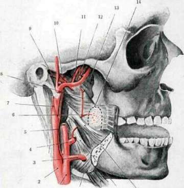
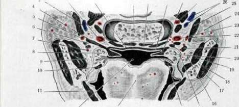
Fig. 4. Relations of blood vessels to the lateral wall of the pharynx: 1 - art. carotis ext.; 3 - art. carotis int.; 4 - art. lingualis; 5 - art. pharyngea ascendens; 6 - art. tonsillaris; 7 - art. maxillaris externa; 9 - art. palatina ascendens; 10 - art. auricularis posterior; 11 - art. tympanica int.; 12 - art. meningea media; 13 - ramus meningeus accessorius; 14 - m. levator veli palatini; 15 - projection of the tonsil; 16 - m. stylo-glossus; 17 - m. stylo-pharyngeus; 18 - m. biventer; 19 - m. hyo-glossus. (According to Spalteholz.) Fig. 5. Cross-section through the pharyngomaxillary space of a 35-year-old man at the level of the II cervical vertebra: 1 - outer wall of the pharynx; 2 and 3 - art. carotis int.; 4 and 5 - v. jugularis int.; 6 - m. stylo-pharyngeus; 7 - art. carotis ext. and processus styloideus; 8 - art. palatina ascendens; 9 and 10 - tonsil capsule; 11 and 12 - pharyngeal process of gl. parotis; 13 and 14 - m. tensor-palatini; 15 - m. buccinator; 16 - m. pterygo-pharyngeus; 17 - tongue; 18 - capsule of the palatine tonsil; 19 - m. pterygoid. int.; 20 - m. stylo-glossus; 21 - gl. parotis; 22 - m. biventer; 23 - art. vertebralis; 24 - m. pterygo-palatinus; 25 - m. constrictor pharyngis superior; 26 - deep cervical lymph nodes; 27 - II cervical vertebra. (According to the author's own preparation.) Pharynx. 385 point, where numerous lymph vessels are directed, collecting lymph from the vault, posterior and lateral wall of the epipharynx, lying behind the Eustachian tube (see figure 6). Lymph vessels penetrate through the posterior wall in the middle or near the midline into
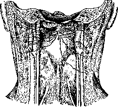
Figure 6. Lymphatic vessels penetrating through the posterior wall of the epipharynx into the retropharyngeal space are directed outward, bypassing the m. rectus capitis anteriorly and posteriorly, to the gl. retropharyngeales mediales, but the greater part of them crosses the gl. retropharyngeales laterales and behind the large vessels and nerves flows into the group of deep cervical lymph nodes. (According to Most.)
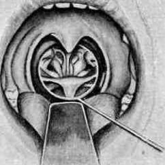
retropharyngeal space, turn sharply outward and, passing behind the v. jugularis interna and a. carotis interna, are directed toward the deep cervical lymph nodes. In doing so, they are interrupted by the gl. retropharyngeales mediales, if they exist, but the greater part of them crosses the gl. retropharyngeales laterales. An insignificant portion of the vessels, bypassing the retropharyngeal nodes, drains directly from the posterior wall of the pharynx into the deep cervical nodes. This circumstance has the practical significance that in infectious processes in the pharynx, the retropharyngeal nodes are comparatively rarely involved, and the infection is for the most part transmitted directly to the deep cervical nodes. The lymphatic vessels of the palatine tonsil region, collecting lymph from the tonsils, palatine arches, and the mucosa of the root of the tongue, penetrate through the capsule and the superior constrictor of the pharynx into the pharyngomaxillary space and are directed from here through the gap between the posterior belly of the m. biventer and the hypoglossal nerve, always in front of the neurovascular bundle, to the upper group of deep cervical nodes. Some of these vessels, bypassing the mentioned nodes, are directed straight to the lower-lying nodes. In case of swelling of these nodes, they can be palpated from the outside on the neck, at the edge of the m. sternocleidomastoideus. Sometimes one of the communicating vessels is directed to a node lying at the posterior edge of the thyroid gland. Furthermore, the efferent vessels drain into the submandibular lymph nodes, located at the outer surface and at the anterior edge of the submandibular salivary gland. The efferent vessels from the lower section of the pharynx are directed anteriorly into the sinus piriformis and, having passed through the membrana hyothyreoidea, drain into the upper anterior group of deep cervical nodes. The innervation of the pharynx occurs mainly at the expense of the branches of the nn. glossopharyngei, vagi, accessorii, and sympathici, which form on the outer surface of the middle constrictor of the pharynx the so-called pharyngeal plexus (plexus pharyngeus), which supplies the pharynx with both sensory and motor fibers. The motor nerves supply the three constrictors of the pharynx, the m. palatopharyngeus, and the m. salpingopharyngeus. The m. stylopharyngeus is supplied directly from the n. glossopharyngeus. The upper section of the pharynx is for the most part supplied with motor nerves from the IX pair [of cranial nerves]. The sensory nerves for the epipharynx originate from the II branch of the n. trigeminus, for the mesopharynx from the n. glossopharyngeus, and for the hypopharynx from the n. vagus (n. laryngeus superior). The m. tensor veli palatini is innervated by the third branch of the n. trigeminus (motor branches through the ganglion oticum). Physiology of the pharynx. Due to its anatomical position—at the crossroads of the respiratory and digestive tracts—the pharynx performs respiratory and swallowing functions. During normal nasal breathing, the palatine velum is freely lowered downward and touches the root of the tongue, and the oral cavity is isolated from the pharynx. The stream of inhaled and exhaled air passes along the entire length of the pharynx and here, just as in the nose, is heated, moistened, and cleaned of dust and bacteria. If there is an obstruction to the passage of air in the nose, then breathing occurs through the mouth, whereby the tongue flattens, and the soft palate, conversely, rises upward. Since the oral cavity, at least when the mouth is widely opened, has larger dimensions in cross-section compared to the nasal cavities, it would seem that the passing air should encounter less resistance here, and oral breathing should be less tiring. However, oral breathing cannot replace nasal breathing. Short-term oral breathing does not have a noticeable effect, but with prolonged mouth breathing, a sensation of dryness and cold appears in the throat, because the inhaled air is not saturated with water vapor and does not have time to warm up, as is the case with normal nasal breathing. Furthermore, oral breathing causes a number of disorders both in neighboring regions and in the entire organism (see Respiration). Normal patency for air through the epipharynx is of great importance for the function of the Eustachian tube and for the ventilation of the middle ear. Snoring during sleep occurs both during nasal breathing and during oral breathing due to the vibrations of the palatine velum by the passing stream of air. During the act of swallowing, there occurs a separation of those sections of the pharynx which serve for the function of respiration. In this process, the entrance to the larynx is closed by the upward pulling of the larynx and the lowering of the root of the tongue, which presses the epiglottis against the entrance to the larynx, and the epipharynx is separated from the mesopharynx by the upward raising of the soft palate, supplemented by the so-called Passavant's ridge. (For the function of the pharynx during singing and speech, see Voice, Vocalization.) Finally, the pharynx performs a gustatory function (taste nerve apparatuses are located on the root of the tongue, soft palate) and a protective one (spasm of the pharyngeal muscles upon the entry of foreign bodies). The view that has prevailed until now regarding the tonsil as a regional lymph node for the mucosa of the nose and mouth, located only superficially, i.e., directly under the mucosa of the pharynx, has been abandoned at the present time. The accumulation of lymphadenoid tissue, united by the general name of Waldeyer's lymphatic pharyngeal ring and especially developed in the palatine tonsils, as well as in the pharyngeal and lingual [tonsils], has nothing in common from a physiological point of view with lymph nodes and belongs, along with Peyer's patches and solitary follicles, to the so-called peripheral follicular apparatus. Their main difference lies in the absence of afferent lymphatic vessels in the tonsils, whereas lymph nodes are inserted along the course of lymphatic vessels and serve as, so to speak, filters for the lymph passing through them. In the tonsils, there are no sinuses of the cortical layer characteristic of lymph nodes, but there are crypts, which are not present in lymph nodes. The network of lymphatic capillaries in the tonsils represents a closed system, ending blindly in the direction of the crypts. The outflow of lymph is accomplished via efferent lymphatic vessels, i.e., centripetally, to the anterior-superior group of lgl. jugulares. There is no movement of lymph directed toward the surface of the tonsil, i.e., centrifugally. From an anatomical and physiological point of view, the palatine tonsils, as well as other lymphadenoid formations included in the composition of the pharyngeal lymphatic ring, possess the same function. The emigration of lymphocytes through the epithelial covering of the tonsil, as pointed out by Stöhr as early as 1882, cannot indicate a similarity to lymph nodes, because a similar phenomenon is observed not only in those places where there is an accumulation of lymphadenoid tissue, but even where it is absent. The palatine tonsils, located at the very beginning of the respiratory and digestive tracts, are in constant contact with the external environment, and mainly with the contents of the oral cavity, which is rich in bacterial flora. The tonsils, more than other sections of the pharynx, are apparently the entry gates for various infections [acute articular rheumatism, endocarditis, pyemia (cryptogenic), latent tuberculosis (scrofula) of the cervical lymph nodes, cerebrospinal meningitis]. Deep, often branching in the depths, crypts serve as hidden foci of local and general infectious diseases. On the other hand, it should be kept in mind that the tonsils, like other lymphadenoid tissue, easily react to various septic conditions arising from the most diverse sources. Examination of the pharynx. Inspection of the fauces and mesopharynx is performed under illumination with a head reflector, whereby if the patient is made to breathe calmly with the mouth widely opened, then, by gently pressing the root of the tongue downward with a spatula, it is possible to inspect the soft palate, the arches, the palatine tonsils, and the posterior wall of the pharynx. Upon pronouncing the sound "eh", the soft palate rises upward, and thereby the degree of its mobility is determined, and it also becomes possible to inspect the greater part of the posterior wall of the pharynx. In some cases, when it is impossible for the patient to relax the pharyngeal musculature, as well as during its reflex spasm, recourse is had to pulling the palatine velum anteriorly by means of a probe bent in the form of a hook or a special hook-shaped spatula. While the mesopharynx is accessible to direct inspection, the epipharynx, the root of the tongue with the lingual tonsil, and the hypopharynx can be inspected only with the help of a laryngoscopic mirror. For inspection of the epipharynx, recourse is had to posterior rhinoscopy (rhinoscopia posterior; see separate table, fig. 3), which is performed in the following manner: the root of the tongue is pressed downward with a spatula, a small, preheated mirror, directed with the mirror surface upward, is carefully introduced into the fauces between the tongue and the palatine velum, slightly to the side of the uvula, without touching them, as well as the posterior wall of the pharynx, in order to avoid gagging movements. The main landmark during inspection of the epipharynx is the posterior edge of the septum, on both sides of which one can see the choanae with the posterior ends of the inferior and middle nasal conchae. By turning the mirror slightly to one side and the other, it is possible to see the lateral walls with the openings of the Eustachian tubes, the tubal tori, and the Rosenmüller's fossae. By rotating the mirror around its horizontal axis, i.e., up and down, it is possible to inspect the vault and the posterior wall. In very sensitive people, posterior rhinoscopy can be performed only after preliminary swabbing of the mucosa of the fauces with a cocaine solution.
Examination can be supplemented by digital palpation and probing. The lower part of the pharynx and the root of the tongue with the lingual tonsil are examined with a laryngoscopic mirror, which is introduced into the pharynx while the tongue is protruded. The mirror, directed with its mirror surface downward, is advanced between the dorsum of the tongue and the palatal arch and is applied to the soft palate at the base of the uvula, which is gently pressed toward the posterior wall of the pharynx. When the sound 'a' is pronounced, the reflection of the root of the tongue with the lingual tonsil first appears in the mirror, then the epiglottis, the entrance to the larynx, and on its sides the piriform sinus. Pathology of the pharynx. Developmental anomalies of the pharynx are observed in the form of the absence of the soft palate or arches. Symmetrical defects in the anterior palatine arches also belong here. When both median frontal processes fail to fuse, median nasal clefts are formed, and when there is a failure of fusion between the median and lateral frontal process, harelip and cleft palate (see) occur. Therefore, these clefts always lie to the side of the midline. Clefts of the soft palate and uvula, on the contrary, are always located along the midline, because the lateral frontal process does not participate in their formation. A minor cleft of the soft palate and uvula in the form of a so-called double uvula is observed relatively frequently. Clefts in the soft palate are always located along the midline. If they are combined with a bilaterally located cleft of the hard palate, then a so-called cleft palate is obtained. The remnant of Rathke's pouch, which continues into the craniopharyngeal canal and has a close connection with the pituitary gland, belongs to the medial cleft. Diverticula develop either in the region of the Rosenmüller fossae, or the palatine tonsils, or the piriform sinus. Cysts, which are encountered in the same places as diverticula, but more often in the piriform sinus, can grow into large tumors located along the inner edge of the sternocleidomastoideus muscle, from the angle of the lower jaw to the sternum. Fistulas of the pharynx usually begin in the region of the palatine tonsil, corresponding to the tonsillar sinus, and open externally at the medial edge of the sternocleidomastoideus muscle, more often closer to the midline. Inflammatory diseases of the pharynx. The clinical picture and course of inflammatory diseases of the pharynx are quite varied depending on the localization of the process and the degree of involvement of the lymphadenoid tissue that makes up the so-called Waldeyer's pharyngeal ring. By angina, one understands acute and chronic inflammatory diseases in the region of the fauces—isthmus of the fauces (see Angina and Tonsillitis). Inflammatory diseases of the remaining parts of the pharynx are united under the general name pharyngitis and are divided into acute and chronic (see Pharyngitis). It is far from always possible, based on the etiological factor, to strictly distinguish non-infectious catarrhal inflammations from infectious ones, in view of the existence of pathogenic microorganisms also in a healthy pharynx. Adenoid growths of the pharynx—see Adenoids. Tuberculosis of the pharynx is almost always of secondary origin, i.e., it develops in tuberculous patients as a result of infection of the pharynx by their own sputum (autoinoculation) or hematogenously. Tuberculous lesions are usually observed in the stage of the ulcerative process, because the initial stage—the formation of subepithelial miliary tubercles—proceeds asymptomatically and escapes the attention of patients, and limited infiltrates (tuberculomas) are extremely rare. The most favorite site for tuberculous ulcers in the mesopharynx is the palatine arches, especially the posterior ones, and the posterior wall of the pharynx. The features of tuberculous ulcers are: 1) uneven undermined edges, 2) their tendency to spread along the surface of the mucosa, 3) pale pink color, 4) sharp pain. The course is chronic, excluding rare miliary forms, which proceed rapidly and for the most part end lethally. The prognosis is generally unfavorable, because usually pain during swallowing (dysphagia), causing insufficient nutrition, worsens the course of the pulmonary process. The differential diagnosis is conducted between syphilis and lupus (see below). The best results from treatment are obtained if, in addition to general measures directed against the pulmonary process, local treatment is applied. Cauterization with trichloroacetic acid proves to be the most effective. - Lupus (lupus vulgaris) is distinguished by a slower course, little pain, and sometimes a complete absence of pain, an insignificant influence on the general condition of patients, and a tendency toward spontaneous healing with the formation of a scar. Manifestations of syphilis in the pharynx are encountered in all three stages. The primary manifestation—a hard chancre—is most often localized on the palatine tonsils (rarely on the posterior wall of the pharynx) and appears as a flat ulcer, protruding above the surface, with a raised, dense edge and a sebaceous coating. The diagnosis is based on 1) the unilateral nature of the process, 2) the sensation of cartilaginous density of the ulcer, 3) the duration of the process (usually 1-2 months), and 4) scleradenitis of the submental and submandibular lymph nodes. Secondary manifestations of syphilis of the pharynx are encountered in two main forms. 1. In the form of erythema—redness of a copper-red color, involving the soft palate, arches, tonsils, and spreading to the larynx. Unlike catarrhal redness, erythema is sharply demarcated from the healthy mucosa, is distinguished by unusual persistence, and is accompanied by swelling of the lymph nodes. 2. Broad condylomas and papules (plaques muqueuses) have the appearance of grayish spots, more or less rising above the surface of the mucosa and surrounded by a red inflammatory border. The picture is almost the same as after painting with silver nitrate. Mucous papules are encountered anywhere in the oral cavity and pharynx, but most often are located in the isthmus of the fauces (angina syphilitica s. papulae syphiliticae). Most often, condylomas occupy the edge of the soft palate, the uvula, and the arches. Individual plaques can merge with each other and involve both tonsils. Exclusive localization of papules on the tonsils is not often observed. It is especially characteristic that the grayish spots spread from the surface of the tonsil to the arches, especially the anterior ones. Condylomas located on the posterior surface of the palatine velum give the edges of the uvula and soft palate a characteristic serrated appearance. Condylomas can exist for a long time, sometimes 1-2 months, without causing any subjective sensations. Sometimes patients complain of awkwardness during swallowing and an unpleasant sensation in the throat. With the passage of time, the condylomas undergo regression. The differential diagnosis is conducted between the following diseases: 1. For leucoplakia oris, the following are characteristic: an opalescent appearance of the plaques, the absence of a red inflammatory rim, and swelling of the lymph nodes. 2. Stomatitis aphthosa is distinguished by an acute onset, often accompanied by a febrile state, sharp pain, and little tendency to increase at the periphery. 3. In herpes, an acute eruption of vesicles occurs in groups and is accompanied by burning, sharp pains and a febrile state. 4. Pemphigus vulgaris is characterized by the appearance of superficial, slightly bleeding erosions, covered with a thin fibrinous coating, resulting from the bursting of vesicles. Erosions suddenly appear and just as quickly disappear in one place, while in others the appearance of fresh eruptions is observed. 5. Membranous-ulcerative angina (angina Vincenti)—see Angina. Syphilis in the tertiary period is encountered in the pharynx either in the form of a widespread infiltrate or in the form of individual nodules, i.e., gummas. Both are distinguished by their instability and tendency to rapid disintegration, as a result of which ulcers of greater or lesser size are formed. In the epipharynx, gummatous processes are not often encountered. Their localization is the posterior wall and the vault. Usually they are accompanied by sharp pains during swallowing, although cases without pain are also observed. They are diagnosed by means of posterior rhinoscopy, which is of especially great importance in those cases when, with the patient's complaints of sharp pain during swallowing, a simple examination of the pharynx does not reveal the cause that could explain these complaints. Posterior rhinoscopy reveals a large ulcer with a yellowish sebaceous base and undermined edges, located on the posterior wall of the epipharynx. Sometimes, even without posterior rhinoscopy, a sharp, pinkish-red edematous infiltration of the posterior wall of the mesopharynx, extending upward into the epipharynx, is noticeable. On the soft palate, the gummatous process usually disintegrates rapidly with the formation of a deep ulcer with sharply cut edges and entails extensive destruction, the victim of which is primarily the uvula, and sometimes the entire soft palate. The diagnosis of gummatous lesions is sometimes extremely difficult; one has to differentiate between tuberculosis, scleroma, and neoplasms. Unlike tuberculosis, gummatous infiltrates, even in the period of ulcerative disintegration, do not cause particular disturbances and are little painful. Syphilitic ulcers are distinguished by their sharply cut edges, redness, and edematous infiltration of the surrounding tissues. Scleroma (see Rhinoscleroma) most often begins in the nasal cavity, but the starting point can also be the nasopharynx.
Scleroma manifests as individual small nodules or a diffuse infiltrate, a characteristic feature of which is cartilaginous density, painlessness, an extremely chronic course of the process, and a tendency toward shrinkage through the development of connective tissue without the formation of suppuration. The nodules and infiltrates are located in the epipharynx in the vault, on the lateral walls, near the posterior edge of the vomer, and in the mesopharynx on the soft palate, palatine arches, and lateral walls. In the further course of scleroma, due to cicatricial shrinkage, the formation of bands, strictures, and adhesions of the most diverse nature occurs (narrowing of the choanae, narrowing of the nasopharynx, pulling upward of the soft palate, etc.). The clinical picture of an ulcerated cancerous neoplasm, especially in the initial stage of its development, may resemble a primary syphilitic lesion. A characteristic distinction is its peripheral border: a primary lesion gradually transitions into the surrounding tissues, whereas cancer is surrounded by a dense, raised edge. The floor of a syphilitic ulcer is smooth and shiny; in cancer, the floor is uneven, nodular, and bleeds easily. The distinction between cancer and a disintegrating gumma is the everted, nodular, dense edges of the ulceration in cancer, whereas in syphilis, the edges are sharply cut and the floor is crater-like and depressed. There are cases where only a trial of specific treatment and a biopsy can provide a final resolution of the question. Neoplasms of the pharynx are divided into benign and malignant. Tumors can be located in the epipharynx, filling its entire cavity, although their point of origin may also be other areas. These include mucous polyps of the nose and choanal fibro-mucous polyps. These are so-called false polyps. Independent tumors of the epipharynx include, besides rarely occurring papillomas and enchondromas, sarcoma and cancer. Among tumors of the epipharynx, a special position is occupied by the true nasopharyngeal fibroma, or polyp of the skull base, which, according to its histological structure, belongs to benign neoplasms, but according to its clinical course, must be classified as a tumor of a malignant nature. Its site of attachment is: 1) The vault of the nasopharynx (a fibrous layer of connective tissue, densely fused with both the bone and the mucous membrane—fibrocartilago basilaris). In such cases, the epipharynx is predominantly affected. 2) The anterior surface of the sphenoid sinus outward and anterior to its opening, or the edge of the choanae, or the base of the vomer. Its growth is directed anteriorly, upward, and posteriorly, i.e., it fills one half of the nose, the epipharynx, and grows into the maxillary sinus, the sphenoid sinus, and the orbit. 3) Fossa pterygopalatina, from where it grows into the epipharynx through the foramen sphenopalatinum. Its growth is directed predominantly toward the cheek and the nasal cavity. Pathogenesis. It occurs predominantly in men (in 87%), most often at the age of 10 to 25 years, and originates from remnants of the chorda dorsalis and perichondral embryonic connective tissue, from which the bony skull is formed from the membranous one in embryonic life. There is a theory according to which the pharyngeal hypophysis is considered the point of origin for these tumors. Pathological anatomy. The size of the tumor depends on the degree of its spread. If it produces outgrowths, its weight can reach 200 g and more. The tumor is covered with stratified columnar epithelium and consists of connective tissue fibers, cellular elements, and a large number of blood vessels. The microscopic picture corresponds to a fibroma, and often, due to edema, the tumor tissue acquires a myxomatous appearance; this includes the "myxomas of the pharynx" of old authors. Sometimes the tumor tissue is very rich in cells, and in such cases, the tumor approaches a sarcoma. Course. A polyp of the skull base possesses enormous growth energy. Having the epipharynx as its site of origin, it first of all fills this cavity, and then grows in the direction of least resistance: it pushes the soft and hard palate into the oral cavity, penetrates into the nasal cavity, expanding its walls and eroding or pushing the septum to the opposite side, and grows into the paranasal sinuses (maxillary sinus, ethmoid labyrinth, and sphenoid sinus). Through the foramen sphenopalatinum, it penetrates into the cheek region and into the temporal fossa. Finally, through the os sphenoidale, the tumor can penetrate into the cranial cavity. The clinical course can be divided into 3 periods: 1) the stage of chronic rhinitis, 2) functional disorders, 3) facial deformity. Symptoms. Depending on the spread of the tumor, a diverse picture of the disease is obtained. The presence of a tumor in the nasopharynx manifests as rhinitis and difficulty in nasal breathing; when the tumor grows into the nasal cavity, the latter becomes impassable, the consequence of which is dryness in the mouth and pharynx, loss of smell and taste, hearing impairment, and a change in the timbre of the voice (rhinolalia clausa post.). When it hangs into the pharynx, swallowing and breathing are impaired. Due to the tumor growing into the orbit, neuralgia of the supraorbital nerve appears, and the eyeball may be pushed outward. Often, due to the pressure of the tumor on the branches of the trigeminal nerve, neuralgic pains appear. Finally, general cerebral phenomena may occur: headaches, a depressed state, dizziness, slow pulse, and papilledema. A very important symptom is bleeding, which occurs for any reason and can weaken the patient. Differential diagnosis. Characteristic of a choanal polyp is its mobility, dense elastic consistency, and the possibility of determining, during examination with a finger, its place of attachment to the edge of the choanae, whereas a polyp of the skull base sits immovably with a broad base in the vault of the nasopharynx. Even with large dimensions, a choanal polyp has a tendency to hang deep into the pharynx, from under the posterior edge of the soft palate. Rare cases include the so-called "retropharyngeal tumors" of Busch, which grow in the retropharyngeal space, where they are mobile and sharply demarcated, such that it seems impossible to pass a finger between the tumor and the posterior wall, which is possible with a polyp of the skull base. Tumors of the nasal cavity, especially malignant ones, often bulge through the choanae into the nasopharyngeal cavity, and therefore, during examination with a finger, if there are no secondary adhesions of the tumor here, we will not find their attachment to the vault or walls of the epipharynx. In far-advanced cases and with large spread of the tumor, facial disfigurement is observed: bulging of the bony skeleton of the nose at the root of the nose. Among malignant tumors in the epipharynx, sarcoma is encountered most often, cancer less often. The differential diagnosis between a fibrosarcoma and a polyp of the skull base is often difficult to establish even under a microscope, and it is even more difficult to draw a sharp clinical boundary. Sarcoma spreads along the same paths as a polyp of the skull base and is distinguished by the rapidity of the neoplastic process. In addition, sarcoma is encountered more often in childhood—up to 6 years of age. Primary cancer of the epipharynx is a great rarity and, unlike a polyp of the skull base and sarcoma, does not fill the spaces open to it, but grows into the surrounding tissues and quickly produces metastases. Prognosis is grave, all the more so because the operation to remove the tumor yields a high percentage of mortality and does not guarantee against recurrence. The unceasing growth of the tumor always eventually entails a fatal outcome from general exhaustion, asphyxia, profuse bleeding, and infection of the meninges. Treatment can only be surgical. Surgical techniques are divided into operations aimed at opening wide access to the tumor and operations per vias naturales. For the removal of the tumor, in view of its hidden position and spread often far beyond the limits of the epipharynx, numerous methods have been proposed, which in most cases consist of two stages—a preliminary operation and the main operation, i.e., the actual removal of the tumor. All these methods, by lengthening the operation, which is usually accompanied by a large loss of blood, leave behind facial disfigurement and far from achieving the goal (29.1% failure—recurrence or incomplete removal). If one adds to this that the operation yields 10% mortality (17% according to the Russian statistics of Kobylinsky), then naturally, preference is deserved by those methods which are dictated, so to speak, by nature itself and are applied per vias naturales, i.e., reaching the tumor per os. With a broad base, the tumor is separated by a finger inserted into the nasopharynx and then removed with forceps (possible only with more or less soft tumors). Removal with a cold snare inserted through the nose is possible only in the case where there are no adhesions and the polyp is small. After the operation, posterior tamponade is necessary; in addition, both halves of the nasal cavity are tamponed through the external nasal openings.
In the mesopharynx, the following benign tumors are encountered: 1) fibrous polyps, sitting on a stalk on the tonsils or soft palate, 2) polyps attaching by a narrow stalk to the palatine tonsil; they usually have the structure of a tonsil (tonsilla pendula), 3) papillomas, having the appearance of small, rarely large, warty tumors; they are located most often on the uvula and on the soft palate, 4) angioma cavernosum in the form of a dark blue, uneven, and soft tumor, 5) mixed tumors, corresponding in their structure to mixed tumors of the parotid gland (see). -Malignant neoplasms of the mesopharynx and hypopharynx are not a rare phenomenon. Cancer begins either in the palatine tonsil, the lingual tonsil, or at the entrance to the esophagus. At first, cancer causes almost no disturbances, as a result of which one usually has to observe patients when the tumor has already managed to spread to the palatine arches, the root of the tongue, or to the sinus piriformis. -Symptoms. One of the early symptoms is swelling of the corresponding lymphatic glands. Another feature of cancer of the mesopharynx is a tendency toward rapid disintegration, as a result of which ulceration of the tumor forms, accompanied by a putrid odor. In the further course, severe pains appear, radiating to the ear, along with difficulty in speech and swallowing. The diagnosis of cancer in the initial stage is difficult without microscopic examination. Upon biopsy, one most often finds squamous cell carcinoma, rarely glandular cancer. Differential diagnosis is conducted between a primary lesion (ulcus durum) and a disintegrating gumma (see above). Sarcoma always begins in the palatine tonsil and differs from cancer by a low tendency toward disintegration and the comparatively late appearance of metastases, which sometimes do not occur at all. Upon examination, it has the appearance of a large, smooth tumor of soft consistency. Histological examination reveals all types of sarcomas, of which round-cell sarcoma is the most malignant. Endothelioma is encountered rarely and develops on the soft palate, on the lateral wall of the Pharynx, sometimes on the posterior wall; it is distinguished by slow growth, rarely produces metastases, and does not grow into the surrounding tissues. In view of the fact that treatment with X-rays and radium is accompanied by striking success and often leads to the complete disappearance of the tumor, endothelioma can be considered a comparatively benign disease. -Regarding congenital polyps of the pharynx, see Epignathus.
Related articles
Cite this page
“Pharynx.” Soviet Medical Encyclopedia. English translation of Bolshaya Meditsinskaya Entsiklopediya, 1st ed. (Moscow, 1928–1936), ed. N. A. Semashko. https://sovietmedicalencyclopedia.pages.dev/article/pharynx/