Sphenoid Bone
Historical document, translated for reference. It reflects medical knowledge of the 1920s–30s and is not medical advice.
Summary
This article from the 1928–1936 Soviet Great Medical Encyclopedia provides a detailed anatomical description of the sphenoid bone, detailing its structure, developmental history, and specific features such as the sella turcica and the sphenoid sinus.
Encyclopedia article (1928–1936)
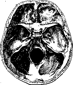
SPHENOID BONE (os sphenoidale, os cuneiforme, alatum, multiforme, pterygoideum, os carinae, os colatorii), or the wedge-shaped bone, in an adult human forms a single whole with the occipital bone (see) and is in this form itself called the "basic" bone — basilare, s. tribasiiare. Usually, however, the Sphenoid bone is understood to be only its wedge-shaped part, i.e., the bone transversely located in the base of the skull, connecting with all its bones — and with the upper jaw, nasal, zygomatic bones and with the vomer. It consists of a body and three pairs of processes. Corresponding to phylogeny, in the human sphenoid bone, the presphenoid is also distinguished, i.e., the anterior part of the body, basisphenoid — the posterior, alisphenoid — the great wings, orbitosphenoid — the small wings, and the pterygoid process, which joins later as a medial plate. The body of the Sphenoid bone (corpus) is of irregular shape; with some approximation it can be compared to the shape of a cube and therefore has six surfaces. The posterior surface connects with the body of the occipital bone by means of hyaline cartilage in youth and by a strong bony synostosis in maturity (ossification occurs in the age of 13–30 years). The upper surface, in its middle part, bears an indentation, variously expressed (fossa hypophyseos). By its appearance it received the name of the Turkish saddle (sella turcica) and contains the brain's appendage. At the bottom of the pit of the saddle, closer to its anterior part, there may be (in adults in 0.3%) an opening of the canal (canalis erano-pharyngeus), penetrating through the body of the sphenoid bone and opening on its lower surface (near the connection of the wings with the vomer) by a "pharyngeal opening". The canal contains a continuation of the dura mater in the form of a fibrous sheath enclosing connective tissue and blood vessels (veins). In front of the pit protrudes a barely noticeable tubercle (tuberculum sellae), on the sides of which are located more or less expressed processes (processus clinoidei medii). In front of these formations a transversely running groove is noticeable, continuing forward and to the sides into the optic foramina (sulcus opticus, foramina optica) and delimited by a relatively sharp edge (limbus sphenoidalis) from the area (jugum sphenoidale) located even more forward; the latter spreads to the sides and continues into the small wings, and forward it projects in the form of a flat, very differently shaped process (spina ethmoidalis), connecting with the spongy plate of the ethmoid bone. The pit of the Turkish saddle is bounded behind by a sharply projecting plate in the frontal plane (dorsum sellae), the posterior surface of which is slightly inclined and continues into the clivus Blumenbachii (see Occipital bone). The upper edge of the plate is uneven and ends on the sides with well-expressed processes (processus clinoidei posteriores). The lateral surfaces of the body of the Sphenoid bone are in most part occupied by the bases of the large and small wings departing here. Only the upper-lateral part of them is free, on which is located an S-shaped curved groove (sulcus caroticus), intended for the internal carotid artery, directing itself from behind and below, forward and upward. The relief of the groove is especially emphasized at the very beginning by a projection on the lateral side (at the base of the large wings) of a process in the form of a bone plate with a sharp edge (lingula sphenoidalis), slightly bent at its end to the medial side. There are no boundaries between the anterior and lower surfaces of the body of the Sphenoid bone: they pass into one another and face the nasal cavity. In the middle of both of them projects a crest (crista sphenoidalis), ending in front in the form of a sharp keel (rostrum sphenoidale). On the sides of the crest vertically located are rough areas (margo ethmoidalis), adjoining the labyrinth of the ethmoid bone. Between the crest and the roughnesses on the anterior and partly on the lower surface are located, "Phc.jI. Base of the skull from inside: connection of the bones of the skull. (According to Spalteholz.) - narrow, curved triangular plates (conchae sphenoidales, s. ossicula Bertini), in essence by embryogenesis belonging to the ethmoid bone and only later fusing with the sphenoid. Above and outside of them there are notches in the form of round openings (aperturae sinuum), leading into the sphenoid sinus (sinus sphenoidalis), located in the body of the Sphenoid bone. The sinus is quite voluminous, varies greatly in shape and size. Its position is determined by the limits of its spread: a strongly developed sinus reaches the for. opticum, surrounding it, as well as reaches the for. ovale and for. rotundum. In childhood the sinus is expressed insignificantly. By the 20th year it acquires definite size and shape, being in essence a double chamber, since a vertically standing partition (septum sinuum) divides it in the sagittal direction into non-communicating cavities. The partition is usually curved and deviated to one side or the other (more often to the right). Three types of sinus are distinguished (Shevchenko). The first — when pneumatization reaches in the anterior-posterior direction to the level of sellae turcicae, so that the hypophysis lies behind the sinus; the second — pneumatization reaches to the level of the posterior edge of sellae turcicae, i.e., two departments are obtained: portio sellaris and portio antesellaris; the third — pneumatization reaches to the level of synchondrosis spheno-occipitalis; then there is still a department of the sinus — portio retrosellaris. The sinus from inside is lined with a mucous membrane penetrating here from the nasal cavity and firmly attaching to the periosteum by a fibrous surface (membrana Schneideriana). Blood and lymph vessels of the mucosa are in connection with those of the posterior part of the nasal cavity. Innervation is due mainly to the p. ethmoidalis post. On the lower surface of the body of the Sphenoid bone, in the posterior part, there is a median groove, on the sides of which in the sagittal direction are located lateral grooves (sulci vomero-basilares) in the form of a gutter open to the median line and supplemented below by vaginal processes (processus vaginalis) of the pterygoid processes. The small wings of the Sphenoid bone (alae parvae) at their base are pierced by an opening (foramen opticum), through which passes the optic nerve. The upper surface of the wings faces the cranial cavity and participates in the formation of the anterior cranial fossa and the posterior part of the orbit; the anterior, serrated edge of them (margo frontalis) connects with the frontal bone, the posterior, smooth one projects freely into the cranial cavity and is the boundary between the anterior and middle cranial fossae (Fig. 1). On the medial part of the posterior edge there is a well-expressed projection (processus clinoideus), hanging over the anterior end of the sulcus carotici. Between this process and the processus clinoideus medius sometimes a bony bridge is thrown, as a result of which an opening (foramen carotico-clinoideum) is formed; this bony bridge can spread to the processus clinoideus post., and since anti-lingula is located opposite lingula, in such a case the artery turns out to be enclosed in a bony semicanal. The outer end of the wings is sharp and reaches the place of connection of the large wings with the frontal bone. The wing as a whole limits above the superior orbital fissure (fissura orbitalis - superior) (Fig. 2).
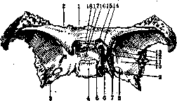
Figure 1. Sphenoid bone from above: 1-corpus; 2- ala parva; 3-ala magna; 4-dorsum sellae; 5-proc. clinoideus post.; 6- sulcus caroticus; 7-lingula sphenoidalis; 8-foramen ovale; 9- foramen spinosum; 10-facies cerebralis; 11-foramen rotundum; 12-proc. clinoideus ant.; 13- fissura orbitalis sup.; 14- proc. clinoideus med.; 15-foramen opticum; 16-sella turcica; 17-tuberculum sellae; 18-sulcus chiasmatis. (According to Spalteholz.)
The large wings (alae magnae) depart from the lower-lateral surfaces of the Sphenoid bone by a wide base, which is pierced by a series of openings (Fig. 3 and 4): in front is located the round opening (foramen rotundum), opening into the pterygopalatine fossa and intended for the second branch of the trigeminal nerve; closer to the posterior edge of the wing is located the oval opening (foramen ovale) for the third branch of nervi trigemini and not 71»
Related articles
Mentioned in
Cite this page
“Sphenoid Bone.” Soviet Medical Encyclopedia. English translation of Bolshaya Meditsinskaya Entsiklopediya, 1st ed. (Moscow, 1928–1936), ed. N. A. Semashko. https://sovietmedicalencyclopedia.pages.dev/article/sphenoid-bone-2/