Ophthalmic Artery
Historical document, translated for reference. It reflects medical knowledge of the 1920s–30s and is not medical advice.
Summary
This article details the anatomy of the ophthalmic artery, including its branches and their distribution within the orbit. It also discusses the clinical implications of trauma to the artery, such as hemorrhage, exophthalmos, and aneurysm formation, as well as the venous drainage of the orbit.
Encyclopedia article (1928–1936)
OPHTHALMIC ARTERY, VEINS. The ophthalmic artery represents a branch of the internal carotid artery and departs from the latter where it makes its last bend posterior to the optic foramen. Passing through this opening, the ophthalmic artery lies external to and below the optic nerve (see figure), then bends upward and lies above the nerve, passing between it and the superior rectus muscle of the eye; further, the ophthalmic artery goes under the superior oblique muscle and breaks up between the trochlea and the medial palpebral ligament into its terminal branches. On its path in the orbit, the ophthalmic artery gives off branches: 1) the lacrimal artery, supplying the lacrimal gland; 2) the central retinal artery, which often departs together with the posterior ciliary arteries and, piercing the sheaths of the optic nerve 7-12 mm posterior to the eyeball,
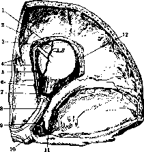
Arteries of the right orbit: 1-supraorbital artery; 2-anterior meningeal artery; 3-frontal artery; 4-anterior ethmoidal artery; 5-posterior ethmoidal artery; 6-posterior ciliary arteries; 7-muscular branch; 8-optic nerve; 9-ophthalmic artery; 10-internal carotid artery; 11-anterior clinoid process; 12-lacrimal artery.
goes along its axis into the retina; 3) short and long posterior ciliary arteries, in the amount of 4-6, breaking up into a series of branches piercing the sclera around the optic nerve and supplying the choroid of the eye; often these arteries begin from neighboring branches; 4) numerous muscular branches, beginning sometimes from neighboring branches and supplying the muscles of the eye; 5) anterior ciliary arteries, in the number of 7-8, which begin most often from the arteries of the rectus muscles of the eye in their anterior part and first in the thickness of the muscle tendons, then the sclera, reach the edge of the cornea (episcleral arteries) and conjunctiva (anterior conjunctival arteries), while partly they pierce the sclera and go into the ciliary body; 6) supraorbital artery, which goes together with the nerve of the same name between the periosteum of the upper wall of the orbit and the levator palpebrae superioris muscle to the supraorbital foramen and exits to the forehead, nourishing the skin and muscles; here it anastomoses with the superficial temporal artery (a branch of the external carotid artery); 7) posterior ethmoidal artery, which through the opening of the same name supplies the posterior sinuses of the ethmoid bone and the border region of the frontal sinus; 8) the larger anterior ethmoidal artery, which through the opening of the same name penetrates to the surface of the cribriform plate of the ethmoid bone, gives off here a branch (anterior meningeal artery) to the dura mater of the anterior cranial fossa and itself through the cribriform plate penetrates into the nasal cavity, branching in the anterior section of its wall. After this, the thinned ophthalmic artery breaks up, as stated above, into 3 terminal branches, of which 9) medial palpebral arteries go externally, one in each eyelid, and, anastomosing with the lateral palpebral arteries (from the lacrimal artery), form the superior and inferior tarsal arches, located in front of the tarsus of the eyelids; thin branches from the upper and lower arterial arch pierce the tarsus and branch in the conjunctiva, giving posterior conjunctival arteries; 10) dorsal nasal artery pierces above the medial palpebral ligament the orbicularis oculi muscle and branches in the skin and muscles of the circumference of the inner angle of the palpebral fissure, anastomosing with the terminal branches of the external maxillary artery (a branch of the external carotid artery); 11) frontal artery goes in the direction of the ophthalmic artery and, exiting together with the nerve of the same name through the frontal notch to the forehead, branches here in the skin and muscles, anastomosing with the superficial temporal artery and the supraorbital artery. Regarding pathological changes of the ophthalmic artery, first of all, one must point out the violation of its integrity by foreign bodies, bone fragments, and fractures in the optic nerve canal. Besides this, many authors recognize the possibility of violation of the integrity of the ophthalmic artery during blunt trauma to the orbit without violation of the integrity of its bones from concussion, from contrecoup. According to Rollet, the branches of the ophthalmic artery are shaken like the branches of a tree whose trunk is firmly compressed in the optic canal, and therefore it is understandable that some branches do not withstand and rupture, especially if one keeps in mind that many of them are connected to the eyeball or to the optic nerve by fibrous strands. The result of hemorrhage from the ophthalmic artery is exophthalmos and—depending on its strength—impairment of eye function up to complete blindness. Cords and others described under the name "pralle Durchblutung" a particularly sharply expressed form of hemorrhage into the orbit, apparently on the basis of damage to the ophthalmic artery, with large exophthalmos (so that the eyelids could not close), impairment of eye mobility, and hemorrhage under the conjunctiva in the form of a dark red ridge around the cornea. The prognosis for orbital hemorrhages is in the majority of cases not bad; they can be reabsorbed with the preservation of eye function. With very large hemorrhages and, mainly, threatening phenomena from the side of the optic nerve and the eye itself, removal of the blood is indicated either by aspiration or—in the case of the formation of cystic hematomas—through a corresponding incision. With trauma, partial and false aneurysms of the ophthalmic artery can also form. Such aneurysms have been found repeatedly during autopsies, and true aneurysms may not give the picture of pulsating exophthalmos during life, whereas false aneurysms give such a picture. False aneurysms can arise also on the basis of spontaneous rupture of true aneurysms. The appearance of a false aneurysm after a wound to the ophthalmic artery was observed during the last war. Aneurysms causing phenomena of pulsating exophthalmos are treated as such. Preceding degeneration (sclerosis) of the ophthalmic artery of course creates more favorable conditions for the violation of its integrity and the formation of aneurysms, causing at the same time a worse prognosis. Vasomotor disorders—vascular spasm—manifest clinically usually on the branches of the ophthalmic artery, namely on the central retinal artery. The same must be said about vascular occlusion—embolism of the central retinal artery. Ophthalmic veins. Blood flowing out of the orbit is collected into 2 large venous trunks; of them, one (superior ophthalmic vein) passes along the upper section of the orbit and corresponds to the branching of the ophthalmic artery, the second, of small caliber (inferior ophthalmic vein), stretches along the floor of the orbital cavity. Both vessels connect at the posterior end of the orbit into a wide trunk, leaving the orbit through the superior orbital fissure, to connect then with the cavernous sinus.
Related articles
Cite this page
“Ophthalmic Artery.” Soviet Medical Encyclopedia. English translation of Bolshaya Meditsinskaya Entsiklopediya, 1st ed. (Moscow, 1928–1936), ed. N. A. Semashko. https://sovietmedicalencyclopedia.pages.dev/article/ophthalmic-artery/