Coloboma
Historical document, translated for reference. It reflects medical knowledge of the 1920s–30s and is not medical advice.
Summary
Coloboma is a congenital or acquired defect in the eye's tissues, often affecting the eyelids, iris, ciliary body, choroid, retina, optic nerve, lens, Zinn's ligament, or vitreous body. It is typically inherited as a genetic trait and can be associated with other ocular anomalies and systemic defects.
Encyclopedia article (1928–1936)
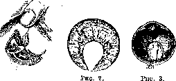
COLOBOMA, coloboma (from Greek kolo-bos - maimed), a defect in the eye's membranes, either acquired or congenital (genotypic). Acquired C. sometimes result from injuries, but most often occur after operations (C. of the iris). C. of the eyelids, iris, ciliary body, choroid, retina, optic nerve, lens, Zinn's ligament, and vitreous body have been described. C. of the eyelid (fig. 1) is a limited defect, mostly triangular, less often quadrangular in shape, with its base on the edge of the eyelid and apex at the orbital edge. The size of C. varies - from a small notch to a complete cleft of the entire eyelid. C. may be present on one or several eyelids; there may be several C. on a single eyelid. There is no typical location for C., but they are often found in the middle of the eyelid; their edges are straight or notched. Eyelashes and Meibomian glands are absent at the site of C. Sometimes a bridge of skin extends from the upper edge of the C., fusing with the cornea. Dermoids are often found at the edges of C., and on the eyes themselves - corneal opacities, C. of the iris, microphthalmia, and other defects. On the body - cleft lip, cleft palate, etc. - C. of the iris (fig. 2) is a defect in the iris in the form of an isosceles triangle with slightly curved edges and apex at the ciliary edge, involving from 1/6 to 1/3 of its entire volume. Rudimentary C. may also exist in the form of small notches on the edge of the pupil, in the form of thinning of the iris stroma, and in the form of stripes devoid of pigment or, conversely, more pigmented. Sometimes the area of C. is divided by a horizontal bridge of preserved tissue. Along the edges of C. for some distance, the iris sphincter can be traced, which is a distinguishing feature of hereditary colobomas from acquired C. The typical position of C. is directly downward or downward-inward, but atypical C. also occur, located in other directions, most often outwardward. C. are more common in one eye, but can be present in both. Very often other anomalies are simultaneously present in the eyes.
Figure 1.
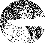
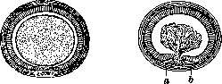
C. of the ciliary body may occur by itself and in connection with C. of the iris and choroid (fig. 3), forming a slit usually directed downward, or in a rudimentary form in the form of pigmented or unpigmented stripes. C. of the ciliary body are difficult to diagnose clinically. The ciliary muscle may remain intact even with a clearly expressed C. of the ciliary body. In certain breeds of chickens (Cochins), C. of the ciliary body regularly occur. C. of the choroid is detected by ophthalmoscopy as a sharply defined white area clearly standing out against the red background of the fundus of the eye (fig. 4), and its edges are often bordered by pigment. The vessels of the choroid are absent in this area; the vessels of the retina run along the edges of the C. or around; existing vessels of the sclera at the C. are often tortuous. Sometimes C. shows a difference in level compared to the rest of the fundus of the eye, bulging backward entirely or partially. Sometimes along the midline of the coloboma, a resemblance to a suture in the form of a strand is noted. The shape and size of C. vary; sometimes C. occupy the entire extent from the optic nerve head, involving the latter to the very periphery of the fundus of the eye. The typical position of C. is downward, corresponding to the site of the embryonic fissure of the eye vesicle (fig. 5 and 6), but atypical C. also occur, located in other directions.
Figure 5.
Figure 6.
Figure 8. On the lower side of the eye vesicle, the fissure is widely open, and mesoderm penetrates inside the eye to the lens. Figure 6. The fissure of the eye vesicle is narrower, and the mesodermal process extends inside the eye. The edges of the eye vesicle have turned in such a way that the inner leaflet has folded outward and only at some distance from the fissure passes into the outer leaflet (in a and b). The fissure preserved in the latter is therefore larger than the fissure of the eye vesicle itself. In the developed eye, therefore, a large area will be devoid of pigment and will appear white. Atypical C. located in other directions. Among such C. is also C. of the macula, located outward from the optic nerve head and in its ophthalmoscopic picture in every way resembling C. of the choroid. - During anatomical examination, in individual cases, C. were found at their site with preserved retina; but most often the area of C. is occupied by connective tissue. Thus, one often has to speak not only of C. of the choroid, but also of C. of the retina. With C. there is a corresponding defect in the field of vision; in addition, visual acuity is usually reduced due to the general underdevelopment of the entire eye. Simultaneously with C. of the choroid or separately from them, C. of the optic nerve (fig. 4) are observed, in which a depression in the form of a pit is found in the lower part of the head, or the entire head is enlarged in volume and deepened. The central vessels of the retina in this case no longer emerge from the center of the head, but appear from below, from above, or along the entire periphery of the depression. If microphthalmia is not simultaneously present, myopia is noted. The usual phenomenon is decreased visual acuity and a defect in the field of vision. Congenital changes in the form of a sickle or cone downward from the optic nerve head, often accompanied by amblyopia, should also be considered as rudimentary C. of the optic nerve. - With C. of the lens, indentations of various sizes and shapes are present at its edge, visible either through C. of the iris or with a dilated pupil. Sometimes instead of indentations there are only seemingly straightly cut off limited places at the edge of the lens. The third type of C. of the lens, according to Hippel, consists in the fact that the lens tapers downward and elongates, while its anteroposterior diameter decreases. In most cases, C. are directed downward, but can also be in other directions. Often C. of the iris, choroid, and optic nerve are simultaneously present. Various forms of lens opacity are common with C. - Defects or C. of Zinn's ligament are observed with C. of the lens and C. of the ciliary body. Their shape is triangular with apex at the edge of the lens. - C. of the vitreous body represents a defect in its lower part or throughout its entire length from the optic nerve head to the ciliary body, or only in the anterior or posterior segment. The height of this defect varies, and it is filled with connective tissue strands containing vessels, which penetrate into the eye from the head or below. - Primary C. exists in the wall of the eye vesicle, i.e., in the retina and pigment epithelium developing from it, and only subsequently the choroid participates in this. Therefore, as Hippel rightly notes, the usual expression "C. of the choroid" is inaccurate, and it is better to replace it with the name "C. of the choroid and retina". To explain those cases where anatomically the presence of retina at the site of C. is proven, Hippel assumes that the role is played by the differently strong development and differently early disappearance of mesoderm. - C. of the optic nerve is the result of insufficient closure of the stalk of the eye vesicle; this is also indicated by the depression that is usually present in the lower part of the head with C. - Among the various explanations for the development of lens colobomas, the views of Hess are most confirmed, according to which the growth of the lens can be hindered by the long preservation of individual vessels of the tunica vasculosa lentis.
A. Pokrovsky. Congenital C. of the iris and choroid, being a genotypic disease, are often observed in members of the same family (see Clausen's summary). There are descriptions of families in which inheritance has been traced for 5 generations (Snell, Loeb). In the latter case, C. of the iris was atypical (directed upward). Since in the same family C. of only the iris (or more often simultaneously of the iris and choroid) is observed (for example, the case of Hessin, who observed in three sisters C. of the iris and choroid, and in their two brothers C. only of the iris), it should be assumed that both are caused by the same gene, varying in its phenotypic manifestation. These variations can be quantitatively very strong, giving either aniridia (complete absence of the iris) or only a small defect of it. (Figure 7 confirms this position.) The phenotypic variation also explains the circumstance that in the same person sometimes C. is observed in one eye and aniridia in the other. Analysis of the pedigrees available in the literature rather suggests a dominant nature of inheritance. Whether this applies to all cases, as well as whether all dominant cases are caused by the same gene, cannot be said at the present time. - C. are not subject to treatment, and one can only speak of correction of refraction anomalies and protection of the eyes from bright light with the help of colored glasses. C. of the eyelids are corrected surgically.
erfKo B Колобома
Аниридия Д
Unknown gender Figure 7. Coloboma of the iris and aniridia, according to De Beck (from Clausen).
Related articles
Mentioned in
Cite this page
“Coloboma.” Soviet Medical Encyclopedia. English translation of Bolshaya Meditsinskaya Entsiklopediya, 1st ed. (Moscow, 1928–1936), ed. N. A. Semashko. https://sovietmedicalencyclopedia.pages.dev/article/coloboma/