Retina
Historical document, translated for reference. It reflects medical knowledge of the 1920s–30s and is not medical advice.
Summary
The retina is the innermost of the three eye membranes, named by the Greek Herophilus for its resemblance to a fisherman's net. It consists of three distinct belts with varying structures and functions, with the posterior belt being the light-sensitive part responsible for visual perception.
Encyclopedia article (1928–1936)
RETINA, the innermost of the three eye membranes, received its name, given by the Greek Herophilus (c. 320 B.C.), from its resemblance to a fisherman's net. Anatomy and histology. The inner surface of the retinal membrane is adjacent to the contents of the eyeball, while its outer surface faces the vascular tract. Corresponding to the three parts of the latter—the choroid, ciliary body, and iris—the retina is divided into three belts that differ greatly in structure and function. The posterior belt, from the opening in the choroid for the exit of the optic nerve (foramen opticum chorioideae) to the so-called serrated edge (ora serrata), constitutes the light-sensitive part of the eye and is called the visual part (pars optica retinae). The middle belt, lining the inside of the ciliary body and about 6 mm wide, is called pars ciliaris retinae. Finally, the most anterior belt represents the posterior leaflet of the iris (pars iridica retinae). Both of these latter belts have a very simple structure, consisting of two layers of cylindrical cells, of which only the cells of the outer layer are pigmented in the area of the ciliary body, while in pars iridica both layers of cells are heavily loaded with pigment. Having no independent physiological significance, these parts are only genetically connected with the retina, but anatomically and functionally they are part of the iris and ciliary body. As for the visual part of the retina, it is the most important part of the eyeball. It serves as the organ for perceiving visual stimulation, consists of nervous tissue that has undergone a high degree of differentiation, and has a very complex histological structure. Macroscopically, the visual part of the retina appears as a transparent, soft, inelastic membrane, the thickness of which at the edge of the optic nerve is 0.4 mm, and decreases toward the periphery to 0.14 mm. Almost homogeneous throughout its entire extent, it presents certain peculiarities in only three places. At the border with pars ciliaris, the retina forms sharp projections directed forward and separated from each other by rounded indentations, so that, especially when the retina becomes cloudy after death or fixation, its edge has a serrated appearance (ora serrata). In position, the serrated edges, orae serratae, correspond to the spaces between the ciliary processes. In the posterior part of the retina, two areas of almost the same size are distinguished. One occupies the posterior pole of the eye and represents a transversely oval, funnel-shaped depression (fovea centralis), the edges of which form a low ridge. The latter protrudes very slightly and without a sharp boundary above the surrounding surface of the retina, and slopes toward the center in the form of a more or less gentle slope (clivus), forming on the bottom of the depression a strongly concave pit (foveola). The diameter of the fovea centralis in the horizontal meridian is about 2 mm, in the vertical meridian 1.6 mm; the diameter of the foveola is 0.2-0.4 mm; the thickness of the retina in the area of the ridge on the nasal side is 0.275-0.41 mm, on the temporal side 0.22-0.35 mm, in the area of the foveola 0.075-0.12 mm. The fovea centralis is often also called the yellow spot (see), but these concepts are not entirely identical. In recent years, especially Vogt has insisted on clarifying the corresponding terminology. The yellow spot (macula lutea) should refer only to the central part of the fovea centralis with a diameter of 0.6-0.7 mm, because it is precisely this area that has an orange-yellow color, depending on the impregnation of the retinal tissue here with a transparent yellow dye (lacquer dye). The foveola is colored relatively weakly, the belt immediately surrounding it is colored very noticeably, and the yellow color gradually disappears toward the periphery. 3.5 mm inward and slightly downward from the fovea centralis lies the papilla, or optic disc (see Fundus of the eye). Microscopic examination reveals in the retina a very typical layering, which was already clarified in the first histological studies of the retina on thin sections (N. Muller, M. Schultze). With ordinary staining, 10 layers are visible in the retina (see Eye and Fig. 2 on separate plate, Vol. 7, pp. 275-276). The morphological and functional significance of these layers was clarified thanks to special research methods for the nervous system introduced by Golgi and Ehrlich. The latter's method was first and with great success applied to the study of the retina by the Russian histologist Dogel. Following him, Ramon y Cajal in the 1890s gave a well-known scheme of the structure of the retina, which has since undergone almost no change (Fig. 1).
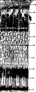
Morphol. and fnkts. zna
The R. as a whole consists of four layers of cellular elements, in turn supplied with characteristic and diverse processes. The outermost layer, when the R. is separated, usually remains in connection with the choroid, but genetically it belongs to the R., and consists of a single row of pigmented cells, sitting on the vitreous lamina of the choroid, and is called the pigment epithelium. The pigment epithelium has a very typical appearance when viewed from the surface. It consists of hexagonal cells, uniformly filled with pigment, more or less concealing the centrally located nucleus; the boundaries of the cells sharply project in the form of colorless narrow strips, the so-called junctions, consisting of neurokeratin. On a perpendicular section to the surface, it is seen that the pigment does not fill the entire cell, leaving completely free the part of the protoplasm directly adjacent to the vitreous lamella. In general, the pigment epithelial cell has the shape of a low hexagonal prism with a diameter of 17 μ and a height of 8 μ, but in the area of the fovea centralis the cells are higher (11-14 μ) and narrower (9-11 μ); in addition, in the area of the ora serrata, very large multinucleated cells are found, up to 60 μ and more in size. From the inner surface of the pigment epithelial cells, numerous processes extend, located between the outer segments of the rods and cones; according to Salzmann, the length of the processes in humans is 5 μ. Both a significant part of the cell and the processes contain pigment granules, more round in the outer part of the cell, more elongated, resembling needles of crystals, in the inner part and in the processes. The pigment grains of this epithelium differ from the grains of other pigmented cells of the eye and are distinguished as a special group of fuscin. It should be noted that not all cells of the pigment epithelium are pigmented to the same degree, which causes a fine mottling of the inner surface of the retina, noticeable even during ophthalmoscopy; the pigmentation is especially uneven near the ora serrata. Behind the pigment epithelium follows the neuro-epithelium, which consists of two kinds of cellular elements—rod and cone cells. These unique elements have considerable length at very small and unequal throughout their width (from 2 to 6 μ) and consist of a protoplasmic process, called, depending on its shape, a rod or a cone, and of an elongated body in the form of a fiber containing a nucleus. A thin glial membrane with holes for the rods and cones (membrana limitans externa), piercing the R., determines the boundary between the process and the cell body. Found in all vertebrates, rod and cone cells differ significantly in form and size in different species. In the human rod cell, the following are distinguished (fig. 2): a) the outer segment, consisting of a doubly refracting substance similar to myelin, contains a transparent purple-colored pigment—visual purple, or rhodopsin; after death and in a solution of table salt, in diluted serum, the outer segments break down into separate flat parallel disks, resembling the shape of coin stacks; b) the inner segment, sharply separated from the outer by a boundary, consists of singly refracting protoplasmic substance; in its outer third lies the so-called ellipsoid, or thread-like apparatus, representing a system of fibers running longitudinally on the surface and intersecting inside at acute angles; the total length of the rod is from 40 to 60 μ, thickness 2 μ; c) the rod fiber, very thin, curved, nodular, ending in a button-like thickening in the outer part of the outer plexiform layer and containing a round, strongly staining nucleus, located at various heights in the outer nuclear layer. The cone cell consists of the same parts as the rod cell, but with the difference that the outer segment has a conical rather than a cylindrical shape, is significantly shorter, and does not contain visual purple; the inner segment is much thicker (6-7 μ), its thread-like apparatus is more sharply expressed, the fiber of the cone cell is also thicker; the oval, more lightly staining nucleus lies immediately inside the membrana limitans externa; the proximal part of the fiber is longer than in the rod, and somewhat thicker and ends in the outer plexiform layer not with a button-like but with a conical thickening, from which thin fibers (cone foot) extend. In amphibians, reptiles, and birds, the inner segment contains at the boundary with the outer an oil drop occupying its entire thickness; these drops are either colorless or colored red, yellow, green, rarely pale blue. The distribution of rod and cone cells is uneven in different areas of the R. With the exception of the central area with a radius of 4-5 mm from the center of the fovea, rod cells significantly predominate over cone cells. On planar sections through the inner segments, it is seen that each cone is surrounded by 3-4 circles of rods. In the central area, the number of rods decreases; on its periphery, each cone is surrounded by one row of rods, closer to the center rods lie singly between cones, and in the area of the foveolae with a diameter of 0.2-0.4 mm there are only cones. In general, the human R. contains about 130 million rod cells and 7 million cone cells. It should be noted that the foveal cones are longer and narrower than the peripheral ones, and in form they more resemble rods. The proximal processes of rod and cone cells pass perpendicularly to the surface of the R. in the outer plexiform layer; only around the fovea they first take an oblique, and then almost parallel to the surface direction, forming here the layer of Henle. In the outer plexiform layer, the proximal ends of rod and cone cells come into contact with the distal branches of the next neuron, called ganglion retinae and consisting of bipolar cells. The latter contain a nucleus poor in chromatin, surrounded by a very thin layer of protoplasm, which sends out two processes: one—distal—into the outer plexiform layer, the other—proximal—into the inner plexiform layer. Two types of bipolar cells are distinguished. In some, the distal process comes into contact with its dendrites with the button-like endings of several rod cells. Unlike these rod bipolars, cone bipolars come into contact with their dendrites with the proximal end of only one cone cell. The proximal process of the rod bipolar envelops with its terminal branches the ganglion cell; cone bipolars come into contact with horizontally branching endings of their proximal process with the dendrites of ganglion cells at various heights of the inner plexiform layer. The last neuron of the R. (ganglion opticum) consists of multipolar cells with a diameter from 12 μ to 30 μ, with a light round nucleus and protoplasm containing neurofibrils and Nissl granules. The numerous dendrites of these cells, called ganglion cells, come into contact as described above in the inner plexiform layer with the neurites of bipolars. From each ganglion cell, one neurite extends, directed into the next layer of nerve fibers. Here these processes, remaining non-myelinated, pass parallel to the surface of the R., connecting into bundles arranged radially with respect to the papilla of the optic nerve. The radial arrangement is disrupted only in the area of the fovea centralis, where the fibers coming from the temporal side bend around the fovea, forming arcs from its upper and lower sides (fig. 3). Only the fibers going from the fovea itself and the area between it and the papilla of the optic nerve have a direct direction here; in the latter they form the so-called papillomacular bundle. In addition to the neurites of ganglion cells, the layer of nerve fibers also contains centrifugal fibers going from some elements in the brain. According to Ramón y Cajal, these fibers are thicker than the centripetal ones. Besides the neurons conducting excitation from the rods and cones to the brain, in the inner nuclear layer there are nerve cells with conductivity in a direction parallel to the surface of the R. This includes horizontal cells,
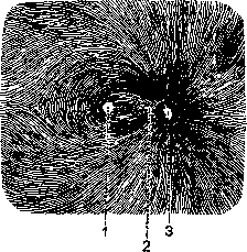
Figure 3. Radial distribution of the fibers of the optic nerve on the inner surface of the retina: 1-fovea centralis; 2-macula lutea; 3-papilla n. optici.
which are arranged in two rows in the outer part of the inner nuclear layer. Their processes have a direction parallel to the surface and partially penetrate into the outer plexiform layer. The inner nuclear layer also contains amacrine cells with a pear-shaped body 10-14 μ in diameter. Among them are distinguished: 1) layer-forming amacrine cells, the processes of which end in branching parallel to the surface in the inner plexiform layer; 2) diffuse amacrine cells with a process that branches multiple times and also ends in the inner plexiform layer; 3) association amacrine cells with a protoplasmic process ending in a branching neurite that runs along the border of the inner nuclear and inner plexiform layers. Amacrine cells come into contact with their processes with the dendrites and bodies of ganglion cells, and association amacrine cells come into contact with centrifugal fibers of the retina. In addition to specifically nervous elements, the R. also contains a supporting framework formed by neuroglia. The latter primarily includes Müllerian or radial fibers. They penetrate the R. in a direction perpendicular to the surface, begin with cone-shaped expansions that merge on the inner surface of the R. into the limiting membrane (membrana limitans interna), give off numerous processes in both plexiform layers, as well as in the inner and outer nuclear layers. At the border between the outer nuclear layer and the layer of rods and cones, the processes of Müllerian fibers, connecting with each other, form a perforated membrane (membrana limitans externa), from which thin fibers extend into the spaces between the bases of the rods and cones, forming fibrous baskets around them. Each Müllerian fiber contains a nucleus located among the nuclei of bipolar cells. Between 12 ganglion cells and in the layer of nerve fibers are also scattered in considerable quantity ordinary glial elements—cells of the astrocyte type with a large number of processes extending in various directions and thin fibers that are in close connection with the protoplasm of the cells. When examining the R. by Hortega's method, elements of microglia (Hortega cells) and oligodendroglia are found in it. The former are mainly found in the inner plexiform layer, the latter in the layer of ganglion cells. The typical histological structure of the R. undergoes changes in the area of the central fovea and at the anterior edge at the border with pars ciliaris. In the central fovea, the R. is greatly thinned (see Yellow spot). The anterior part of the R. is characterized by the gradual disappearance of neuroepithelial cells as well as other nerve cells, and an increase in neuroglial elements. Of the neuroepithelial cells, rods disappear first, then cones. The layers of nerve fibers and ganglion cells near ora serrata are greatly reduced. The inner nuclear layer persists longer than others, but it also sharply ceases at the border with pars ciliaris, as a result of which the edge of pars optica forms either a rounded or a sharp-edged step (fig. 4). In the thickness of the peri
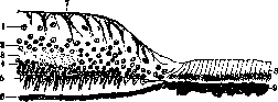
fig. 4. Section through ora serrata: 1-ganglion cells; 2-inner granular layer; 3-outer retinal layer; 4-outer granular layer; 5-layer of rods and cones; 6-pigment epithelium; 7-Müllerian fibers; 8-cylindrical epithelium of orbiculus ciliaris; 9-continuation of the pigment layer.
In the peripheral part of the R. in many eyes there are cavities of various sizes, the so-called lacunae of Blessig (see below). Following Schwalbe's proposal, the R. is distinguished into an outer neuroepithelial layer, which includes rod and cone cells with their processes and the pigment epithelium adjoining them from the outside, and an inner neural layer, which includes the other two neurons of the R. In addition to the main functional difference, these layers also differ from each other in terms of their blood supply. The neuroepithelial layer does not contain blood vessels, including capillaries; in the neural layer, a system of central vessels branches out. The larger branches, arteries and veins, lie in the layer of nerve fibers and the layer of ganglion cells, while narrow capillaries penetrate deeper, but not beyond the outer surface of the inner nuclear layer. The vascular system of the R. is represented by the central artery and central vein, the distribution of which can be clearly determined during ophthalmoscopy (see Fundus of the eye). The circulation in the R. is generally independent and isolated from the circulation in the vascular membrane. Only near the optic nerve head are there usually capillary connections between the two systems. According to Behr, the lymphatic pathways in the R. consist of two systems isolated from each other. On the one hand, the veins and capillaries of the R. are surrounded by perivascular spaces containing lymph; on the other hand, neuroglia in the R. and optic nerve is an independent lymphatic pathway that transports nutrients and removes metabolic products. Development of the R.-see Visual organs. Physiology. The significance of the R. in the act of vision was understood only in the early 17th century. Until then, the lens was considered the light-sensitive part of the eye. The new doctrine was proclaimed by Kepler, who finally established that during vision a reduced and inverted image of visible objects is formed on the R. Since the time of H. Müller, the layer of rods and cones has been considered the light-sensitive layer of the R. But while the light-perceiving role of rod and cone cells themselves is not currently in doubt, unity of views regarding the mechanism of converting light energy into nerve excitation cannot be considered achieved. The most widely accepted view is that of the photochemical nature of the processes occurring in the rods and cones under the influence of light waves (see Vision, Ionic theory of excitation, Light perception, Color perception). Pathology of the R. is very diverse. Among congenital anomalies, medullated nerve fibers (fibrae medullares) are relatively common. Ophthalmoscopically, they appear as white spots adjoining the optic nerve head, usually to its upper or lower edge, or to both together, much less frequently to the medial edge, and extending into the retina for a distance, mostly not exceeding one diameter of the nerve head. The edge of the spot adjoining the nerve head is usually sharply defined, while the periphery shows divergent fiber structure, resembling the pattern of a flame or a comet's tail. The surface has a silky luster. The vessels of the R. are partly visible in the area of the spot, and partly obscured by it. Functionally, a small defect in the visual field (enlargement of the blind spot) is found, while central visual acuity is not reduced. Pathological-anatomical studies have shown that in the corresponding cases, the nerve fibers of the optic nerve, as in normal conditions, lose their myelin at the posterior edge of the lamina cribrosa, and when passing from the nerve head to the retina, part of the nerve fibers again acquire a medullary sheath, retaining it for a greater or lesser extent. This anomaly is mostly found in one eye and is often accompanied by other congenital eye defects. Aplasia of the macula is of more serious significance. Studies by Seefelder, Elschnig, and others have clarified that as a developmental defect of the R., the absence of differentiation of the central fovea occurs. In it, the layers of the R. are not reduced, so no typical depression is formed, and at the same time the neuroepithelium in the center does not consist solely of cones, as normally, but also contains rods. Such underdevelopment of the macula is usually combined with other congenital eye defects, which has been anatomically found in albinism, aniridia, congenital nystagmus, and fully explains the accompanying amblyopia. In congenital colobomas of the fundus of the eye, the condition of the R. varies. In some cases, above the defect in the vascular membrane, changes in the R. affect only the pigment epithelium, which is either completely absent or consists of scattered islands of cells that do not contain pigment. More often, the R. in its entire thickness shows abnormal structure in the area of the coloboma: irregular alternation of layers, absence of rods and cones, proliferation of glia, and fusion of the outer surface of the R. with the sclera. Complete absence of the proper R. (inner leaf of the secondary eye vesicle) above the coloboma is very rare. Among colobomas of the fundus of the eye, coloboma of the macula is of particular interest (see Coloboma). Changes in vessels and circulatory disorders. In recent years, fine ophthalmoscopic changes in the vessels of the R. in connection with hypertension and arteriosclerosis have attracted increased attention. According to Guist, based on the condition of the vessels on the fundus of the eye, it is possible to differentiate essential hypertension from secondary or toxogenous. In the first form, the veins retain their elasticity, and the increase in pressure causes a pronounced corkscrew-like tortuosity, especially noticeable on the small macular veins. In secondary hypertension, the walls of the veins are altered, greatly thickened, and as a result do not show pronounced tortuosity. In essential hypertension, the transparency of the arterial wall is early impaired, as a result of which the color of the arteries becomes darker (copper-wire arteries), and the reflective strip on them is interrupted. With more pronounced sclerosis, the picture first noted by Gunn of silver-wire arteries is obtained: the reflective strip on the vessel is very narrow and bright, and the entire artery appears lighter with a special luster. The rigidity of the altered arterial wall and the thickening of the surrounding lymphatic sheath cause compression of the vein at the crossing where an artery passes over it, and the blood flow in the vein at the crossing appears interrupted (Gunn's phenomenon). Narrowing of the arteries and unevenness of their caliber are also signs of arteriosclerosis. The ratio of the diameters of arteries and veins, normally 2:3 (Leber), shifts to 2:4. On such an overall narrowed artery, limited areas with an even narrower lumen may be noticeable, and between them slight expansions up to small aneurysms. Varicose protrusions are found on the veins. It should be noted that the sclerotic process can involve the trunks and separately the branches of the central vessels independently of each other. The clinical significance of the ophthalmoscopically detectable arteriosclerotic changes in the vessels of the R. lies in the fact that they provide information about the condition of the cerebral vessels. There is no doubt that despite severe damage to the cerebral vessels, the vessels on the fundus of the eye may have a completely normal appearance, all the more so that pathological-anatomical changes are not always found in the ophthalmoscopic picture (Hertel). But, on the other hand, a positive result of ophthalmoscopic examination is highly pathognomonic for the presence of sclerotic changes in the cerebral vessels. Arteriosclerosis of the vessels of the R. affects the function of the eye only if the circulation is disturbed or stopped or if hemorrhages occur from the affected vessels. Two main types of circulatory disturbance in the R. are distinguished. Depending on the obstruction in the artery, the blood flow stops with the onset of ischemia, or due to obstruction in the vein, the outflow of blood is impaired or stopped with the development of stasis and its further consequences. The rapidly occurring ischemia of the R. shows a very typical clinical picture described in 1859 by Graefe under the name of embolism of the central artery of the R. The affected eye suddenly becomes blind. Sometimes the loss of vision is complete, up to the absence of light perception; in other cases, part of the visual field on the temporal side is preserved, and the eye is able to count fingers at the closest distance. On ophthalmoscopic examination, the vessels of the R., especially the arteries, appear greatly narrowed; the latter are noticeable only in the immediate vicinity of the optic nerve head and, due to the dark color of their blood, differ little from the somewhat wider veins. Often, a breakdown of the blood column in the veins into separate segments is noticeable, the gaps between which contain only plasma. The immobility of these segments of the blood column definitely indicates the cessation of blood circulation in the vessels. The resulting ischemia quite quickly leads to diffuse clouding of the R., especially noticeable around the optic nerve head and in the area of the macula; in the center of the latter, on a milky-white background of clouding, a red spot with a diameter of 1/5 of the nerve head stands out, corresponding to the depression of the R.
The thin layer of the retina in this place remains transparent and through it the choroid is clearly visible, the red color of which is especially sharp against the surrounding cloudiness due to contrast. The optic nerve head is pale, its boundaries are barely noticeable. The cessation of blood flow may not occur in the trunk of the central artery of the retina itself, but in one of its branches. Objective changes then develop only in a limited part of the retina, supplied with blood from the affected arterial branch. Functional examination reveals a corresponding sector-shaped defect in the field of vision, while central vision is largely preserved. On the other hand, there are cases where even in so-called trunk embolism, a small remainder of central vision is preserved, which is explained by the presence of a fairly large cilio-retinal artery. The area of the retina supplied by the latter between the optic nerve head and the macula does not cloud over and is clearly noticeable by its normal color. The resulting cloudiness of the retina lasts for several weeks and then gradually disappears. The fundus of the eye takes on its usual color, but an atrophic white optic nerve head with sharp boundaries and very narrow arteries remains forever.-Patho-anatomically, the closest consequence of the resulting ischemia is edema of the nerve fiber layer of the retina, most sharply expressed around the optic nerve head and central fovea, where the thickness of this layer is greatest. The cloudiness of the retina depends mainly on this edema. At the same time, degenerative changes occur in the nerve elements of the ganglion cell layer and the inner nuclear layer. Subsequently, the edema fluid and breakdown products are absorbed. In the retina, only the nuclei of Müller's supporting fibers remain, in addition to the outer nuclear layer with rods and cones, which is nourished from the choriocapillaris. The disintegration of ganglion cells leads to ascending atrophy of the optic nerve. As for the pathogenesis of the ischemia of the retina itself, this question cannot be considered fully clarified. The original opinion of Graefe that embolus is the usual cause of ischemia still has a number of very authoritative supporters at present. It does indeed satisfactorily explain the clinical signs of the disease, especially the sudden development of blindness. However, patho-anatomical studies have given rise to significant doubts regarding the embolic nature of the lesion. In many cases, searches for an embolus were fruitless. On the other hand, plugs found in a number of cases in the lumen of the central artery consisted of an amorphous mass with an admixture of a small number of cells and showed no signs of organization, despite the fact that from the onset of blindness to the death of the patient 2 months or more had passed. Scheerer is inclined to consider these plugs, interpreted by authors as emboli, as postmortem plasma clots. In his opinion, there are also insufficient grounds to consider these clots thrombotic. At one time, Haab recognized the cause of the cessation of blood flow as obliterating endarteritis of the central artery on the basis of general arteriosclerosis. In a number of cases, corresponding changes in the artery were indeed found. Scheerer attaches particular importance to the circumstance that the closure of the arterial lumen is almost exclusively localized in the region of the lamina cribrosa, where the central artery and vein lie close to each other, surrounded by a dense ring of connective tissue; here, due to the mutual pressure of the vessels, local circulatory disorders can easily arise, completely independently of embolism. He does not exclude vaso-motor disorders in the form of spasm or paralysis, causing a stoppage of blood circulation.--The blindness developing in so-called embolism of the central artery usually has a persistent and incurable nature. In isolated cases after massage of the eye or puncture of the anterior chamber, improvement or complete restoration of vision occurred, and this effect, possibly without sufficient grounds, was considered the result of displacement of the plug due to a decrease in intraocular pressure. The clinical picture of embolism of the central artery of the retina is observed at all ages. Both sexes are affected almost equally often. In the vast majority of cases only one eye is affected, but bilateral embolism has also been described. Spasm of the retinal arteries. In contrast to the persistent blindness in embolism, recurrent attacks of suddenly occurring and rapidly passing blindness are observed, the occurrence of which is attributed to spasmodic contraction of the retinal arteries. Due to the short duration of the attacks, it was only in rare cases that the fundus of the eye could be examined during an attack, and the retinal arteries were not always found narrowed; often they were normal, sometimes even dilated. No signs of retinal edema were observed either. Leber admits the possibility of spasm of the central artery in its proximal part, and the absence of edema explains by the short period of cessation of blood flow. The attacks last from 5 to 15 minutes, can often repeat during the day, sometimes the intervals between attacks are long and are measured in weeks, months, and even years. That vascular motor disorders underlie them is proven by the simultaneous observation in some patients of migraine and Raynaud's disease. It is characteristic that recurrent cloudings of vision of this kind occur more frequently in younger age (up to 40 years) than in later years, and usually both eyes are affected. In some cases, after repeated attacks ending with complete restoration of vision, as a result of a subsequent attack, persistent blindness or persistent decrease in vision develops with signs of retinal ischemia. Whether in such cases there is thrombosis on the basis of changes in the arterial wall, as Leber assumes, or whether ischemia is based on vascular spasm here, as Scheerer is inclined to admit, remains unexplained. Therapeutically, the attacks in some cases were well controlled by inhaling amyl nitrite, taking nitroglycerin. The significance of paracentesis or iridectomy, recommended for cases with longer attacks, remains doubtful, all the more so since a favorable result was obtained not only on the operated eye but also on the unoperated eye. Cessation of outflow of blood from the retina gives a typical ophthalmoscopic picture, which first bore the name hemorrhagic retinitis, later under the influence of research by Michel (v. Michel) renamed thrombosis of the central vein of the retina. With signs of rapidly developing very significant decrease in vision (rare complete blindness, often the ability to count fingers at close distance remains), sharply expressed changes are found on the fundus of the eye: almost the entire retina is strewn with numerous hemorrhages of various sizes, partly strip-like of elongated shape, partly more solid and round. Veins are noticeably dilated and tortuous, arteries are narrow and near the optic head are hardly noticeable due to edematous cloudiness of the retina; somewhat later in the retina, white nests of degeneration are visible in places (see Retinitis). The optic head stands out little in color from the surrounding retina, its boundaries are indistinct, on its surface there are individual strip-like hemorrhages. The changes in the retina, having developed rapidly, usually persist in one place for a long time. Only in rare cases do hemorrhages completely resorb, more often during the process fresh hemorrhages are observed; often secondary glaucoma develops in the affected eye. A more favorable outcome is given by thrombosis of a branch of the central vein. Hemorrhages and nests of degeneration appear only in a limited area of the retina, causing a corresponding defect in the field of vision. The changes subsequently resorb, the remainder of vision is preserved and secondary glaucoma does not develop. The disease affects mainly older age. In Coats' 35 cases, 7 were under 50 years, 6 from 50 to 59, the rest older. Most often one eye is affected, but bilateral lesions are also occasionally observed. As patho-anatomical studies show, the basis of the stagnant process in the retina is a disease of both central vessels at the place of their passage through the lamina cribrosa or in its immediate vicinity. In both the artery and the vein, narrowing of the lumen occurs due to proliferation of the endothelium and thickening of the entire intima. In addition, according to Scheerer, due to sclerosis of the connective tissue ring surrounding both vessels and their adventitia, mutual compression of the latter occurs. As a result, the rigid artery compresses the more pliable vein; the lumen of the latter narrows, which, in connection with the presence of subendothelial proliferation, leads to more or less complete obstruction. Prognostic significance of changes in retinal vessels. The question has been discussed more than once whether the arteriosclerotic changes observed in the retinal vessels indicate similar changes in the cerebral vessels and to what extent one or another circulatory disorder in the retina is a precursor of hemorrhage or softening in the brain.
As can be seen from the observations of Geis, in patients over 40 years of age suffering from general arteriosclerosis, the presence of pronounced sclerotic changes in the arteries of the R., hemorrhages into the latter, or sudden blindness of the eye in the picture of embolism of the central artery has a very poor prognostic significance, serving as a harbinger of a fairly rapidly approaching cerebral stroke. The prognostic significance of thrombosis of the central vein is less definite, since only 50% of such patients are subsequently affected by cerebral stroke, and the latter occurs much later. Hemorrhages into the R. that are not of arteriosclerotic character. In addition to arteriosclerosis, hemorrhages into the R., as is known, are a common symptom of retinitis of various origins (see Retinitis). But besides this, they often appear on their own, and are caused by the most diverse causes. It is self-evident that hemorrhages form with direct damage to the eyeball. Cases of general venous stagnation accompanied by hemorrhages into the R. are of greater interest. With very severe traumatic compressions of the chest, and sometimes with compression of the abdomen, when blood is quickly squeezed out of these parts of the body, hemorrhages into the R. occasionally appear. Hemorrhages of the same origin are those that arise under the influence of strong straining during coughing, vomiting, difficult defecation. About a quarter of newborns show hemorrhages into the R. of various sizes in the first days of life, mainly in the posterior pole of the eye, around the optic nerve head and in the area of the central fovea. These hemorrhages quickly resorb and disappear without a trace by the end of the first week. According to some, they arise from compression of the skull during childbirth, according to others - asphyxia. The pathogenesis of hemorrhages into the R. that appear in certain infectious diseases, in general as rare complications, is far from clear. These include hemorrhages in influenza, malaria, in typhus, typhoid fever and other infections. Hemorrhages into the R. are relatively rare in various pathological forms previously grouped under the name of hemorrhagic diathesis. Hemorrhages are rare in scurvy, in hemophilia, in Werlhoff's disease, they are more often described in purpura haemorrhagica in cases with a generally severe course. Finally, various anemic conditions can cause hemorrhages into the R., both depending on blood loss and on the basis of a violation of the dynamics of hematopoiesis. Repeated profuse bleeding from the stomach, nose, genital tract, very profuse bloodletting in some cases are the cause of hemorrhages into the R., sometimes accompanied by very significant visual impairment, up to complete blindness. Of the so-called primary anemias, chlorosis rarely causes changes in the fundus of the eye. Pagenstecher found only 3 cases of hemorrhages into the R. out of 246 cases of chlorosis studied. On the contrary, hemorrhages into the R. are very characteristic of malignant anemia and can serve as a diagnostic sign. According to Hesse's statistics, hemorrhages into the R. were found in 47 out of 50 cases of pronounced malignant anemia. In all these diseases, the basis of hemorrhages lies not only in the decrease in the amount of blood, but also in the slowing of its flow due to the resulting weakness of the heart; the resulting incomplete stagnation in the veins of the R. causes a disturbance in the nutrition of their walls, making them permeable to erythrocytes. In general, hemorrhages from the vessels of the R. most often occur per diapedesin and very rarely per rhexin. The ophthalmoscopic picture of hemorrhages into the R. depends primarily on which layer they appear in. In the layer of nerve fibers, the blood is located between the fiber bundles in the form of elongated or spindle-shaped strips; in the middle and deep layers of the R., hemorrhages more often take a round shape. The size and number of hemorrhages are subject to large fluctuations. Most often, the blood that has come out of the vessels remains inside the R., but it can penetrate to its outer surface and separate it from the choroid, or by rupturing the membranae limitans interna, pave its way into the vitreous body. The extravasate of the R. acquires a special characteristic appearance when a more or less significant amount of blood, reaching the limitans interna, does not rupture the latter, but accumulates between it and the layer of nerve fibers. Such a preretinal hemorrhage appears ophthalmoscopically as a large, sharply delimited dark spot, which at the time of its formation has the shape of a complete circle or oval. Subsequently, due to the settling of erythrocytes in the upper part of the extravasate, only a clear serum remains, and the boundary of the lower dark red part is strictly horizontal, changing its position with different tilts of the patient's head. Preretinal hemorrhage is often located in the area of the macula, has a fairly significant size (2-4 diameters of the optic nerve head) and can develop under the influence of any of the causes that generally cause hemorrhage into the R. The degree of visual impairment in hemorrhages into the R. depends on their number, size, and mainly on their location. Extravasates in the periphery of the R. usually have little effect on vision; on the other hand, even a small central hemorrhage can significantly reduce visual acuity. Since in preretinal hemorrhage the light-sensitive layer of the R. is not disrupted and continues to function, patients perceive the shadow from the blood mass as a positive scotoma, and depending on the color of the blood, they see everything as if through a red mist.--The course of hemorrhages into the R. is not always the same and depends largely on the underlying cause that caused them. Small hemorrhages quickly resorb; preretinal extravasates resorb slowly, often over several months, but in general their course is relatively favorable. More significant and especially recurrent hemorrhages often lead to the formation on the anterior surface of the R. of connective tissue and glial strands and membranes protruding into the vitreous body (retinitis proliferans - see Retinitis). Degenerations of the R. A number of diseases of the R. known under the name of retinitis, in essence, have not an inflammatory but a degenerative character, especially the so-called pigmentary retinitis (see Retinitis). A special place is occupied by degenerations of the R. localized in its central part, in the area of the macula and its surroundings. This includes above all the familial degeneration of the macula, described by Best, Stargardt, Behr and others. The disease affects several members of one family and is manifested by the appearance of grayish, initially barely noticeable spots in the area of the macula, to which small accumulations of pigment then join. The initial focus gradually increases, but usually does not exceed 1½-2 diameters of the optic nerve head. Both eyes are always affected, and according to Scheerer, the ophthalmoscopic picture in both eyes often completely coincides. Central vision is significantly reduced. The disease begins at different ages. Behr distinguishes a number of forms according to the time of onset of the disease - congenital, childhood, juvenile, adult age and presenile - and especially emphasizes that in members of the affected family, the disease begins at the same age. Behr is inclined to include the 'senile disease of the macula' (senile Maculaerkrankung) described by Haab in the group of familial degeneration of the macula. Both diseases have many similarities in ophthalmoscopic picture. In senile degeneration in the area of the macula, a faintly expressed light or dark mottling, yellowish-red or yellowish spots are visible in connection with pigment accumulations. Both eyes are most often affected in elderly people, vision is strongly reduced. Familial degeneration of the macula is a rare disease, while the senile form is relatively common. In one case, Behr's path-histological study found a degeneration originating from the neuroepithelium without reactive phenomena from the glia and connective tissue. For old age, another degenerative change of the R. is also characteristic, known under the name of cystoid degeneration. It usually develops in the anterior part of the R. near its ora serrata and is not clinically recognized due to its peripheral position, but is only noticeable upon histological examination. It was first discovered by Blessig (1855) and is characterized by the appearance in a somewhat thickened R. of numerous small, partly merging cavities, or lacunae (Blessig's lacunae), located mainly in the inner nuclear layer, in the outer plexiform layer, and sometimes extending to the outer nuclear layer as well. Cystoid degeneration of the anterior part of the R. undoubtedly represents a widespread change, apparently beginning around the age of 40 and subsequently progressive.--Detachment of the R. (ablatio, amotio or solutio retinae) is a violation of its connection with the pigment epithelium. There are various types of detachment.
Separation of the R. from the pigment epithelium can be caused by a new growth of the vascular membrane that has grown through the lamina elastica of the pigment epithelium, a subretinal cysticercus, hemorrhage, or an inflammatory exudate located under the R., but in these cases the detachment is merely a symptom or complication of the underlying disease. These forms are distinguished from genuine, or spontaneous detachment, which most often develops on the basis of high myopia, but is not so rare in non-myopic eyes, especially in old age. In the following discussion, only the so-called genuine detachment of the R. is meant. Symptomatology. The first subjective disorders in the beginning detachment of the R. consist in the appearance of peculiar light sensations - flickering, fiery balls, sparks. These so-called photopsias are the result of irritation of the light-sensitive layer of the R. Metamorphopsia is also often found: objects appear curved, bent, interrupted, especially in the case of detachment of the central part of the R. Most often the patient clearly feels a dark cloud before the eyes, approaching from the edge of the visual field opposite to the site of detachment. Functional examination reveals a defect in the visual field, the size of which depends on the size of the detached part of the R. Central visual acuity is reduced in the case when the detachment spreads to the area of the yellow spot. Ophthalmoscopically, the detachment is detected by a number of characteristic signs. The corresponding part of the R., when illuminated with an ophthalmoscope, has a gray, not red, color with a greenish or bluish tint. In flat detachment, the R. remains so transparent that its red color is almost completely preserved. In such cases, an excellent distinguishing sign is the indistinguishability of the pattern of the vascular membrane under the detached part compared with the clear visibility of it in other parts of the fundus. Usually it is easy to confirm the protrusion of the affected part above the level of the rest of the R., either by determining the refraction in the direct view or upon examination in reverse on the basis of parallax displacement at the edge of the detachment. The surface of the detached part is sometimes flat, tense, but more often folds are visible in it, oscillating with eye movements. To confirm the detachment, it is necessary to be convinced of the presence on the visible surface of the characteristic branching vessels of the R., which usually can be traced to the optic nerve head. In the presence of folds, the vessels make numerous bends, sometimes disappearing in the depressions and again appearing on the elevations. In addition, in detachment the vessels have a dark, almost blackish tint, depending both on the fact that they are partly seen in transmitted light and on the contrast with the grayish surrounding background. In recent years, Gonin has drawn special attention to an objective change that was already known earlier and the frequency of which was particularly noted by Wecker, Leber, Horstmann, and others. This concerns a hole, i.e., a rupture in the retina, having the form of a triangular, round, horseshoe-shaped slit, through which the red background of the vascular membrane is visible. The edges of the rupture are uneven, often turned inward, its size is very different; very small ruptures are found, but they can also reach the size of two or more diameters of the optic disc. Sometimes there is not one but several such holes in the R. Ruptures are observed in various parts of the R., but more often in its upper half, especially in the upper-outer quadrant. As for the frequency of ruptures of the R. in spontaneous detachment, Leber already found them in more than 50%, and in fresh cases in 73%, and admitted that in fact they occur even more often, but cannot be recognized either due to their peripheral position or due to interfering opacities of the refracting media. According to the latest data of Gonin, Vogt, and others, ruptures are diagnosed even more often. Course. Detachment most often begins in the upper part of the R., and in the first period the visual disturbance consists mainly of a defect in the lower part of the visual field. In most cases, in the coming weeks or months, the subretinal fluid descends downward, obeying gravity. Then at the site of the original detachment of the R., it may again adhere, even with complete restoration of function. If during this movement of fluid the area of the yellow spot previously occupied by the detachment is freed, a significant improvement in vision may occur. Thus, limited detachments almost always occupy the lower part of the fundus. In rare cases, such a partial detachment does not progress and remains stationary. Even more rarely the R. spontaneously adheres again (according to Uhthoff in 8%), and then at the site of the former detachment white or yellowish-white streaks (striae retinae) and small pigment nests are visible. But more often the amount of subretinal fluid simultaneously with its descent increases, as a result of which the detached area increasingly enlarges, and the detachment gradually becomes total, causing complete blindness of the affected eye. In such a eye blinded by detachment, regressive changes usually subsequently occur: intraocular pressure greatly decreases, the lens becomes cloudy. Often inflammation of the iris develops with all its severe consequences. Pathogenesis. The question of the pathogenesis of genuine detachment of the R. has been the subject of numerous studies, but even at present it cannot be considered finally resolved. According to modern views, spontaneous detachment of the R. is most often observed in high myopia. About 5% of myopes above 10 diopters suffer from detachment. In second place in frequency is detachment in the aged eye, developing primarily without pronounced preceding diseases. Finally, the same symptoms and course can have detachment developing in a previously healthy eye after an injury not accompanied by either rupture of the eyeball or intraocular hemorrhage. Lebel believed that such traumatic detachment occurs because a rupture occurs in the retinal membrane, usually in its anterior part, through which fluid gradually penetrates from the vitreous body and detaches the R. from the vascular membrane. Vogt fully joins this opinion of Leber. In his cases he saw the formation of a blister with a hole on its top. The rupture forms in the periphery of the R., because there it is thinner and contains fewer nerve fibers that give it a certain strength. But such a rupture, as was indicated above, is very often also observed in genuine detachment, and it is more than probable that it also serves as its immediate cause here. The mechanism of the origin of the rupture is not yet sufficiently clarified, and here there are different views (Leber, Vogt, Hansen, Lindner, and others). Most often ruptures are associated with degenerative changes in the R. in myopia. Strong fluctuations of the R. during movements of the body and eyeball, caused by the specific weight of the R. being greater than that of the vitreous body, contribute to the occurrence of ruptures. The same role is played by the traction exerted on the internal limiting membrane by the tangled masses and threads of the disintegrating vitreous body directly connected with it. During movements of the eyeball, the latter move very strongly, possibly due to a decrease in the viscosity of the vitreous fluid, and this constant pulling of the R. can easily cause its rupture. In old non-myopic people, the anterior part of the R. also shows signs of degeneration, and this explains why in old age genuine detachment often arises independently of myopia in emmetropic and even hypermetropic refraction. Treatment. Experience shows that in rare cases spontaneous adhesion of the detachment or a long-term arrest of the process occurs (according to Leber in 3-5%). The conservative treatment previously used in the form of bed rest, pressure bandage, diaphoretic agents, subconjunctival injections of salt, as well as surgical in the form of punctures of the subretinal space, injections into the vitreous body, cauterization of the sclera both in the area of detachment and in the area of Schlemm's canal (colmatage according to Lagrange), does not give reliable results. If improvement occurs during treatment, it is again replaced by deterioration after some time. More favorable results have been obtained in recent years using the surgical treatment method proposed by Gonin. The latter's method aims to close the rupture and thus stop the penetration of vitreous fluid under the R. and cause its adhesion. For this, Gonin, after puncturing the sclera and vascular membrane with a Graefe knife corresponding to the site of the rupture, performs cauterization through and through of both the sclera and choroid, as well as the edges of the rupture in the R. with a thermocautery needle. For the success of the operation, it is necessary to find the rupture, which often proves to be very difficult, and to accurately localize it on the surface of the eyeball to determine the site of puncture. The high figures of cures achieved by Gonin (according to 1931 data 53% in 300 cases) have attracted great attention to the method he proposed, which has also given encouraging results in the hands of other ophthalmologists.
In the last 5 years, reports on operations for detachment have occupied a prominent place at meetings of ophthalmological societies and congresses. Methods for localizing the rupture are being developed, and modifications of the method using diathermy or chemical cauterization of the choroid with caustic potash after multiple trepanations of the sclera are being proposed (Guist, Lindner). Thanks to Gonin, there has been a significant shift in the treatment of detachment, the results of which have not yet been fully determined. New growths. The characteristic new growth of the R. is glioma. Sattler estimates the frequency of this disease as 2-5 cases per 10,000 eye patients. Glioma of the R. usually develops in early childhood. About a third of cases occur in the first year of life, 80% in the first 4 years. In only isolated cases was the disease observed in patients older than 10 years. The basis of the disease is a congenital, apparently hereditary factor. Glioma often affects several children in one family. Direct hereditary transmission has also been observed. In 20% of cases, glioma affects not one but both eyes. In the course of glioma of the R., 4 stages are distinguished. In the early stage, a rounded, irregularly delimited focus of whitish or yellowish-gray color, slightly protruding above the surrounding retina, is visible at the fundus; the retinal vessels either pass over it or are submerged in it. When the other eye is healthy, such a focus usually remains undetected. Relatives become aware of the disease only when they notice a yellowish-golden reflex from the pupil, which depends on the fact that the tumor, which protrudes strongly into the vitreous body, reflects the rays falling on it in a widely diverging direction. Due to the glowing pupil, this condition was named by Beer (1817) amaurotic cat's eye. At this time, the eye is already blind, the pupil is immobile, and examination reveals a bumpy tumor behind the lens, covered with numerous thin, tortuous vessels. The first stage of the disease proceeds without signs of irritation. At the end of it, changes in the anterior segment of the eye sometimes appear in the form of deposits on the posterior surface of the cornea and the anterior surface of the iris of small cellular clusters detached from the tumor, which can also be suspended in the anterior chamber fluid and form a sediment at its bottom resembling hypopyon. Usually, the second stage soon follows - an increase in intraocular pressure with all the symptoms of subacute glaucoma. The still pliable walls of the child's eye undergo stretching, and the eyeball gradually enlarges. The third stage is characterized by the growth of glioma into the optic nerve (Fig. 5) and through the sclera, often at the edge of the cornea, as a result of which the orbit is filled with tumor nodules, the eye protrudes, cannot be closed by the eyelids, its anterior surface ulcerates and bleeds. In the fourth stage, metastases form in the lymph glands, in the brain and its membranes, as well as in the bones of the skull. Death usually occurs 1-1/2 years after the disease is detected by relatives. Pathological anatomy. In most cases, glioma develops in the posterior part of the R., much less frequently in the equatorial region, or in front of it. The tumor nodes either grow from the R. into the vitreous body (glioma endophytum) or push the R. forward and grow backward from it (glioma exophytum). On microscopic examination, glioma of the R. presents a very characteristic picture. It consists of cylindrical, partly branched and anastomosing strands of cells with intensely staining nuclei. Along the axis of each strand runs a blood vessel, surrounded as if by a sheath of cells; the latter are often located inside the strand, forming peculiar figures resembling sections of tubular glands and known under the name of rosettes. The spaces between the strands are filled with an almost homogeneous mass containing shrunken pyknotic nuclei and in a state of more or less pronounced necrosis.
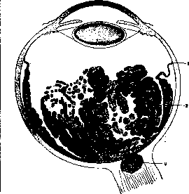
Figure 5. Spread of glioma to the optic nerve: 1-orra serrata; 2-tumor masses; 3-optic nerve.
The nuclei of the glioma cells are mostly round or oval in shape, with a diameter of 4-8 μ, surrounded by very narrow rims of protoplasm forming thin processes. The cells that make up the rosette are conical in shape, 15-20 μ in length, the broad base contains a nucleus, the same as in other cells of the tumor; the narrow apex adjoins a small cavity around which the cells are arranged in a wreath. Some researchers (Flexner, Wintersteiner), finding similarities between rosette cells and neuroepithelium, concluded from this that glioma originates from the first neuron of the R. and proposed the name neuroepithelioma for this tumor. At present, this view can be considered refuted. Histological studies of gliomas in the early stage showed that the development of the tumor begins in the inner layers of the R. (layer of nerve fibers, ganglion cells, or inner nuclei) and that its source is exclusively the glial elements of the R. (Watzold); thus, this new growth belongs to pure gliomas, it is entirely of ectodermal origin. In diagnostic terms, it sometimes presents certain difficulties to differentiate glioma of the R. from the picture of pseudoglioma, which most often develops as a result of metastatic ophthalmia after meningitis, as well as some acute exanthems. In these cases, a fibrino-purulent exudate forms in the vitreous body, followed by the proliferation of connective tissue and glia and detachment of the R., as a result of which a picture of amaurotic cat's eye is also obtained. Similar pictures can be given by injuries, especially with the penetration of a foreign body, and other inflammatory conditions of the retinal and choroidal membranes (tuberculosis, exudative retinitis). For differential diagnosis, the state of intraocular pressure (reduced in pseudoglioma), the presence of signs of a preceding inflammatory process, etc., are taken into account. Prognosis and treatment. The only means to preserve life is the timely removal of the affected eye. The result of the operation depends to a high degree on whether the tumor has already grown into the optic nerve or through the sclera. If infiltration has occurred, a recurrence with a fatal outcome almost inevitably occurs in the orbit, which usually cannot be prevented by subsequent exenteration. Experience has shown that if a recurrence does not appear within 2 years after the operation, it need not be feared in the future. In bilateral glioma, after the removal of the more severely affected eye, attempts are now made to influence the new growth with radiant energy (X-rays, radium). In a number of cases, it was possible to temporarily delay the growth of the tumor, but with the cessation of treatment, development resumes. Injuries. Bruises of the eyeball can cause, in addition to transient edema of the R. (see Berlin's clouding of the retina), hemorrhages into the R., traumatic detachment (see above), sometimes perforation of the R. in the region of the macula lutea, which forms not immediately after the injury, but as a more or less distant consequence of it, apparently on the basis of a preliminary cystoid degeneration of the R. as a result of edema. The region of the macula lutea can also be damaged by the action of sunlight. Such a burn of the R. often develops when observing a solar eclipse. Subjective disorders consist in the appearance of central scotoma with a decrease in visual acuity to 1/3 and below. Objective changes are sometimes so insignificant that they are not detected ophthalmoscopically, but in many cases, a small focus from yellowish to red color is noted in the center of the macula lutea. Subsequently, vision often recovers, but with severe damage to the R., a small atrophic nest develops and the decrease in vision remains permanent. In view of the fairly numerous diseases observed during solar eclipses, preventive measures in the form of wide recommendation of dark protective glasses are necessary in appropriate cases.
Related articles
Mentioned in
- Adaptation (a3136)
- Adaptometer
- Alvar Gullstrand
- Amaurotic Cat's Eye
- Amaurotic Family Idiocy
- Amblyopia
- Ametropia
- Amorphus
- Animal Electricity
- Aniridia
- Astigmatism Of The Eye
- Bardet Syndrome
- Berlin's Retinal Clouding
- Binocular Instruments
- Binocular Vision
- Brain (cerebrum, a comprehensive term for the entire)
- Burners
- Capillaries
- Cataract
- Chorioiditis
Cite this page
“Retina.” Soviet Medical Encyclopedia. English translation of Bolshaya Meditsinskaya Entsiklopediya, 1st ed. (Moscow, 1928–1936), ed. N. A. Semashko. https://sovietmedicalencyclopedia.pages.dev/article/retina/