Astigmatism Of The Eye
Historical document, translated for reference. It reflects medical knowledge of the 1920s–30s and is not medical advice.
Summary
Astigmatism of the eye is a refractive condition where rays from any point cannot form a focal point on the retina due to irregular corneal curvature. It can be classified as regular or irregular, with various subtypes including simple, compound, and mixed astigmatism, and can be corrected using cylindrical lenses.
Encyclopedia article (1928–1936)
ASTIGMATISM OF THE EYE (from Greek a- negative particle and stigma-point), such a refraction (see.), under which rays emanating from any point in space, with any setting of the eye, cannot give a point focal image on the retina. In most cases, astigmatism of the eye is associated with irregularity in the structure of the cornea, which in astigmatic eyes does not form a proper spherical surface, as in normal cases, but is more or less irregular. In cases of A. g., a distinction is made between correct and incorrect astigmatism. In the first case, the cornea has the form of not a segment of a sphere, but a segment of a rotation ellipsoid. Rays passing in the direction corresponding to the short axis of the ellipse experience the greatest refraction and converge at one point closer than rays passing through all other meridians of the cornea. Rays passing through the meridian corresponding to the position of the long axis of the ellipse, i.e., having a direction perpendicular to the short meridian, converge farthest. Thus, the image of a point in an astigmatic eye is never point-like but takes the form of a strip, ellipse, or circle, depending on where the points of intersection of rays passing through the most and least refracting meridians of the cornea are located in relation to the retina (see figure 1). An astigmatic eye, therefore, always sees unclearly. - From individual
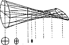
Figure 1. Diagram of ray path in astigmatism. forms of correct A., first of all, simple A. should be noted, in which the eye in one of the meridians is emmetropic, while in the perpendicular direction there is hypermetropia (simple hypermetropic A.) or myopia (simple myopic A.). In those cases where rays passing through both the short and long axes of the cornea converge either behind or in front of the retina, but at different distances from it, we are dealing with a compound (hypermetropo-hypermetropic or myopo-myopic) astigmatism. Finally, if rays passing in the direction of the smaller axis of the ellipse converge in front of the retina, while rays having a direction perpendicular to it converge behind it, we are dealing with mixed (myopo-hypermetropic) astigmatism (ast. mixtus). Where the most refracting meridian is vertical, we are dealing with direct astigmatism. Where the refractive power of the horizontal meridian is higher, there is the inverse type of astigmatism (Astigm. perversus). In a number of cases, the most refracting meridian of the cornea is located in some lateral position. In such cases, the meridian of least refraction also assumes an oblique position. In such cases 14 speaks of A* with "oblique axes". All these forms of A. belong to cases of regular astigmatism. In irregular A., the cornea has a complex irregular surface. While regular A. in the vast majority of cases is a congenital and hereditary anomaly in the structure of the cornea, irregular A. is almost always an acquired condition, in which scars of the cornea play the main role in its formation. In these cases, visual impairment can be very significant and is caused not only by A. itself but also by the scattering of light as it passes through the uneven and partly scarred-degenerated cornea. Such cases are extremely difficult to correct. On the contrary, in most cases of regular A., correction of the eye with glasses leads to significant and even complete correction of vision. It is only necessary to timely begin wearing corrective glasses, since otherwise the unclearness of the image forces the astigmatic person to bring the object being examined close to the eyes when working, which can cause the appearance of severe asthenopic phenomena, extremely distressing for the patient, who often does not even suspect the causes of these pains. There are several methods for detecting A., based both on the statements of the patient himself and on the objective examination of the eye. Already during the usual determination of visual acuity using test fonts, A. can be suspected in those cases when the patient, quite confidently recognizing some letters even in small fonts, at the same time incorrectly names individual large letters in the first rows of the chart. This occurs because the astigmatic person still sees quite well lines parallel to the meridian that gives the greatest deviation from emmetropic refraction, and conversely, sees lines perpendicular to this meridian in large circles of light scattering. This feature of the astigmatic eye forms the basis of the subjective method for determining astigmatism using a stenopeic slit. When conducting this examination, the eye is covered with an opaque shield with one slit-like opening. By rotating the movable shield, the direction of the slit is changed, and thus the position can be found in which vision of the examined eye is best. This, precisely, position of the slit will correspond to the position of the eye meridian that deviates least from normal. By turning the slit by 90 degrees, it is set in that meridian of the eye that gives the t Z.__j 2. Cylindrical glasses. 90'
90'
Fig. 3. Designation of axis direction in astigmatism. worst vision. Having determined by the usual subjective method the refraction of the eye in both these meridians (by applying corrective glasses to the eye), the degree of A. of the examined eye can be directly established in diopters (see.).--Besides this subjective method for determining A., there are also objective methods for its determination. Thus, the degree of A. can be determined by means of skiascopy (see.). Since A. is associated primarily with irregularity in the structure of the cornea, the main moment in determining it is establishing the radius of curvature of the cornea in its various meridians. This determination is made in a completely objective manner by means of ophthalmometers (see.). The best is the Iaval-Schiotz ophthalmometer.--For the correction of A., cylindrical glasses, simple or in combination with spherical glasses, are used. A simple cylindrical glass represents a segment of a cylinder (see, fig. 2). Along the axis of the cylinder, such a glass is optically inactive. In the direction perpendicular to the axis, the optical power of the cylindrical glass is, on the contrary, maximal. When: prescribing cylindrical glasses, it is always necessary to indicate the direction of the axis of the cylindrical glass. The direction of the axis, of course, corresponds to the location of that meridian of the cornea, the refraction of which deviates least from normal. With oblique positioning of the axes, it is necessary to indicate the direction of the axis of the prescribed cylinder in arc degrees. For this purpose, a special scheme, now adopted in most countries, for designating axes when prescribing astigmatic glasses has been established; the count of angular degrees is made from the horizontal meridian, the count of arc degrees is in the direction opposite to the clockwise movement (see figure 3), with the calculation being made from 0° to 180°.
Related articles
Cite this page
“Astigmatism Of The Eye.” Soviet Medical Encyclopedia. English translation of Bolshaya Meditsinskaya Entsiklopediya, 1st ed. (Moscow, 1928–1936), ed. N. A. Semashko. https://sovietmedicalencyclopedia.pages.dev/article/astigmatism-of-the-eye/