Cataract
Historical document, translated for reference. It reflects medical knowledge of the 1920s–30s and is not medical advice.
Summary
Cataract is a disease of the lens characterized by its cloudiness. The article discusses the historical understanding of cataracts, including ancient references, medieval terminology, and the eventual scientific recognition that cataracts are lens opacities. It also describes false cataracts such as black cataract and double-focus lens.
Encyclopedia article (1928–1936)
473 Cataract, cataracta (from the Greek kata-rhaktēs-waterfall) (synonym-grey opacity, gutta opaca), a disease of the lens, clinically manifested by its cloudiness. C. is one of those diseases that have been known since very ancient times. References to C. and to operations for it are already found in the oldest monument of writing-the laws of the Assyrian king Hammurabi. In Egyptian and ancient Hebrew monuments, no strict distinction is made between C. and clouding of the cornea (opacity). The Greeks knew of C. and distinguished it from glaucoma, calling it hypophyma. The Greek word hypophyma was translated by the Romans as suffusio, and by the Arabs as 'descent of water'. In the Middle Ages, the terms 'gutta opaca' and 'cataract' appeared. All these terms contain a definition of the essence of the disease, as it was understood in those times. Since, according to the concepts of that time, the lens was the main organ of vision, it was of course impossible to see the cause of blindness, which could be eliminated by operation, in the disease of the lens itself. Therefore, the cause of C. was seen in the effusion of fluid between the iris and the lens, which descends from above like a waterfall. The protein fluid clots and forms a film that destroys vision. In the operation, this film was pushed deep into the eye and thus the lens was exposed. Only at the end of the 17th century, Rolfink showed by examining several eyes with C. that the condition was a clouding of the lens itself. This was finally proven by Brissaud in a work presented to the Paris Academy of Sciences in 1705. The doctrine of Brissau, initially met with skepticism and repeatedly disputed, gradually gained general recognition. Thus, at the beginning of the 18th century, the true nature of cataract was discovered. False cataracts. At present, only clouding of the lens itself or its capsule are considered true cataracts. But in a broader sense, the term cataract also includes certain changes in the lens that lead to a sharp decrease in vision without the formation of the typical greyish-white opacities of cataract. Among these changes should be mentioned the so-called black cataract (c. nigra) and the lens with a double focus. In black cataract, the lens, or more precisely its nucleus, acquires a very dark brown, almost black color. The pupil appears black; with side illumination, a dark brown mass of the nucleus is visible; the latter is usually of very large size in this condition. On ophthalmoscopic examination of the fundus, a weak red reflex is obtained, which indicates the transparency of the lens; however, due to its black color, it absorbs almost all the light rays entering the eye, allowing only a small part to pass to the retina. Black cataract occurs mainly in very old people and rarely in relatively young subjects between 40 and 50 years of age. It is often associated with high myopia. The cause of the peculiar coloration of the nucleus is not entirely clear. Black cataract cannot be considered as a far-advanced process of sclerosis of the lens, in which it is completely transformed into a single nucleus, because the color differs from that of the senile nucleus, no matter how darkly the latter is colored. Apparently, it is a matter of deep cleavage of the lens protein molecule, one of the breakdown products-tyrosine-under the influence of oxidase acquires a dark color, passing almost completely into melanin (Fürth and others). In the second of the mentioned anomalies (lens with a double focus), there is a strong difference in refraction between the nucleus of the lens and the cortical layers. The difference is easily detected in the skiascopic determination of refraction: in the central layers, more or less high myopia is determined, in the peripheral layers-weak myopia or more often slight hypermetropia. The reason lies in the large difference in the magnitude of the refractive index of the nucleus and cortical layers, and probably also in the greater than usual curvature of the anterior surface of the nucleus. The anomaly leads to a sharp decrease in vision, so that surgical removal of the lens may become necessary despite its transparency. A lens with a double focus, clinically, resembles posterior lenticonus (see Lens). The main difference is the size of the mirror image from the posterior surface of the lens (the third Purkinje figure). In lenticonus, its size changes depending on whether the image is obtained from the posteriorly protruding or non-protruding part of the lens surface; in a lens with a double focus, the size of the image is the same on any part of the posterior surface of the lens. CD'
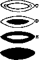
True cataracts are characterized by the appearance of grayish-white opacities in the substance of the lens or on its capsule. By location, true cataracts are divided into capsular (cataracta capsularis) and lenticular (catar. lenticularis). Capsular cataracts. Of capsular cataracts that develop in isolation, without opacification of the lens itself, the anterior polar and posterior polar cataracts are of clinical significance (figure 1 A). In the first (cataracta polaris anterior), on the anterior capsule of the lens, usually at its anterior pole, i.e., opposite the center of the pupil, a sharply limited, rarely larger than a pinhead, Figure 1. Schematic representation of various forms of cataracts: A - anterior and posterior cataract; B - central and spindle-shaped; C - lamellar; D - nuclear; E - cortical; F - complete. intensely white opacity is observed (see separate table, fig. 1); sometimes the latter projects conically into the anterior chamber (s. pyramidalis). The basis of the cataract is limited proliferation of the capsule epithelium and the formation of rudimentary fibers that resemble connective tissue in appearance (fig. 2). Sometimes this cataract occurs as a congenital change, representing the result of an abnormally long-lasting adhesion between the capsule and the posterior surface of the cornea. But more often it develops during life as a result of perforation of the cornea by ulcerative processes. After the escape of aqueous humor through the perforation opening, the lens adheres to the posterior surface of the cornea. After the chamber is restored, when the lens moves back to its place, an opacity forms on its capsule in the area of the pole, and it is not necessary that the pole of the lens itself was in contact with the perforation opening. This cataract particularly often develops after perforating ulcers of the cornea in gonorrhea of newborns. By itself, the anterior polar cataract, due to its small size and sharp outlines, does not lead to any noticeable decrease in vision and therefore usually does not require special intervention. The visual impairment observed with it depends less on the cataract itself than on the opacities of the cornea that remain after the ulcer. Only with large sizes of the cataract, especially if it has led to the formation of folds on the capsule, does vision suffer, and surgical intervention may be necessary - usually optical iridectomy.-In posterior polar cataract (fig. 1 A), a limited, usually round opacity is observed at the posterior pole of the lens, detectable only upon examination in transmitted light.-The change is always congenital. In most 2. Anterior [polar cataract. cases, it is not a matter of capsule opacification, but of the deposition on it of a tissue layer consisting of elongated cells with a large number of vessels and representing remnants of the embryonic vascular capsule of the lens. Very often, remnants of the embryonic hyaloid artery (a. hyaloidea persistens) are encountered in the form of a more or less long strand extending from the posterior pole into the vitreous body. Sometimes congenital opacities of the capsule itself or the adjacent layers of the lens are also encountered. Essentially, they represent a rudimentary form of lamellar cataract and are often encountered in combination with it. It is almost impossible to distinguish between deposition on the capsule and its opacification using ordinary research methods; for this, examination with a slit lamp is required. This form of cataract does not require intervention, as it itself does not affect vision. As an acquired change, posterior polar cataract is encountered in pigmentary retinitis and in chorioiditis of various origins. It may remain stationary, but often, especially in chorioiditis, it passes into posterior cortical and even complete cataract. Cataracts of the lens itself are divided by degree of spread into partial and complete, and by course into stationary and progressive. It is clear that progressive cataracts in their initial development are partial; but unlike stationary ones, the opacification gradually increases until it captures the entire lens or at least all its cortical layers. In stationary cataracts, the opacification as a rule remains limited throughout life. An idea of the various forms of partial and complete cataracts is given in fig. 1. Stationary cataracts The most common form is zonular, or lamellar cataract (c. zonularis, s. perinuclearis). It is characterized by the presence between the nucleus and the cortical layers of a cloudy layer that covers the nucleus both in front and behind, like a skin covering a nut (fig. 1 C). The nucleus is either transparent or more often slightly cloudy, the cortical layers are always transparent. Clinically, lamellar cataract, upon examination with side illumination, appears as a cloudy gray disk lying somewhat behind the plane of the pupil (see separate table, fig. 2). The center of the disk is less cloudy than the periphery. Often along the periphery of the opacity there are small radial projections resembling the spokes of a steering wheel. Each projection represents a small loop, one limb of which goes to the anterior surface of the disk, the other to the posterior, so that the loop sits as if astride the disk (riders). Upon examination in transmitted light, the disk appears dark, its center usually slightly transmits a faint red light. At the periphery, the disk is surrounded by a ring of normal red reflex of the fundus (see separate table, fig. 7).-The size of the opacity varies, usually its diameter is 4-6 mm, rarely larger; but even with a diameter of 4 mm, the cataract covers the entire pupil and causes a decrease in vision to some extent, depending on the intensity of the opacity. With strong opacification, vision is rarely better than 0.1. Sometimes rudimentary forms of cataract are observed, in which the opacity does not capture the entire layer, but only a more or less extensive sector of it; often the opacity is particularly pronounced in that part of the sector that lies behind the nucleus, while the opacity of the anterior part of the sector is significantly weaker. Lamellar cataract is almost always bilateral and in most cases genotypic, hereditary disease. Acquired lamellar cataract is rare. This cataract develops after some disease (iritis, corneal ulcer) or injury to the eye, but sometimes without a demonstrable cause, especially in early childhood. Lamellar cataract is most often found in people who suffered from rickets and convulsions in childhood. The convulsion-induced shaking of the lens and the resulting loosening of the connection between the nucleus and the cortical layers is, according to previous authors, the moment that causes the development of cataract. According to this view, lamellar cataract should be an acquired, though at a very early age, change. However, it is now proven that in most cases it is a congenital disorder. With lamellar cataract, there are almost always other changes in the body - rachitic bone lesions, hypoplasia of tooth enamel, tetany. All this forces one to seek the cause of development in changes in the body itself, especially in disorders of the endocrine system. In view of the undoubted connection between lamellar cataract and tetany on the one hand, and between tetany and insufficient function of the epithelial bodies on the other, it is very possible that the cause of the cataract lies precisely in the hypofunction of these bodies. This is all the more likely that experimentally - upon removal of the epithelial bodies, and clinically - upon their hypofunction, cataract often develops, and in the eye the same changes are found as in lamellar cataract, namely changes in the ciliary epithelium, which plays a major role in the nutrition of the eye. Obviously, in this case, a change in the composition of intraocular fluids occurs, which leads to opacification of those layers that developed precisely during this disorder. It is hardly possible to think that this change is qualitative; rather, it is only a quantitative change in the concentration of salts in the intraocular fluids, which disrupts the processes of osmosis and causes opacification of the lens.-Lamellar cataract is almost always stationary. The prognosis for it is favorable in most cases. If the visual acuity is sufficient for reading and other fine work (not less than 0.3), there is no need for surgical intervention. With a more severe decrease in vision, it is necessary. If adequate vision is obtained upon dilation of the pupil, one can limit oneself to iridectomy, the advantage of which is that the eye retains the ability to accommodate. In this case, the peripheral, flatter and also always more or less astigmatic parts of the cornea are used for vision; therefore, vision never becomes very good. In view of this, the most indicated operation will be the removal of the lens by discission with subsequent paracentesis or directly by linear extraction. Vision after the operation is good in most cases. Only rarely, even after a successful operation, vision does not improve noticeably, obviously due to insufficient development of the light-perceiving apparatus of the eye functionally. Usually in such cases, nystagmus is observed. Close to lamellar cataract are central and spindle-shaped cataracts (fig. 1 B). In c e n t r a l cataract, the opacity captures the central parts corresponding to the nucleus of the lens, which is often displaced somewhat posteriorly.
Central cataract occurs either in isolation or in combination with lamellar cataract; like the latter, it is often genotypic, inherited as a dominant trait. When combined with lamellar cataract, the most pronounced opacity is noted in the center of the disc—whereas in ordinary lamellar C., the center, conversely, is relatively transparent. - In spindle-shaped C., along the axis of the lens there is an opacity having the appearance of a more or less thick strand connecting the anterior pole with the posterior. In the middle of the strand, corresponding to the nucleus of the lens, there is a bulge surrounding the nucleus. The change is always congenital and, like central C., develops as a result of a disruption in the normal course of lens development. In these forms of partial C., the cause is very likely due to a disruption in the process of the lens detaching from its vesicle. In at least some cases, complete congenital soft C. (Fig. 1 F) is in rather close relation to lamellar C. The entire lens is found to be uniformly opaque at birth, without any indication of the radial arrangement of fibers. Anatomically, the changes are completely identical with those observed in lamellar C. This similarity of changes, due to the fact that lamellar C. can soon after the child's birth transform into a complete form, brings these two forms closer together, between which there is only a quantitative difference in the sense that complete C. represents only a more advanced stage of lamellar cataract. But of course this position holds true only for some cases, since undoubtedly in another, perhaps even more significant group of cases, there are differences between the two types of C. First of all, functionally, the eye with complete C. is always underdeveloped, and in most cases after the operation, vision is not very satisfactory, whereas with lamellar cataract, vision is good. Additionally, very often with complete C., there are other changes in the eye indicating deep developmental disorders—remnants of a. hyaloideae, opacities of the cornea, a mild or even pronounced degree of microphthalmia, traces of past iritis, etc. All this suggests that in such cases, the cause of the development of C. is different than in the lamellar form. Here it is necessary to consider disorders in the development of the lens (a delay in its detachment from the ectoderm) or in the reverse
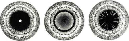
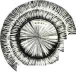
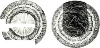
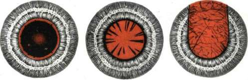
1jmc. L Anterior polar cataract*. Layered cataract with lateral illumination, 4. Developing cataract with reflected light*, 4. Immature cataract, a. Mature cataract. ft. Secondary cataract with reflected light*, See Cataract, development a. hyaloid., as well as intrauterine inflammations, most likely on the basis of syphilis. Complete cataract can be both bilateral and unilateral. Very often due to resorption of cortical layers, wrinkling and deposition of lime salts, it turns into a b. or m. dense, dry, intensely white membrane covering the pupil - membranous C. (c. membranacea). Sometimes the process occurs still in utero, and the child is already born with a membranous C. Treatment - surgical removal of C. (discission with subsequent paracentesis, linear extraction, extraction of the membrane); but it must be borne in mind that the prognosis in terms of vision is by no means always favorable. Progressive C. Depending on whether the entire lens or only its cortical layers become cloudy, a complete C. (c. totalis, fig. 1 F) and cortical (c. corticalis, fig. 1 E) are distinguished. By consistency, progressive C. are divided into soft (c. mollis) and hard (c. dura). Consistency is determined mainly by the presence of a nucleus, which forms relatively early; by 25-30 years there is usually a distinct nucleolus in the lens, but its consistency is still very soft, it noticeably hardens only by 40-45 years, and only after this age can one speak, in case of lens clouding, of a hard C. Consequently, all C. developing after the age of 40-45 years belong to hard cataracts, while C. in younger individuals will be soft. Senile C. (c. senilis) - a typical representative of progressive forms. All those C. in elderly people for which no general or local cause can be found are included here. From this it is clear that the concept of senile C. is rather vague and is based mainly on the age of the patients; when it is difficult to determine the true cause of C. development, C. that are caused by some other change in the body may and often do fall into the group of senile ones. E.g. when C. develops in an elderly diabetic, it is not always possible to decide whether to attribute it to diabetic or senile C. Patho-anatomically in c. senilis, first of all, degenerative changes of the capsule epithelium are found - its cells increase in size, their protoplasm is stained weakly, vacuolizes, while the nuclei are poorly stained or, conversely, turn into shrunken, intensely stained clumps. Sometimes limited proliferations of epithelium are encountered, which under the capsule form the formation of a b. or m. thick layer of tissue consisting of elongated fibers resembling connective tissue. The formation of epithelium on the posterior capsule, which is normally devoid of it, is also often observed. Here this false epithelium forms a layer of enlarged, swollen, often vacuolized cells, loosely connected with the cortical masses. Extensive changes are observed in the cortical fibers; they lose their regular outlines, appear swollen, the boundaries between them disappear. Slits form between the fibers, filled with protein fluid, first transparent, then cloudy. As the fibers break down, they turn into a homogeneous or crumbly mass. The edges of the still preserved fibers are eroded, between them lie coagulated protein fluid and large round formations, the so-called myelin balls. In the end, all cortical layers turn into a uniform, fine-grained or liquid mass. The pathogenesis of senile C. is not yet sufficiently clear. The theory of Becker (Becker), who explained the origin of C. by shrinkage of the nucleus and tension and layering of the lens layers as a result, was quite widely accepted. The tension should be particularly strong at the equator of the lens, where younger and therefore more loosely connected fibers lie, and where moreover Zinn's ligament counteracts the tension from the nucleus and thus promotes the layering of the fibers. The first slits and the first breakdown of fibers appear in the area of the equator, followed by clouding of the entire lens. As simple and illustrative as this explanation may be, it cannot be generalized for all cases of C., since the reduction and shrinkage of the nucleus, which is a necessary prerequisite for the theory, is not always present. Besides, it is completely incomprehensible why the first cloudings appear in most cases subcapsularly near the equator of the lens, and not in those layers that are first subjected to tension and detachment from the nucleus, i.e. in the ones closest to it. Becker himself noted that shrinkage of the nucleus alone is not sufficient; he particularly emphasized that C. is a bilateral disease and that the cause of its development must lie in the entire organism.-If we set aside the unsuccessful attempts to explain the development of C. by nephritic processes and sclerosis of the internal carotid artery, then among the theories connecting cataract with general changes in the entire organism or at least the entire eye, the views of Peters, Romer, Golovin and Hess deserve attention. Peters thinks that due to senile changes in the ciliary epithelium, which is the organ producing intraocular fluid, the composition of the latter changes in the sense that the concentration of salts contained in it increases. For the nutrition of the lens, which occurs by osmosis, it is necessary that the concentration of salts in the lens be higher than in the fluid surrounding it. With an increase in the concentration of the latter, the difference in osmotic pressure between it and the lens fluid decreases, and equilibrium can even be established. This disrupts the intake of nutrient fluid, leads to a disturbance of lens nutrition and can give rise to its clouding. However, verification of this view by Romer showed that in senile C. there is no prolonged increase in the molecular concentration of intraocular fluid; if fluctuations in this concentration are observed, the lens responds with a corresponding change in its own concentration. Hess, based on his anat. studies in C., in which he found the degenerative changes of the capsule epithelium described above, sees in these changes the cause of clouding. The degenerated epithelium can no longer regulate the intake of nutrient fluid into the lens; it lets through all substances, including those toxic to the lens, which leads to its clouding. In confirmation, Hess cites the similarity of anat. changes in the epithelium with those observed in naphthalene and other toxic C.-Romer sees the cause of the changes in the epithelium and clouding of the lens in the action of special harmful substances of the cytotoxin type, formed in the body; usually they do not reach the lens, as they are retained by the ciliary epithelium; but in senile and other changes they apparently get the opportunity to act freely on the lens. A similar view on the origin of senile C. was expressed by Golovin even before Romer, but not developed by him in all details.-In connection with the increasingly revealed role of the endocrine system in the life of the organism, attention was also paid to this aspect. First of all, it is necessary to point to the attempt to connect C. with obvious or latent tetany and disturbance of calcium metabolism. According to the studies of Triebenstein, latent tetany is present in 88.2% of patients with senile C. However, verification of these data on a large material, and the studies were not limited only to the symptoms of latent tetany, but the amount of calcium in the blood was also determined, gave no indications of disturbance in the parathyroid glands in senile C. In a broader form, the question of the dependence of senile C. on changes in the function of the endocrine apparatus is raised by other authors, who assign a major role to the senile involution of the endocrine glands, especially the sex glands. The question still needs further development.-In recent times, approaches to the pathogenesis of C. have also been made from the point of view of physicochemical processes taking place in the body. Jess by means of microchem. analysis established that throughout life there is an increase in the weight of the lens, which depends mainly on an increase in protein content (in youth 32% protein and 67.5% water, in old age 38% protein and 61.5% water). In old age among proteins albuminoids predominate, whereas in youth crystallins. In C. a loss in weight of the lens is observed, the amount of water and the amount of proteins decrease, from the latter crystallins decrease mainly, while the amount of albuminoids even increases. This sharp shift in protein content in the cataractous lens, in which insoluble proteins begin to predominate, Jess explains by the action of enzymes released from the cells of the capsule epithelium during their senile death. The enzymes first destroy the superficial, and then the deeper layers of the lens fibers.-Gold Schmidt (Goldschmidt) developed a purely chemical theory of the origin of senile C. According to him, C. develops due to a disturbance of the internal respiration of the lens.
The oxidation processes in the lens occur with the help of glutathione contained in it, which gives up its hydrogen to oxygen, turning into oxidized glutathione. A special thermostable reducing system restores the oxidized glutathione, and the process begins anew. For the proper course of the process, a certain concentration of hydrogen ions in the aqueous humor is necessary. With an increase in this concentration, the process of internal respiration of the lens is disrupted, which leads to its clouding. This is also caused by a deficiency of glutathione and the reducing system, either together or separately. Some physical theories explain the development of C. by the action of ultraviolet rays. This interpretation was prompted by observations of the widespread prevalence of C. in countries, especially rich in sunlight (India), as well as experimental data on the effect of these rays on the isolated lens. Schanz sees the cause of C. in the direct harmful effect of ultraviolet rays on the lens fibers. But according to the opinion of van der Hoeve, the rays cause C. indirectly, through the ciliary body. The rays absorbed by the lens undergo diffuse scattering within it, and part of them falls on the ciliary body, causing changes in its epithelium. As a result, a disruption of its function occurs, a change in the composition of the intraocular fluid, and from this, C. Finally, according to Vogt's observations with a slit lamp in the lenses of very young people, cloudings appear, located in the form of a wreath around the equator of the nucleus (c. coronaria). These cloudings are the first sign of senile decay of the lens. In the future, they increase in number and lead to complete C. If not all people develop C., it is only because not all people live equally long. The difference in the time of onset of development of these cloudings depends mainly on the genotypic features of the organism.
* As can be seen, the pathogenesis of senile C. is still very far from a unified explanation. The course of senile C. In most cases, senile C. begins at the periphery of the lens, in the cortical layers closest to the capsule. The appearance of opacities is often preceded by the development of myopia, which may depend both on an increase in the general refractive index of the lens due to sclerosis of the nucleus, and on an increase in the convexity of the lens due to some swelling of it. The appearance of true opacities often begins with the formation of so-called water clefts. Due to the splitting of lens fibers or lamellae, clefts filled with transparent fluid form between them. When examined in transmitted light with a strong magnifier placed behind a plane mirror, the clefts appear as light, stripe-like bands, which, if the mirror is moved slightly, sometimes flare up with bright light, sometimes become dark, and sometimes disappear. Sometimes these clefts remain transparent, and the condition does not progress to C. But in most cases, the clefts are followed by the appearance of true opacities. In accordance with the structure of the lens, which consists of radially arranged lamellae made of cemented together lens fibers, the opacities appear as radially running streaks directed with their pointed apexes toward the anterior and posterior poles. Most often, the first opacities appear in the lower periphery of the lens. Less frequently, the first opacities appear not in the subcapsular, but in the layers closest to the nucleus (supranuclearis). Here too, they initially appear as radial bands starting from the equator of the lens and forming a narrow circular line around it; later the bands merge and form a thin cloudy haze lying at the boundary between the nucleus and the cortical layer. In very rare cases, senile C. begins with diffuse thin opacification of the nucleus itself (nuclearis, see Fig. 1D), while the cortical layers remain more or less transparent. All initial opacifications of the lens, when examined with side illumination, appear grayish-white against the black background of the pupil (see separate table, Fig. 3), and when examined in transmitted light, they appear dark against the red background of the fundus reflex (see separate table, Fig. 8). The very earliest stages of opacification require dilation of the pupil for their detection, as they are mostly located at the extreme periphery of the lens. The examination of lens opacifications is greatly aided by corneal microscopy with a slit lamp, with which the finest opacifications can be detected. Observations by Voigt showed that in most people, very early in life, in the 3rd-4th decade, small opacifications of the lens appear surrounding its equator like a crown (coronaria).- The subjective complaints of patients with incipient C. are very varied. With peripheral opacifications, there may be no complaints of visual impairment, and C. is discovered during accidental examination, or patients may complain of the development of myopia or of polyopia, which manifests mainly when looking at bright, luminous points. This phenomenon is explained, firstly, by irregular lenticular astigmatism due to the formation of clefts in the lens, but mainly by the fact that radial opacities extend into the pupillary area and divide the lens into a number of transparent and opaque segments that disrupt the proper refraction of light. A noticeable decrease in vision is observed when cataract development begins in the nucleus or in front of the nucleus, as here the opacifications are located directly opposite the pupillary area. With further development of C., the number of opacifications increases (see separate table, Fig. 4); they merge with each other and form a more or less uniform opacity with a clearly expressed radial structure, located at some distance from the capsule (nondum matura). The opacity is more pronounced at the periphery, less intense at the poles of the lens. The red reflex of the fundus is obtained in places. Vision decreases significantly. Simultaneously with the increase in opacifications, there is an increase in the volume of the lens, which is clinically detected by a noticeable flattening of the anterior chamber (intumescens). Eventually, all cortical layers become opaque, C. passes into the mature stage (matura; see separate table, Fig. 5). At this time, all cortical substance of the lens appears uniformly opaque, grayish-white in color. The radial structure, sectors, and the seams between sectors that form the pattern of the lens star are clearly distinguishable at first. The opacity extends right up to the capsule, so that the pupillary edge of the iris lies flush against the opaque surface of the lens, whereas in the preceding stage there was always a transparent gap between it and the opacity. The volume of the lens decreases due to the excretion of water, and the chamber regains its original depth. Vision in this period is reduced to the perception of light, and the patient not only perceives light but can correctly indicate from which direction light is falling on the eye at any given moment (correct light projection). If left to its own course, senile C. undergoes further changes, which are designated as overripe C. (hypermatura). Most often, wrinkling of the opaque masses is observed due to the continuing excretion of water. The volume of the lens decreases, the opaque cortical layers turn into a dry, patternless mass, through which the yellowish nucleus is slightly visible. The depth of the anterior chamber increases, and the iris, not finding proper support in the shrunken lens, trembles with eye movements. On the capsule, due to the proliferation of its epithelium, opacities appear that stand out against the general background of the opaque lens with their intensely white color. Less frequently, in overripe C., the opaque masses liquefy, forming a uniform milky fluid-liquid, or Morgagnian C. (fluida, s. Morgagniana). The nucleus, due to gravity, sinks downward, and in the pupillary area its upper arched edge is visible; when the head is tilted forward, the nucleus sinks to the anterior pole and becomes visible in the pupillary area. With further progression, the opaque liquid masses may completely disappear, the pupil becomes black again, and only the nucleus remains in the capsular cavity, lying in its lower part. Vision returns and, with correction by appropriate glasses, can be good. Such cases of spontaneous healing of C. are, however, very rare.-Senile C. is almost always a bilateral disease, but both eyes are not always affected simultaneously, and the rate of development of C. in both eyes can be different. The interval between the involvement of both eyes also varies, and often in one eye C. is already mature, while in the other it has not yet developed or there are only insignificant peripheral opacities. Regarding the speed of maturation of C., large fluctuations are observed. Sometimes C. matures very quickly, within several weeks or months, but more often its course is slow, and individual peripheral opacifications may for years or even decades not change their appearance. In general, it can be said that the younger the subject, the faster the maturation proceeds. Among the progressive C. for which the general diseases of the body causing them have already been clarified, one of the most common forms is diabetic C. In appearance, it presents nothing specific that would allow it to be immediately distinguished from senile C. In young diabetics, it begins to develop near the equator and very quickly, sometimes within several weeks or days, involves all layers directly under the capsule, forming a pearl-gray opacity. In older diabetics, C. develops more slowly, and if it appears after the age of 50, it is not always possible to determine with certainty whether it is a diabetic or senile C. C. develops only in severe cases of diabetes and, with rare exceptions, affects both eyes. It occurs in approximately 5% of diabetics. An increase in sugar content in the aqueous humor plays no role in its development, as the amounts penetrating into the lens are too insignificant to cause its opacification. The opacification is probably caused by the toxic effect on the capsular epithelium and then on the subcapsular layers of abnormal metabolic products in diabetics. In very rare cases, clearing of C. has been observed, occurring when sugar in the blood and urine decreased. Since such clearing is very difficult to expect, the only method of treatment remains surgery. Due to the great tendency of wounds in diabetics to suppurate, surgery requires great caution and prior treatment of diabetes. Among other C. associated with general changes in the body, those developing in severe ergot poisoning and myotonic dystrophy are significant. All of them are associated with disorders of the endocrine system, and in character and course they do not differ from senile C., and their etiology is established only through general examination of the body.-Traumatic C. also belongs to the progressive C., developing after damage to the capsule in eye injuries. Initially, partial opacification usually involves the entire lens, and cases where the development of opacification stops and C. remains partial are relatively rare.
The clouded masses of the lens gradually dissolve, and in young subjects the condition may end in complete dissolution or the formation of a membranous cataract. In older age, complete dissolution does not occur. A traumatic cataract can develop without disruption of the integrity of the membranes in cases of contusion of the eye. In such cases, the condition is either a rupture of the capsule somewhere in the equatorial region, or a strong shaking of the lens, which has caused degeneration of its epithelium and shifts in the arrangement of the fibers and their mutual connection. The prognosis of traumatic cataract depends on the nature of the injury. If it is limited only to the peripheral parts of the cornea and the lens and is not accompanied by infection, then vision after dissolution or surgical removal of the cataract can be very good. But often the condition involves serious damage to the deep tissues of the eye, accompanied by inflammation, which significantly darkens the prognosis. Complicated cataracts are those opacities of the lens that develop as a result of some eye disease. In pigmentary retinitis and in high myopia, opacity of the posterior cortical layers (c. corticalis posterior) is often observed, having the appearance of either a large or small gray disk lying deep in the layers of the lens or a radiant star with its concavity forward. In chronic iridocyclitis leading to destruction of the eye, a cataract also usually develops, first posterior cortical, then involving the entire lens. Very often in this case, degenerative changes are noted in the lens, deposits of cholesterol crystals, lime, etc. The cataract is often adherent to the pupillary border of the iris (c. accreta). Professional cataracts. These include first and foremost opacities of the lens in glassblowers. In workers in glass factories working near glass-melting furnaces at very high temperatures, a round-shaped opacity of the lens located at the posterior pole is sometimes observed. Over time, the opacity spreads forward and involves the entire lens. The cataract usually develops in workers with many years of experience and rarely occurs before the age of 40. If in the early development the characteristic form of the opacity allows establishing the etiological dependence on the professional hazard, then in more developed opacity it is not always easy to speak of a causal connection between the opacity and the profession, since it concerns already elderly people, over 50 years old, in whom senile cataract cannot be ruled out. The cause of the cataract lies in the effect on the lens of thermal rays, mainly infrared, the focus of which falls near the posterior pole of the lens. A cataract from the action of thermal rays can also develop in other workers forced to work for a long time at high temperatures, e.g., in workers in metal factories, at smelting furnaces, at metal autogenous welding, etc. For prophylaxis, it is important to protect the workers' eyes from the harmful effect of these rays with the help of glasses made of glass that does not transmit thermal rays. For this purpose, glasses with an admixture of iron salts (e.g., so-called Robon glass) are recommended. To professional cataracts also belongs the cataract from the action of high-voltage current that has passed through the worker's body (short circuit). The cause lies in the death of the capsule's epithelium under the action of the current and the resulting disturbance of the lens nutrition. The shaking of the lens is also of importance; the cataract sometimes develops quickly after the injury, and sometimes after many weeks and even months. Similar to this cataract is the one that develops after lightning has passed through the body. Finally, to professional cataracts belong those that sometimes develop after prolonged work without proper protective measures with X-ray and radium rays. Here, the cause lies in the harmful effect of rays with very short wavelength. This form of cataract can also develop in persons who for therapeutic purposes were subjected to irradiation with X-rays or radium in the head area. The diagnosis of cataract, especially if it is a partial opacity of the lens, is made mainly on the basis of examination in transmitted light. Examination with the naked eye or with the help of side illumination is not always sufficiently convincing, since even with a transparent lens an impression of its opacity can be obtained. In a well-developed nucleus, due to enhanced reflection of light from its surface, the pupil has a fairly noticeable grayish tint, similar to that obtained in true opacity. However, the nucleus remains transparent and, if such an eye is illuminated with a beam of light with an ophthalmoscope, the pupil lights up with an even red light. In the presence of true opacities, they will not allow through themselves the light reflected from the fundus, and will stand out as dark bands or spots on the red background of the rest of the fundus. Complete opacity of the lens does not present difficulties for diagnosis: when examined with the naked eye, with side illumination, and in transmitted light, the pupil appears equally grayish or bluish-white. The only thing that can be confused with true cataract in such cases is the closure of the pupil with a film of organized exudate. However, films are always adherent to the pupillary border of the iris, whereas in cataract the pupillary border is free and easily glides over the surface of the cloudy lens. Treatment of cataract. In relation to mature, almost mature, or such immature cataracts that significantly reduce vision and make the eye unfit for work, the only option is surgical removal of the clouded lens, since it is impossible to achieve dissolution of the opacities by medicinal means. In relation to beginning opacities, attempts have been made for a long time to cause their dissolution or at least stop their progression by the application of various "medicinal substances." Previously, mercury, Sulfur aurant., calcium chloride, etc., were used. At present, almost exclusively preparations of iodine are used, mainly potassium iodide, in the form of eye drops, baths (2-10%) or internally. However, dissolution of the opacities does not occur with this treatment. As for stopping further development of the opacities, in beginning cataract, especially senile, they very often for a very long time, sometimes for decades, remain in one position without showing a tendency to progress, without any intervention. Therefore, it is very difficult to link this stationary state of the opacities with the action of iodine. The same can be said about attempts to treat cataract by introducing iodine through iontophoresis. Here too, reports speak only of stopping the development of opacities, not of their disappearance. And if one takes into consideration the results of experimental research by Stemdorff, which showed that with therapeutic doses of iodine does not penetrate the lens during iontophoresis, then great hopes cannot be placed on this treatment either. In the very recent past, attempts have been made to treat cataract with preparations of endocrine glands, especially the thyroid gland. Siegrist proposed "Euplakin," which is a mixture of hormone extracts from the sex, thyroid, and parathyroid glands. With this preparation, he in a fairly large number of cases noted absence of increase in opacities and sometimes improvement in visual acuity. However, the number of cases and the duration of observation are not yet large enough to be able to speak of the advantages of this method of treatment over potassium iodide. Improvement in visual acuity in the absence of objective signs of dissolution of opacities cannot serve as a reliable criterion, since visual acuity, especially in elderly subjects, often fluctuates within fairly wide limits depending on the most diverse factors (degree of illumination, width of the pupil, degree of attention of the patient, etc.). All these considerations make it necessary to still rather cautiously relate to the results of medicinal treatment of cataract, for which, as before, surgery remains the only radical method. Of operations for cataract, discission, linear, and flap extractions are used. Discission is indicated for soft cataracts, capable of independent dissolution after opening of the capsule. It is performed with a thin discission needle or a discission knife by Knapp or Kunt. The knife passes through the cornea near the limbus into the anterior chamber and widely opens the capsule of the lens. Coming into contact with the aqueous humor, the masses of the lens swell, become cloudy, and gradually dissolve. To accelerate this very slow process, when the lens has swollen enough, paracentesis is resorted to: with a spear-shaped knife, a small incision, 4-5 mm long, is made in the cornea near the limbus, and by pressing one lip of the incision backward with a spatula, the cloudy masses are expelled from the chamber, gliding them along the cornea with another spatula or the convex surface of Daniel's cataract spoon. Linear extraction is indicated mainly for soft cataracts, while for hard ones it is done only with small nuclei, i.e., in relatively young patients. The incision (its shape and size are visible in Fig. 3) is made with a linear or broad spear-shaped knife. Then the capsule is opened with a cystotome or a piece of it is torn out with a capsule forceps, and the lens masses are expelled as described above. The iris is reposited so that the pupil becomes round again. Flap extraction is indicated exclusively for hard cataracts.
The incision is made with a Graefe linear knife. The size of the corneal flap formed (Fig. 3) should be not less than */z-a/s. Fig. A-linear, its circumference. After the corneal incision, the knife is passed a little further under the conjunctiva and it is incised to form a conjunctival flap to cover the corneal wound. Then an iridectomy is performed, or if the operation is done with a round pupil, proceed to opening the capsule with a cystotome, which is introduced flat into the anterior chamber at the level of the lower edge of the pupil, it is turned with its point toward the capsule and it is opened with two cross-cutting incisions or one circular incision. Instead of incising the capsule, many tear out the capsule in its middle portions with a special capsule forceps. Then the nucleus and cloudy cortical masses are removed with two or Daviel's spoons, as in linear extraction. The edges of the coloboma are tucked into place with a spatula to correct the flap incision, or, if the operation was done without iridectomy, the iris is placed in its position with a spatula (round pupil), the eye is cleared of blood clots, and the conjunctival flap is spread out. With normal healing, this occurs in 10-12 days. Besides these most common methods of removing C. without the capsule, there are operations for C. with the capsule; but these methods, though theoretically the best, have not yet become widespread due to technical difficulties and the danger of complications. Such are the methods of Smith, Stanculeanu-Torok. The operation for C., being one of the most responsible for an ophthalmologist, as it were, a touchstone of his technical maturity, requires very careful preliminary examination of the patient both in regard to his general condition and in regard to the condition of the conjunctiva and tear ducts. Bacteriological examination of the flora of the conjunctival sac is one of the guarantees of a successful outcome of the operation. When pneumococci and other species close to them are found, which are quite common even in the clinically normal condition of the conjunctiva, the operation is contraindicated until the conjunctiva is freed from them by appropriate treatment (washing with a 1:5,000 solution of mercuric cyanide, 3% collargol, 1:500 rivanol, 1% optochrome, etc.). In performing the operation, immobilization of the eyelids, reliable fixation of the eye, and elimination of any pressure on it are of great importance. Immobilization of the eyelids is achieved by so-called akinesia or blockade of the facial nerve, for which 3-5 cm3 (depending on the thickness of the subcutaneous fat) of 1% novocain with 3-4 drops of adrenaline is injected into the circular muscle of the eyelids 5 minutes before the operation, keeping closer to the periosteum. The injection is made at the inner and outer corners of the eyelids. The most reliable way to fix the eye is with a bridle suture applied to the tendon of the superior rectus muscle. The suture during the operation is handed to an assistant, who at the same time expands the palpebral fissure with Demar's eyelid retractors. This method of expanding the palpebral fissure is preferable to the commonly used spring eyelid retractors, as it eliminates pressure on the eye, which could lead to undesirable complications in the form of prolapse of the vitreous body. The bridle suture is better than fixation with a fixation forceps, which even in experienced hands still exerts some pressure on the eye. A forceps is needed for the suture only during the making of the incision; all other acts of the operation are performed only with suture fixation. With all these precautions and with proper technique, the operation in most cases gives a good result, and the percentage of eyes lost from infection does not exceed 1-2. As a result of the C. operation, the eye loses its lens, its refraction becomes highly hypermetropic, and the ability to accommodate is lost. This condition of the eye is called aphakia (see). Clear vision is possible only with the help of strong biconvex lenses. For an emmetropic eye, a lens of +10.0 to -11.0 D is required with the addition of a cylindrical lens to correct the astigmatism that results after the operation. For a myopic eye, a weaker lens is needed, for a hypermetropic eye, a stronger one. For clear vision at close range, due to the absence of accommodation, a second lens is needed, usually 3-4 D stronger. And ordinary biconvex lenses give good vision; however, with them, the so-called oblique astigmatism is strongly manifested, i.e., the incorrect refraction of those rays that fall on the lens at some angle to its axis. This astigmatism is especially noticeable if the eye looks without turning the head through the side or lower part of the lens, e.g., when reading or when climbing stairs. This defect is largely eliminated in the newest aspherical optical lenses (see), the best of which are the Gullstrand's cataract lenses, manufactured by Zeiss.


With the described methods of C. operation, the lens is removed from the eye without the capsule; at most, the middle part of the anterior capsule is removed. The peripheral parts of the anterior capsule and the entire posterior capsule remain in the eye along with part of the closely associated cortical layers. From these remnants, a new opacity, the so-called secondary C. (s. secundaria), can develop over time. The clinical picture of this C. is very diverse. Since the capsule always remains in the eye, a thin, more or less folded membrane with a transparent opening in the center, corresponding to the torn or incised part of the anterior capsule, is always found in the area of the pupil and coloboma, if the operation was done with iridectomy. In such cases, vision is not impaired or is only slightly impaired. A severe decrease in vision occurs if the membrane covers the entire pupil, especially if it is gathered into folds. On examination with side illumination, a more or less thin grayish membrane with sharply raised folds in the form of white stripes is found in the area of the pupil and coloboma (see separate table, Fig. 6). In transmitted light, the pupil gives a red reflex, but it is intersected by a network of dark lines (see separate table, Fig. 9), corresponding to the folds of the capsule and its thicker parts. With a more developed membrane, the fundus reflex may be absent. This is especially observed in cases where the operation was not performed cleanly and much cortical mass remained in the eye, or if the postoperative period was complicated by iritis, as a result of which a membrane of organized exudate covering the pupil was formed. On anatomical examination of secondary C. (Fig. 4), a more or less closely adjacent anterior and posterior wall with proliferated epithelium, rudimentary newly formed, highly vacuolated fibers and remnants of cortical masses are found. The latter are present in particularly large quantities behind the iris, in the area of the former equator of the lens. Here they often form a ridge in the form of a ring behind the root of the iris, the so-called lens ridge of Soemmering.

Fig. 4. Secondary cataract. Pupil and coloboma closed by the capsule; between the anterior and posterior walls of the capsule-granular mass and remnants of cortical masses (left half under the iris).
Treatment of secondary C. can only be surgical. In most cases, discission is sufficient, i.e., incision of the membrane perpendicular to the direction of its greatest tension. In the case of a very dense membrane that does not yield to incision, it is either extracted from the eye through a small corneal incision with a strong capsule forceps, or it is incised with iris scissors or special instruments (piece-emporte).
Related articles
Mentioned in
Cite this page
“Cataract.” Soviet Medical Encyclopedia. English translation of Bolshaya Meditsinskaya Entsiklopediya, 1st ed. (Moscow, 1928–1936), ed. N. A. Semashko. https://sovietmedicalencyclopedia.pages.dev/article/cataract/