Ophthalmological Instrumentation
Historical document, translated for reference. It reflects medical knowledge of the 1920s–30s and is not medical advice.
Summary
This article provides an overview of the specialized surgical instruments used in ophthalmology during the early 20th century. It details the design and application of various eyelid retractors, fixation forceps, cataract knives, discission needles, and iris instruments.
Encyclopedia article (1928–1936)
OPHTHALMOLOGICAL INSTRUMENTATION. The first to be mentioned are the most commonly used Desmarres eyelid retractors. They consist of a wide plate with a saddle-shaped notch, set at an angle on a long handle, which is convenient for placing under the eyelid (Fig. 11). These retractors come in various sizes due to the difference in the eyelid fissures of children and adults. They serve not only for opening the eyelids and holding them during examinations and operations, but they can also be used for double eversion of the eyelid and examination of the transitional fold and the conjunctiva of the upper eyelid, thereby replacing special instruments. Even more frequently used in operations are eyelid retractors of the second type—b l e p h a r o s t a t s—by which the eyelids are held with the help of two plates connected in one instrument in various ways. These instruments are required to have plates that grip the edges of the eyelids well, to open quickly, to be removable quickly, and at the same time to firmly hold the expanded eyelids. Among eyelid retractors of this kind, spring-loaded ones are in great use, an example of which is the Graefe b l e p h a r o s t a t. In such a blepharostat, the spring shape is given to the entire instrument by making it from thick, appropriately bent wire. The degree of divergence of the retractor branches is regulated by a device in the form of a plate inserted between the branches with a screw for fixing the latter at various distances from each other (Fig. 57). Furthermore, blepharostats with separate springs inserted between the branches are widely used due to their convenience. Such are the Mellinger blepharostat (Fig. 58) and the Panas blepharostat (Fig. 55). In the Mellinger blepharostat, a spiral-shaped spring encompasses a plate with teeth inserted between the branches perpendicular to them; by applying pressure to the handles of the instrument, it is possible to spread the branches, and the cessation of pressure firmly fixes the retractors in a certain position.
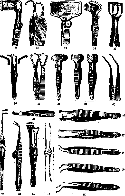
Forceps for fixing the eyeball are an essential instrument in various eye operations. Usually, forceps with relatively wide tips are used, equipped with shallow teeth at the end, with some of these forceps equipped with a lock (Fig. 47) and others not (Fig. 46). Various types of knives are used for making incisions on the eyeball during various operations. The classic instrument in operative ophthalmology is the Graefe cataract knife. This knife belongs to one of the sharpest, most delicate instruments known to surgery. It has a length of 3–3.5 cm with a width usually of 2 mm; its handle is 10 cm long; the point of the knife gradually transitions into its body; this creates the possibility, when inserting the knife for incisions, to avoid premature leakage of the anterior chamber fluid (Fig. 9). There are various modifications of the Graefe knife; in particular, some prefer significantly wider knives than the typical Graefe knife. Such is the knife used by Elschnig. The basic requirement for any cataract knife is the correctness of its manufacture and the good quality of the material (in this regard, the knives of the Collin factory in Paris have been highly valued until now). Another type of knife, very often used in eye surgery, especially for iridectomy, is the triangular spear-shaped Jager knife, bent at an angle (Fig. 18). Such knives have various sizes; the width usually fluctuates from 0.5 cm to 1.2 cm; the height of the blade reaches 1 cm, and the angle of inclination of the blade is on average about 50°. A very convenient instrument is the spear-shaped knife by Landolt, which differs from ordinary spear-shaped knives by its shape, resembling a tree leaf, and mainly by the fact that its blade is not bent at an acute angle to the handle but forms a slight arc (Fig. 13). In recent years, the Treutler double spear-shaped knife has been proposed and is used by some for cataract operations. The knife consists essentially of two half-spears with a slit in the middle (Fig. 17). Besides curved spear-shaped knives, straight ones are also used, which are especially useful for incisions of the intermarginal space during various operations on the eyelids (Fig. 19).
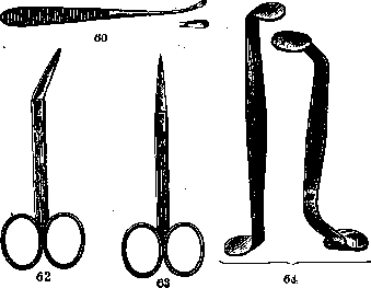
Necessary instruments for operations on the lens are discission needles. They serve mainly for cutting the anterior capsule during operations for soft cataracts or for cutting secondary cataracts. The most frequently used is the straight Bowman discission needle, the sharp end of which is flattened on both sides and sharpened (Fig. 3). For the same purpose, the Knapp needle-knife is used, representing a narrow spear-knife having one side sharpened (Fig. 5), or a similar knife-needle, only bent at an angle to the handle—the Kuhnt knife (Fig. 6). The instrumentation for cataract and iridectomy operations includes various forceps for the iris, most often in the form of narrow curved forceps equipped at the ends with low hooks (Fig. 48) or flat notches, as in anatomical forceps. For operations with basal iridectomy, the Hess iris-forceps with a double curvature of the branches is convenient (Fig. 50). A small hook, sharp according to Jager or blunt according to Himly (Fig. 4), can serve for grasping the iris. For cutting out the iris, various scissors are used; among them, the most commonly used are the Wecker pince-ciseaux (pince-ciseaux de Wecker), consisting of two plates about 15 cm long on average, having the shape of forceps branches set at an angle to each other. At the ends of the branches, at an angle of 35°, narrow little knives less than 2 mm wide are located, with one having a blunt end and the other a sharp end. By a special device, the knives can be brought together by the branches like scissors (Fig. 43). For iridectomy, ordinary small-sized scissors are also used, which are often used in eye surgery during various operations. Such scissors can be straight or curved, sharp-pointed, or blunt-pointed (Fig. 62 and 63). For cutting the anterior capsule during cataract extraction, cystotomes or capsulotomes are used. They have the appearance of sharp triangular-shaped hooks, usually set at a right angle to the shaft of the instrument (Fig. 7). Instead of cystotomes, many use capsule forceps for tearing out the capsule. These forceps are curved along their length and are equipped at the ends with a row of teeth located along the edge of the end of each branch of the forceps. Such is the Schweigger capsule forceps (Fig. 49). Removal of a cataract by expression requires the use of a Daviel spoon, which has a history of about 200 years (Fig. 8). The spoon is often connected to a cystotome or a narrow spatula, usually used as a special instrument for repositioning the iris (Fig. 26) and in a number of other cases. Recently, the instrumentation for cataract operations has also included a special forceps for grasping the anterior capsule of the lens in a fold, which is then removed according to the Stanculeanu-Török method.
Stanculeanu-Török). An example of such a forceps is the Elschnig forceps (Fig. 40). The Weber loop for extracting the lens in appropriate cases (Fig. 28) also pertains to cataract surgery. The loop is replaced by flat spoons with a diameter the same as that of the Weber loop; such is the Pagenstecher spoon (Fig. 29) or the Jaeger-Elschnig spoon (Fig. 27), which in certain cases have an advantage over the Weber loop. The spoons come in various diameters and are separate for each eye. The instrumentation for antiglaucomatous operations, in addition to those mentioned, includes the Elliot trephine of various diameters, most often 1.5–2.0 mm (Fig. 23), and then instruments for cyclodialysis according to Heine; a special spatula proposed by Elschnig and called a cyclospatula (Fig. 12) deserves attention, with which it is more convenient to perform the detachment of the ciliary body than with ordinary narrow spatulas. Instruments used in the treatment of trachoma and its complications. The expression of trachomatous follicles is performed with various forceps, but the most commonly used are the Bellarminov forceps (Fig. 37), Dohnberg forceps (Fig. 32), the Knapp roller forceps (Fig. 35), and the Kuhnt lattice forceps (Fig. 39). The Bellarminov and Dohnberg forceps differ in the shape given to the ends of the forceps, but in their concept, they have much in common: the operation is performed by them through uniform squeezing of the conjunctiva captured between the branches of the forceps. The Knapp roller forceps deservedly enjoys wide distribution due to the convenience of its use. It is equipped at the ends of the branches with rollers with blunt longitudinal grooves, and the rollers rotate on an axis, thanks to which this forceps allows for the removal of pathological products of the trachomatous conjunctiva with less damage to the integrity of the tissue. The Kuhnt expressor forceps has lattice plates at the ends; in some, both plates have a lattice, in others, one plate is solid. Here, the effect is achieved by simple compression of the conjunctiva between the branches. When removing follicles, rasps, curettes, and brushes are also used. Such is, for example, the Dohnberg rasp (Fig. 10) or the Schroeder metal brush (Fig. 24). During operative procedures on the conjunctiva, a special Graefe or Demarres scarifier is sometimes used to make incisions (Fig. 21). Diverse instrumentation is required for operations on the eyelids. Here, a Jaeger plate (Fig. 22) is often needed, on which it is possible to flatten the eyelid and protect the eyeball during incisions through the entire thickness of the eyelid. The Jaeger plate, metal or horn, has a somewhat convex anterior surface and a slightly concave posterior surface, and rounded edges, about 2–2.25 cm wide, thanks to which it is conveniently placed under the eyelid and, with a certain tension of the eyelid, in addition to flattening it, counteracts bleeding during eyelid incisions. For the same purposes—to flatten the eyelid, ensure against bleeding, and protect the eyeball—various fenestrated forceps are used even more often. Of such forceps, the Demarres forceps (Fig. 38) is widely used. Such forceps come in various sizes. The Snellen forceps (Fig. 34) is often used, which is especially convenient for operations for entropion. It is constructed separately for the right and left eye and allows the eyelid to be held in the forceps along its entire length. The Knapp forceps (Fig. 31) is built on the same principle, differing mainly in the shape of the plates. The instrumentation for operations on the eyelids with mucous membrane transplantation must include a forceps for application to the lower lip for bloodless cutting of the mucous membrane. Such is the Dohnberg forceps (Fig. 33). For operations on the eyelid cartilage—during its extirpation, during inversion according to Blaskovics, etc.—it is useful to use the Burchardt forceps (Fig. 44). For the removal of improperly growing eyelashes, epilation forceps are used. An example is the forceps shown in Fig. 41, which has well-fitted flat ends. For operations on the lacrimal organs, first of all, various probes are required; Bowman probes for insertion through the lacrimal passages are commonly used. They are of various diameters—from 0.5 mm to 1.5 mm; usually, 6 numbers of probes are in use, differing from each other in diameter (Fig. 45). To facilitate the insertion of Bowman probes through the lacrimal puncta and canaliculi, a conical thin probe is introduced beforehand (Fig. 45). The splitting of the canaliculi in necessary cases requires a Weber knife, a narrow knife with a small button at the tip (Fig. 1). Forced dilation of the nasolacrimal duct is carried out by some using probes with a significantly larger diameter (7–9 mm according to Golovin). The instrumentation for diseases of the lacrimal organs also includes the Anel syringe with hollow probes of various diameters for irrigating the lacrimal passages (Fig. 51). For the operation of removing the lacrimal sac, in addition to ordinary surgical instruments, a Müller wound speculum is used to expand the wound, arranged according to the type of spring eyelid retractor, but having hooks at the ends for grasping the edges of the wound (Fig. 59). For the same purpose serves the Axenfeld retractor, a more cumbersome and powerful instrument, allowing the wound to be expanded particularly well, stopping bleeding during the extirpation of the sac (Fig. 56). Axenfeld also proposed special spoons for scraping the mucous membrane of the nasolacrimal duct during the removal of the lacrimal sac (Fig. 60). For the Toti operation, in addition to a number of general surgical instruments, special narrow chisels, grooved, 2–2.5 mm wide, and a hammer are required, and in the modification according to Dupuy-Dutemps—a special curved needle on a long handle for suturing the nasal mucosa to the sac mucosa. For operations on the muscular apparatus of the eye, Graefe tenotomy hooks (Fig. 16) are required, as well as a Prince-type forceps, equipped at the ends of its branches with teeth on one and holes on the other, into which these teeth enter when the branches are closed, thereby allowing the captured muscle to be well fixed (Fig. 36). The Stroschein forceps (Fig. 42) serves the same purpose. For evisceration of the eyeball, Bunge spoons are used—sharp, round, flat spoons of various diameters (Fig. 14). Among the instruments for enucleation, one should point out the Wells spoon (Fig. 20), which some use to protect the eyeball when cutting the optic nerve. For operations on the orbit and especially for the Krönlein operation, in addition to general surgical instruments, special Axenfeld wound retractors are used (Fig. 64). Necessary instruments for the removal of foreign bodies from the cornea are the needle and chisel for foreign bodies (Fig. 30 and 15). The keratoplasty operation used at the present time is most often performed with Hippel trephines. For the appearance of such a device, see the article Keratoplasty, Figure 2. As individual instruments, one should mention the needle for corneal tattooing (Fig. 25), the needle-knife for vitreous aspiration according to zur Nedden (Fig. 54), the Heurteloup artificial leech for bloodletting (Fig. 52), and finally the galvanocautery (Figure 53) and thermocautery with tips appropriate for various interventions on the eye (Fig. 61).
Related articles
Cite this page
“Ophthalmological Instrumentation.” Soviet Medical Encyclopedia. English translation of Bolshaya Meditsinskaya Entsiklopediya, 1st ed. (Moscow, 1928–1936), ed. N. A. Semashko. https://sovietmedicalencyclopedia.pages.dev/article/ophthalmological-instrumentation/