Iridectomy
Historical document, translated for reference. It reflects medical knowledge of the 1920s–30s and is not medical advice.
Summary
Iridectomy is a surgical procedure involving the removal of part of the iris. Once widely performed, its indications have narrowed with advances in ophthalmology, primarily for corneal opacity, glaucoma, pupil closure, and preparation for cataract surgery.
Encyclopedia article (1928–1936)
IRIDECTOMY, iridectomia, an operation consisting of the excision of a portion of the iris. Previously, I. was very widespread, but with the improvement of ophthalmology, the indications for this operation have narrowed and are now limited to the following. 1. Opacification of the cornea (formation of an artificial pupil for optical purposes - optical I.). 2. Glaucoma (with the aim of reducing intraocular pressure - antiglaucomatous I.). 3. Closure of the pupil, ingrowth of the iris into the cornea (I. for the purpose of preventing an increase in intraocular pressure and at the same time with an optical purpose). 4. Preparation for the operation of lens removal (preparatory I.). The most important of these indications are increased intraocular pressure and cessation of light access while preserving all or some part of the transparent cornea. Preparatory iridectomy is resorted to in cases of complicated cataract, especially when one eye has already been lost. The main indication for optical I. is given by scars on the cornea (leucoma corneae), when they cover the central part of the pupil. Closure of the pupil, limited opacification of the lens and its dislocation - are also indications for optical I. It is unnecessary to resort to optical I. too early, since opacifications that have developed as a result of deep keratitis, especially parenchymatous in young people, often clear very slowly, and an iridectomy done too early may not give a positive result here. I. should be proposed only when the inflammatory process of the cornea has already completely ended. The place for optical I. should be chosen especially carefully, t. 1S. the artificial pupil should lie opposite the transparent cornea. For this, the cornea must be examined carefully with a binocular loupe or preferably with a slit lamp. Often, diffuse, delicate opacifications are found in places that seemed completely transparent upon examination with the naked eye. Only after I. does the corneal opacification catch the eye against the black background, whereas before it escaped the attention of the examiner. Small opacities missed during examination significantly interfere with the access of light in general and its proper passage into the eye. In addition to the transparency of the cornea, its curvature should also be taken into account, as often a transparent area lying near an opacity has a somewhat curved surface, which completely distorts the path of the rays, and the undertaken optical I. does not give the proper effect. In cases of widespread opacification of the cornea, there is no need to especially carefully choose the place for optical iridectomy; it should be done opposite the remaining transparent cornea. A central position of the opacification with transparency of the entire peripheral part of the cornea presents a free choice of place for iridectomy.

Figure 1.
One should strive to make the artificial pupil as close to the natural one as possible, opposite the center of the cornea, directed to one side or the other. Usually I. in these cases is made inward and somewhat downward, t. ch. the visual line b. ch. passes through the cornea somewhat inward and downward from its center. This position of the artificial pupil when viewing objects at close distance requires less convergence than in other positions, especially outward. Rothmund places the position of the artificial pupil outward in second place, Desmarres downward, and then downward-outward; Arlt places the direction inward-upward on a par with inward-downward. Most ophthalmologists avoid choosing the place for iridectomy outward and upward: the first because of strong convergence and the possibility of diplopia, the second because of the covering of the coloboma by the upper eyelid. Some authors recommend making iridectomy outward in order to expand the field of vision, which in their opinion is often more important than increasing visual acuity. An artificial pupil made for optical purposes should be as small as possible (figure 1). Deprived of the ability to react and thereby regulate the access of light into the eye, it should, to avoid blinding in bright light, let it through in small amounts. Before proposing optical I., it is necessary to examine the other parts of the eye as well, paying attention to the depth of the anterior chamber, pressure, function of the retina and visual nerve. Technique of optical I. The incision is made with the help of a spear-shaped knife, unless the chamber is too shallow. Otherwise, it is necessary to use a Graefe knife. The point of entry should lie approximately 1 mm behind the edge of the cornea, in extreme cases on the limbus. The position of the incision in the cornea itself often with a narrow transparent part of it leads to opacification of the latter, which interferes with the access of light, and optical I. does not achieve its purpose. From the position of the entry, as well as from the depth of the anterior chamber, depends the further guidance of the knife. Entry closer to the cornea and a deep anterior chamber make it necessary to hold the knife more vertically at first, entry further from the limbus and a shallow chamber - more flatly. Subsequently, the knife is guided parallel to the surface of the iris, until an incision of the required size is obtained (fig. 2). The size of the incision in optical I. is not important, since the size of the coloboma does not depend on the size of the incision. Desmarres (1847) makes an incision 5-6 mm in length, Panas (Panas; 1844) - 2 mm. A serious act in optical II. is the withdrawal of the knife, which is done slowly with special attention from the surgeon. The latter should watch the end of the knife, k-ry turns so that the end 78S

Figure 3.
Wecker's,
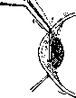
Fig.
ne edge of it passed along the iris, not the pupil, in order not to injure the capsule of the lens and not to cause traumatic cataract (fig. 3 and 4). It is not recommended to increase the size of the wound with the side of the knife, as it loses its correctness and the astigmatism of the wound is of greater magnitude. After the incision, an iris forceps is introduced into the wound, k-ry grasps the iris at the pupillary edge (fig. 5), it is pulled out of the wound and cut off with scissors, and for the formation of a smaller opening the branches are held perpendicular to the corneal incision (fig. 6). Pulling the iris with a blunt hook makes it possible to make a smaller opening. The next act is the repositioning of the iris with a spatula
z. and then the toilet of the wound. One of the unresolved questions in optical I. is the question of the operation in the presence of an absolutely good other eye. Old ophthalmologists in such cases usually did not undertake I. At present, although the condition of the second eye is taken into account, there are no contraindications to I. with a normal other eye. The results of optical I. in cases of corneal opacities are quite moderate. According to Haas, it gives an improvement in visual acuity in 70-75%, according to Hallauer (1903) - in 65-87%. Antiglaucomatous I. The reduction in intraocular pressure during optical I. and the decrease in staphyloma, noted by Graefe, led him to the idea of applying I. to reduce intraocular pressure in glaucoma. The indications for antiglaucomatous P., established by him in 1857, its application and results to this day remain almost unchanged. The meaning of glaucomatous I. lies in the liberation of a large space of the angle of the anterior chamber and in the restoration of the normal outflow path from the eye, which is difficult in glaucomatous eyes due to the adhesion of the iris to the posterior surface of the cornea and the obliteration of Fontana's space. In view of this 1) the incision should open the anterior chamber in the very angle, i.e. it should not lie corneally, 2) the iris should be cut off at its very root to free the angle of the anterior chamber, and 3) it should be cut off over a large extent (fig. 6). To avoid blinding, I. is usually made upward. The upper eyelid covers it, reduces the access of light into the eye and corrects the cosmetic defect. A coloboma downward can always be closed by hyphaema (hyphaema), which is 4. Difference-positions of the knife: 1-when cutting the cornea; 2-when penetrating into the anterior chamber; 3 - when extracting. (From Elschnig.) Gal

Figure 5.
Figure 6. quite a frequent complication in operations, especially on glaucomatous eyes. The difference in the purposes of optical and antiglaucomatous I. has also led to differences in technique. Technique of antiglaucomatous I.: the incision is made b. ch. with a spear-shaped knife; a wide knife is taken to avoid deep entry into the anterior chamber. The incision should pass 2 mm above the edge of the cornea. The line to it (fig. 7) shows the direction
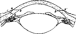
Figure 7. Anterior part of the eye in cross-section to demonstrate the conditions of the position of the angle of the chamber and limbus. The latter is located 2 mm in front of the angle of the chamber; aЬ and atbi-limbus. Graefe's knife, which is inserted in the cataract incision on the limbus in o, perforates the posterior wall of the cornea in b. For the knife to pass exactly on the limbus in a„ the entry should already begin in (>!, i.e. at the point which the operator sees 1 mm inside the limbus. (From Meller.)
incision, if the knife is placed at an angle of 40-45° to the curvature of the sclera. After the knife enters the anterior chamber, it is guided parallel to the iris, for which the handle is lowered slightly. During the incision, attention must be paid to the following: the external incision should remain parallel to the limbus, and when it reaches 7-8 mm, the knife is slowly withdrawn to avoid rapid emptying of the chamber. The cutting of the iris is also performed in a somewhat different manner. To obtain a larger coloboma, the iris forceps is opened by about 7 mm and widely grasps the iris. It is pulled out 5 mm and, in contrast to optical iridectomy, is cut in two moments parallel to the incision, i.e., first one leg is cut, then the other (Fig. 8). Pressure with scissors on the wound cuts the iris at its very root. Graefe, who proposed iridectomy as an antiglaucomatous operation, did not attempt to explain its action, since the question of fluid exchange in the eye was too little studied at that time. Subsequently, the action of I. began to be seen 1) in filtration through the scar and 2) in the liberation of the angle of the anterior chamber due to the excision of the root of the iris. Histological studies of iridectomized eyes played a major role in resolving this question. In most cases, there was a dense scar showing no signs of filtration, which led most ophthalmologists to see the influence of I. not in filtration through the scar. Only a poorly performed but effective I. can find in this an explanation of its action. The main role in I. is played by the excision of a piece of iris and thereby the liberation of the angle of the anterior chamber and the restoration of the normal outflow path of fluid from the eye. Therefore, I. should be made as large as possible, and the iris should be excised at its very root (Fig. 1). The condition of the angle of the anterior chamber strongly influences the outcome of the operation. The best effect, which is observed in acute glaucoma and in the earlier period of the disease, can be fully explained by the condition of the angle of the anterior chamber, i.e., by the pressing of the iris against the cornea when the obliteration of Fontana's space has not yet had time to develop. Antiglaucomatous iridectomy is more often applied in the acute form of glaucoma, where it gives 80-100% good results. In chronic glaucoma, the results are less favorable (50%). As for simple glaucoma, observations of recent years in the Meesmann clinic showed that a good result in this form of glaucoma is obtained when a fistulous scar forms, i.e., when the iridectomy was not performed lege artis.
Figure 8. Antiglaucomatous iridectomy is more often applied in the acute form of glaucoma, where it gives 80-100% good results. In chronic glaucoma, the results are less favorable (50%). As for simple glaucoma, observations of recent years in the Meesmann clinic showed that a good result in this form of glaucoma is obtained when a fistulous scar forms, i.e., when the iridectomy was not performed lege artis.
Related articles
Mentioned in
Cite this page
“Iridectomy.” Soviet Medical Encyclopedia. English translation of Bolshaya Meditsinskaya Entsiklopediya, 1st ed. (Moscow, 1928–1936), ed. N. A. Semashko. https://sovietmedicalencyclopedia.pages.dev/article/iridectomy/