Staphyloma
Historical document, translated for reference. It reflects medical knowledge of the 1920s–30s and is not medical advice.
Summary
Staphyloma is a protrusion of the cornea and sclera in ophthalmology, occurring after penetrating corneal ulcers with iris prolapse. It can be complete or partial, leading to decreased visual acuity and potentially serving as an entry point for intraocular infection.
Encyclopedia article (1928–1936)
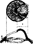
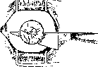
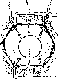
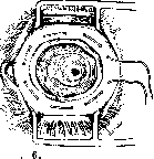
STAPHYLOMA (from Greek staphyle- grape), in ophthalmology, a protrusion of the cornea and sclera. S. of the cornea forms after penetrating ulcers of the cornea from remnants of it and the prolapsed iris. In the process of healing of the ulcerated cornea, scar tissue arises in the anterior part of the eye, which has great pliability and, under the influence of increased intraocular pressure, stretches and protrudes. A complete S. (staphyloma corneae totale) is distinguished, when the protrusion occupies the entire area of the cornea (fig. 1), and a partial one, when the protrusion is limited to only one area of the cornea (staphyloma corneae partiale) (fig. 2). With complete staphyloma, we are dealing with a scar-filled sac, filled with aqueous fluid and occupying the anterior part of the eye. The inner surface of this sac is almost always lined with atrophied iris (fig. 3). Sometimes the cavity of the sac is divided by adhesions of the iris with the lens into a number of separate cavities, and the surface of the S. becomes uneven due to the simultaneous development of new protrusions, thus obtaining a lobular S. (staphyloma racemosum). A long-existing S. is covered with rather large blood vessels; yellow spots - hyalin-degenerated areas and white necrotized foci appear on its surface. S. of the cornea acquires either a conical shape (staphyloma conicum) or a hemispherical one (st. sphe-ricum). Depending on the thickness of the wall of the S., its color changes from white and ashen-gray to bluish-black. A bluish-black S. resembles a grape, hence the name. Often the S. protrudes from the palpebral fissure so much that it becomes impossible to close the eyelids. In this case, the surface of the S. becomes dry and uneven, its apex acquires a skin-like appearance and ulcers may appear on it. S. leads to a significant decrease in visual acuity, sometimes only to light perception, often even the latter ceases. In the process of formation of S., the angle of the anterior chamber closes, which causes an increase in intraocular pressure, glaucomatous pains, an increase in protrusion and thinning of the walls. The latter may burst, the S. collapses, and then again fills with fluid. Thin walls of S. often serve as an entry gate for intraocular infection, the outcome of which is shrinkage of the eye. Partial staphyloma develops from partial prolapses of the iris in the absence of increased intraocular pressure. In this case, in the peripheral zone of the cornea there is a sharply limited whitish-gray or ashen-gray hemispherical elevation. Visual acuity is reduced due to significant astigmatism as a result of the curvature of the part of the cornea adjacent to the S. Partial S. can also serve as a gate for intraocular infection, since the epithelium covering it is thinned and easily desquamates. The formation of S. can be imagined as follows. After perforation of the ulcer, the iris prolapses, which is also involved in the inflammatory process and is covered with a fibrinous exudate. The pupil becomes occluded, the prolapsed iris adheres to the edges of the ulcerated cornea. The anterior and posterior chambers become separated. The former gradually decreases and completely disappears due to the pressing of the iris to the angle of the anterior chamber, which already leads to the closure of the outflow pathways of intraocular fluid. At this time, the posterior chamber significantly expands. In the early development of S., its pliable walls stretch, thereby compensating for the increase in intraocular pressure. Later, when the prolapsed iris with remnants of the cornea turns into dense scar tissue, signs of glaucoma appear with all its characteristic consequences: excavation and atrophy of the optic nerve head. Keeping in mind the occurrence of S. after perforation of corneal ulcers and prolapse of the iris, to which increased intraocular pressure especially contributes, preventive measures should be directed at eliminating the causes of increased intraocular pressure. Usually, when treating ulcers of the cornea, atropine is used, and the state of intraocular pressure and the depth of the ulcer are monitored, and if the former increases and there is a tendency for the ulcer to perforate, atropine is canceled and replaced with eserine. The patient is prescribed rest, timely emptying of the bowels is monitored, and any physical exertion is avoided. A moderately tight bandage is applied to the eye. If, despite these measures, there is a tendency from the cornea and iris to form S., a wide iridectomy is indicated. However, the latter is quite difficult to perform due to the severe inflammation of the conjunctiva and the significant flattening of the anterior chamber. Of the surgical methods for treating S., the simplest is the removal of the entire protrusion (staphylotomy), and Beer after such an excision of S. did not apply sutures, removed the lens, and left the exposed vitreous body to heal on its own. This method requires strict postoperative rest, long-term use of a tight bandage, and does not guarantee against recurrence and intraocular infection (fig. 4).
Figure 1. Complete anterior staphyloma
Figure 2. Partial staphyloma
Figure 3. Complete staphyloma of the cornea: 1 - conjunctiva; 2 - sclera; 3 - ciliary body; 4 - ciliary processes; 5 - Zinn's ligament; 6 - lens; 7 - staphyloma.
Figure 4. Complete staphyloma of the cornea: 1 - conjunctiva; 2 - sclera; 3 - ciliary body; 4 - ciliary processes; 5 - Zinn's ligament.
Figure k. Staphylotomy according to Beer.
Figure 5. Staphylotomy according to Critchett. Critchett proposed his method for removing S. It consists in excising S. with sutures applied to the scleral wound: first, 4-5 needles with threads are passed from top to bottom into the base of S., and the needles are not brought out but remain under S., then the latter is removed, the lens is also removed, the needles are pulled out, and each thread is tied in a knot, whereby the scleral wound is closed (fig. 5). Critchett's method does not guarantee against recurrences, and passing sutures through the ciliary body leads to its inflammation and even to the occurrence of sympathetic inflammation. To eliminate the possibility of sympathetic inflammation, de Wecker modified Critchett's method: he dissected the conjunctiva around the cornea, applied a purse-string suture to it, which was tightened after excising S. (fig. 6). Kuhnt recommends closing the gaping wound after staphylotomy with a fascia lata flap. Elschnig for the same purpose transplants mucous membrane from the lip. Kankrov recommends his keratoplastic method of staphylotomy. Its essence is that the gaping wound after excision of staphyloma is closed by means of a flap from the densest part of the staphyloma. This flap is excised
Figure 6. Staphylotomy according to de Wecker.
PlIC


Staphylotomy according to Wüsker is performed in such a way that it remains in one place in connection with the limbus of the cornea, and in three areas it is sutured with three sutures passing through it and brought out through the scleral attachments of the three rectus muscles and the scleral conjunctiva. The previously dissected conjunctiva is drawn together with a purse-string suture so that it covers the edges of the flap. The above-mentioned surgical methods for treating S. are not always accompanied by the desired success, and sometimes one has to face the risk of recurrence, panophthalmitis, or sympathetic ophthalmia. This compels many to offer patients enucleation or evisceration of the eyeball instead of staphylotomy. However, patients often refuse to undergo such radical operations. Both of these together have been the reason for proposing a method of bloodless treatment of anterior S. by injecting 10% sodium chloride into the vitreous body. Usually, a day after the injection, in the absence of any irritation, a significant decrease in intraocular pressure is observed, and after several days, flattening of the S. occurs (Koyanagi, Solovyev).-Partial S. can be excised and Kalt sutures applied to the corneal wound, which capture only part of the thickness of the cornea.-According to Berberov, complete S. and corneal scars are found: after smallpox-in 49%, ophthalmoblennorrhea-in 48%, corneal diseases-in 33%, and trachoma-in 31% of all cases of bilateral blindness on the basis of the indicated diseases (each separately). According to Krivetsky, scar changes and staphyloma are observed in 23.99% of all blind in both eyes. The prolonged existence of S. of the cornea in combination with increased intraocular pressure leads to the appearance of protrusions of the sclera, called scleral ectasias (staphyloma sclerae anticum). The thinned wall of the sclera becomes translucent when strong illumination is directed at the S. According to the location of S. of the sclera, they are distinguished: S. limbal (s. intercalate), located in the circumference of the cornea; S. ciliary (s. ciliare), developing in the area of the ciliary body, and S. equatorial (s. aequatoriale), forming in the area of the equator of the eyeball. The described S. of the sclera belong to the so-called secondary scleral ectasias and develop in the anterior part of the sclera in connection with previous inflammation of it (scleritis) and increased intraocular pressure.-Primary scleral ectasias arise without increased intraocular pressure and previous inflammation. They are localized in the posterior part of the sclera. An example of such a posterior S. is staphyloma verum, which occurs in high degrees of myopia and is visible on ophthalmoscopy. Two forms of staphyloma verum are distinguished: 1. On the nasal side of the optic nerve head, a sharply defined arcuate line, concentric with respect to the head, is visible on ophthalmoscopy. This line sharply limits the area of ectasia, the presence of which is proven: a) by the bending of vessels at the arcuate line, b) by the difference in refraction, with myopia being several diopters higher on the ectatic area, and c) by the lighter color of the ectatic area with translucency of the choroidal vessels. 2. In the second form, the line limiting the

Figure 7. Keratoglobus

Figure 8. Keratoconus.
area of ectasia closes into a complete circle surrounding the optic nerve head and the area of the yellow spot. If staphyloma verum is a sharply limited ectasia, then the so-called staphyloma posticum is an expression of uniform stretching of the entire posterior part of the sclera (see Myopia). The above-mentioned S. of the cornea are as a rule accompanied by more or less significant clouding of it. However, there is a group of transparent S. of the cornea (staphyloma pellucidum), in which the cornea protrudes sharply forward, entirely or partially preserving transparency. To this group should be assigned two classic forms: 1) conical enlargement of the cornea (keratoconus) and 2) spherical protrusion of it (keratoglobus) (fig. 7 and 8). In keratoconus, the cornea takes the form of a blunt cone, on the apex of which there is usually a cloud-like opacity. Keratoconus is an acquired condition, developing slowly between the ages of 20 and 30 years, mostly in women and in both eyes. Some believe that keratoconus results from congenital thinness of the central parts of the cornea. Due to this defect, it easily stretches under the influence of even normal intraocular pressure. Some associate the development of keratoconus with disorders of the internal secretion organs (thyroid, pituitary gland, ovary).-In the early stages of the disease, diagnosis is difficult, and a change in the shape of the cornea can be suspected by the presence of astigmatism, the degree and direction of the axes of which change. Of course, the existing irregular myopic astigmatism in this case sharply reduces visual acuity. In keratoconus, the prognosis is unfavorable both in terms of working capacity and in terms of the possibility of complications - corneal ulceration. Treatment of keratoconus is conservative-organotherapy aimed at stopping the process, and surgical-cauterization of the center of the cornea with a paculum, the task of which is to form a dense opacity preventing further stretching. Transverse cauterization of the apex of the keratoconus or its excision followed by covering with the conjunctiva is also recommended. Improvement of vision is achieved by directly applying so-called contact glasses to the cornea, the posterior surface of which has a concavity corresponding to the conical shape of the cornea. The anterior surface of these glasses is provided with a regular spherical curvature. The space between the cornea and the glass is filled with tears, as a result of which a homogeneous medium with a proper refracting surface is obtained. For the purpose of improving vision in keratoconus, a hydriasiscope is also used (see). Keratoglobus develops with congenital increased intraocular pressure. In it, along with an increase in the diameter of the cornea, an increase of the entire eye is observed-hydrophthalmus, buphthalmus; there are also other symptoms of glaucoma: high intraocular pressure, glaucomatous excavation. The anterior chamber is enlarged, the cornea is cloudy, and on it there are striae-like opacities as a result of ruptures of Descemet's membrane. Increased intraocular pressure ultimately leads to loss of vision. Treatment is little reliable: medicinal remedies are almost ineffective, and operations in the best case only slow down the process.--Keratoglobus must be distinguished from a congenitally enlarged cornea (megalo-cornea), in which no increase in intraocular pressure is observed, and such a condition should be considered as a congenital anomaly of the cornea. Megalocornea is inherited, like hemophilia and color blindness. Partial or complete protrusion of the cornea is observed on the basis of a long-existing trachomatous pannus (keratoectasia ex pannu). The scarred cornea loses resistance even to normal intraocular pressure, as a result of which such keratoectasias develop, accompanied by a severe violation of vision.
Related articles
Mentioned in
Cite this page
“Staphyloma.” Soviet Medical Encyclopedia. English translation of Bolshaya Meditsinskaya Entsiklopediya, 1st ed. (Moscow, 1928–1936), ed. N. A. Semashko. https://sovietmedicalencyclopedia.pages.dev/article/staphyloma/