Macula Lutea
Historical document, translated for reference. It reflects medical knowledge of the 1920s–30s and is not medical advice.
Summary
This article from the 1928–1936 Soviet Great Medical Encyclopedia describes the anatomy and histological structure of the macula lutea, the most sensitive part of the retina responsible for central vision. It details the specific features of the fovea centralis, including the arrangement of photoreceptors and the absence of retinal blood vessels in the center, which allows for maximum visual acuity.
Encyclopedia article (1928–1936)
MACULA LUTEA, the most sensitive part of the retina; it is the organ of central vision. It lies approximately on the optical axis and enters the field of vision when the patient looks directly into the aperture of the ophthalmoscope. [For its more precise location, color, size, and general appearance during ophthalmoscopy, see Fundus of the eye; see separate table (Vol. VII, pp. 303-304), Figs. 4 and 5.] The fovea centralis, i.e., a depression 0.2–0.4 mm wide in the center of the macula lutea, is surrounded by a low ridge, which on the periphery merges imperceptibly with the normal level of the retina, and toward the middle of the fovea descends somewhat more steeply, in the form of a gentle slope (clivus). In the middle of the fovea there is a flat area (fundus foveae), in the center of which is a small concave pit (foveola). Due to the ridge and the depression in the region of the fovea, the retinal reflexes are formed in a peculiar way.
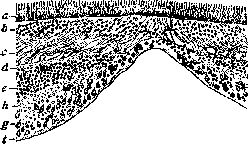
Histological picture of the macula lutea: a - layer of rods and cones; b - outer nuclear layer; c - fibrous layer of Henle; d - outer plexiform layer; e - inner nuclear layer; h - inner plexiform layer; g - layer of ganglion cells; r - layer of nerve fibers.
In its ring-shaped form, it produces a reflex of the same appearance, which has received the name of the macular reflex. The foveal reflex, however, has the shape of a sickle and moves in the opposite direction when the ophthalmoscope is moved. The macula lutea differs sharply in its structure from the rest of the retina. The fovea centralis consists only of cones, which are longer and narrower than in other parts of the retina. These foveal cones resemble rods more closely, and they can be identified only by the absence of visual purple. The greater length of the cones leads to an indentation of the membrana limitans externa in the center of the fovea. The outer plexiform layer in the circumference of the fovea changes sharply. The rod and cone fibers take on a more oblique direction and, in the circumference of the center of the fovea, lie almost parallel to the surface, forming the so-called outer fibrous layer of Henle, which occupies the space around the fovea for 8 mm. The cerebral layer of the retina spreads apart, and if one disregards the circumstance that individual cells of the second neuron may be found at the bottom of the fovea, it turns out that it consists only of elements of the first (see figure). Each cone of the macula lutea has its own bipolar cell and ganglion cell; therefore, the cerebral layer, pushed to the sides, forms a ridge around the macula lutea, which consists mainly of ganglion cells arranged in 8 rows. In addition to the cerebral layer, retinal vessels are also absent in the middle of the fovea. The nutrition of the fovea centralis occurs at the expense of the choriocapillary layer. The disappearance of the cerebral layer in the middle of the fovea, the absence of retinal vessels, and the presence of only cones lead to an increase in function to the greatest possible limits and make the fovea centralis the site of the highest function of the eye.
Related articles
Mentioned in
Cite this page
“Macula Lutea.” Soviet Medical Encyclopedia. English translation of Bolshaya Meditsinskaya Entsiklopediya, 1st ed. (Moscow, 1928–1936), ed. N. A. Semashko. https://sovietmedicalencyclopedia.pages.dev/article/macula-lutea/