SKIOSCOPY
Historical document, translated for reference. It reflects medical knowledge of the 1920s–30s and is not medical advice.
Summary
Skioscopy, also known as the shadow test or retinoscopy, is an objective method for determining eye refraction developed by Cuignet in 1873. This technique allows for determining refraction independently of the subject's responses, making it particularly valuable for examining children, malingerers, and individuals with reduced intellectual capacity.
Encyclopedia article (1928–1936)
SKIOSCOPY, or "shadow test" (from Greek skia-shadow and skopeo-I examine), is an objective method for determining eye refraction, proposed in 1873 by Cuignet under the name "keratoscopy"; it is also called "retinoscopy." This method, by its simplicity and accuracy, has great practical value, allowing determination of refraction regardless of the responses of the subject being examined. Therefore, S. is especially valuable when examining refraction in children, malingerers, and subjects with reduced intelligence. The essence of S. is as follows. If, by means of a flat or concave mirror, a beam of light is directed into the pupil of the subject's eye, the latter will illuminate a certain area of the fundus of the eye, corresponding to the shape and size of the pupil. The pupil will appear to the observer to be colored with a uniform red color. If now, without ceasing observation of the pupil through the opening in the mirror, the latter is slowly rotated around its vertical axis, the illuminated spot on the bottom of the eye will shift to one side, which will manifest itself as the appearance of a crescent-shaped shadow from the opposite edge of the pupil, which, with further rotation of the mirror, will gradually expand until the entire pupil becomes dark. At the same time, if the rotational movements of the mirror are performed quickly enough, the observer will see some shadow moving across the pupil, which in some cases moves in the same direction as the mirror, in others-toward it. The basis of the method is the observation of the nature of the movement of this shadow, since the direction of this movement itself is in close dependence on three factors: 1) the refraction of the subject's eye, 2) the properties of the mirror used, and 3) the distance between the subject and the examiner. The dependence of the nature of the movements of the mentioned shadow on refraction and the properties of the mirror used can be formulated by the following basic provisions: 1. When examining an eye with a flat mirror, the shadow moves in the opposite direction if its far point of clear vision is located in the space between it and the examiner's eye; it moves in the same direction as the mirror if the far point of clear vision of the subject's eye lies either behind it or behind the examiner's eye. 2. When examining with a concave mirror, the results are the opposite of what has been said. 3. If the examiner's eye is placed exactly at the far point of clear vision of the subject, the appearance of a shadow is not observed at all, or it is indefinite, whatever mirror is used for the examination.
The theoretical justification of S. can be presented as follows: if the examination is carried out with the help of a concave mirror with a focal distance of 15-20 cm and from a distance of 1 m, the rays falling from the light source onto the mirror are reflected from it and converge in front of it at the focus, where an aerial, inverted image of the light source is obtained, which in turn becomes a source of illumination for the subject's eye. The rays of light coming from here pass through the circular opening of the pupil and illuminate a part of the fundus of the eye in the form of a small circle. When the mirror is turned in one direction or another, the image of the flame, located in the focus, will move in the same direction, while the illuminated area on the bottom of the eye, due to the reversibility of optical refraction, will move in the opposite direction, and a shadow will approach in its place. This is what actually happens; the movement of light and shadow will appear to the observer differently depending on the refraction of the subject's eye. The path of the rays in myopia is shown in Fig. 1. The rays coming from the light source A fall on the mirror in position 1, are reflected from it and converge at its focus a, from where they go into the subject's eye, where they illuminate a certain area b. Since the subject's eye is nearsighted, the rays of light, upon exiting the eye from b, will take a converging direction and converge at the far point of clear vision of this eye, for example at c. From c they will go in the form of a diverging beam through the opening of the mirror into the observer's eye and will illuminate area c' on its retina. If now the examiner moves the mirror from left to right (it is necessary to imagine all parts of the drawing arranged not in a vertical, but in a horizontal plane), i.e., gives it position 2, then the following will result: the image of the flame a will move to a', the illumination in the subject's eye will move from b to b', the aerial image of the illuminated area from c to c', while the image of the illuminated area in the observer's eye (due to optical refraction) from d to d'. To project in space the movement of light (and shadow) that occurred in the observer's eye, the latter will, as always, be in the opposite direction, i.e., to c', so it will seem to him that the shadow (illumination) moves in the same direction as the mirror. This case corresponds exactly to the situation when the far point of clear vision of the subject's eye is between the subject and the examiner, which is possible only if the subject's myopia is stronger than 1.0 D (Fig. 2). Figure 2 illustrates the path of the rays in emmetropia, hypermetropia, and myopia weaker than 1.0 D.
Figure 1. Scheme of skioscopy. Subject's eye-myopic. 6 71
With the primary position of the mirror at 1, the rays from the light source, as in the previous case, converge at a and from there go into the subject's eye, where they illuminate area b. If the subject's eye is farsighted, the rays of light, upon leaving it, will take a diverging direction; if the subject is emmetropic, the rays will take a parallel direction, and thus in both cases the rays will not converge anywhere in the space between the subject and the examiner. If, however, the subject's eye has myopia weaker than 1.0 D, the rays, upon leaving it, although they will take a converging direction, will be able to converge only behind the examiner's eye. This implies that in these three cases an aerial image of the illuminated area cannot form in the space between the mirror and the subject's eye: the rays will go directly into the observer's eye and illuminate area d there. If now the examiner moves the mirror from left to right, i.e., to position 2, then a will move to a', b to b' and d to d'. The observer will again project the new position of the illuminated area in the opposite direction, i.e., from right to left to d'. Consequently, it will seem to him that the shadow (illumination) moves in the direction opposite to the movement of the mirror. This case already corresponds to the situation when the far point of clear vision of the subject's eye is generally outside the space between both eyes. Then one must assume one of three possibilities: either the far point of the subject's eye lies behind the examiner at infinity, if the subject is emmetropic, or it lies behind the examiner at a distance further than 1 m, if the subject has myopia less than 1.0 D, or finally it lies in the negative space behind the subject's eye, if he is farsighted. When using a flat mirror, all relationships will be reversed. In practice, S. is performed under the same conditions as ophthalmoscopy (see Ophthalmoscope). It is only necessary to relax the accommodation of the subject's eye (usually atropinization is used) and a flat mirror is more often used. To determine the degree of refraction, it is necessary to keep in mind the basic provision of S.: the disappearance of the shadow occurs if the examiner's eye is at the far point of clear vision of the subject. Based on this, S. is performed either by the so-called neutralization of shadows or by finding the neutral point. In the first case, S. is conducted from a certain distance, most often 1 m. At a distance of 1 m, the disappearance of shadows can be observed either if the subject has myopia of 1.0 D or if any refraction is brought to myopia of 1.0 D by applying appropriate optical glasses. In the latter case, the degree of refraction is determined from the calculation of the power of the applied glasses and the correction for distance (with myopia over 1.0 D, 1.0 D is added, and with other types of refraction, 1.0 D is subtracted).
The method of "finding the neutral point" is used less frequently. Its essence is that the disappearance of shadows is achieved here not from a certain distance, but, on the contrary, by changing the distance from the subject, one tries to find the point at which the shadow disappears. This method is convenient for determining only weak and moderate degrees of myopia, and therefore in high myopia, preliminary partial correction with concave glasses is resorted to. The degree of refraction is also determined from the calculation of the power of the applied glass and the distance. Astigmatism is determined by S.
Fig. 3. Skia-


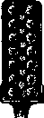
by way of studying the scanto-refraction in each principal meridian. For performing S., ophthalmoscopes with concave or flat mirrors are usually used, attaching optical glasses from eye sets when determining the strength of refraction. In addition, for this same purpose there also exists a whole series of special instruments—skiascopes—mirrors with measuring tapes, with sets of correcting glasses in special frames, mounts. Such are the skiascopes of Antonelli-Lavrentiev (Antonelli) (fig. 3), Roth, Hess, etc.
Related articles
Mentioned in
Cite this page
“SKIOSCOPY.” Soviet Medical Encyclopedia. English translation of Bolshaya Meditsinskaya Entsiklopediya, 1st ed. (Moscow, 1928–1936), ed. N. A. Semashko. https://sovietmedicalencyclopedia.pages.dev/article/skioscopy/