Fundus Oculi
Historical document, translated for reference. It reflects medical knowledge of the 1920s–30s and is not medical advice.
Summary
The fundus oculi refers to the parts of the eye visible during ophthalmoscopy, primarily the retina, optic nerve head, and choroid. It is the only location in the human body where blood vessels and nerves are directly observable, providing valuable diagnostic information about systemic diseases.
Encyclopedia article (1928–1936)
FUNDUS OCULI, a term used in clinical practice to designate those parts of the eye that can be examined by ophthalmoscopy, i.e., primarily the retina, optic nerve head, and choroid. The fundus oculi is the only place in the human body where blood vessels and nerves lie exposed and accessible to observation. Therefore, we are justified in looking for changes in the fundus oculi in diseases of the central nervous system, general infections, and disorders of other organs, in one way or another associated with changes in the blood and vascular wall. During ordinary ophthalmoscopy, the red color of the fundus oculi first strikes the eye, against which the optic nerve head (papilla n. optici), yellow spot (macula lutea), and retinal vessels stand out. The color of the fundus oculi is extremely varied and depends, first, on the source of light used for ophthalmoscopy (kerosene lamp, electric, gas, and Auer burners give different nuances of the red color of the fundus oculi), and second, on the rays reflected from the fundus oculi (namely: from the retina, pigment epithelium, choroid, and sclera). Rays from the retina and sclera are completely lost in the large number of red rays reflected from the choroid, for which reason the fundus oculi appears red. The pigment epithelium, covering the choroid like a cloudy sheath, makes the individual vessels of the choroid indistinguishable and determines both the uniformity of the coloring of the fundus oculi and its brightness. In brunettes, the pigment epithelium hardly allows the red color to show through, and the fundus oculi takes on a dark gray tone. The less pigmented a person is, the more intensely red the fundus oculi. The fundus oculi can be, first, uniformly red, if the pigment epithelium is heavily pigmented and does not allow the choroid to show through; second, mottled, when there is little pigment epithelium, while the spaces between the choroidal vessels are particularly strongly pigmented (stromal pigment), and the vessels appear as red stripes dividing the fundus into pigmented diamonds (such a fundus has been called 'parquet'); third, albinotic, in which the pigment epithelium and choroid are so poor in pigment that the sclera shows through and the entire network of choroidal vessels is visible in the form of multi-layered plexuses, standing out on a pale red background. The spaces between the loops are white due to the sclera showing through. Above them lie the retinal vessels, which are easily distinguished from the former. The choroidal vessels are wider, appear flat, less sharply defined, and have no reflex on them. In contrast to the tree-like, non-anastomosing retinal vessels, they form a dense network with the help of numerous anastomoses. Ophthalmoscopy, both direct and indirect, does not make it possible to obtain a picture of the entire fundus oculi at once; therefore, usually the optic nerve head with vessels is examined, then the central part of the fundus oculi with the yellow spot, and the periphery of the retina. On the bright red background, the optic nerve head sharply stands out in the form of a light pink disk (see separate table, fig. 4). The color of the optic nerve head is composed of the brilliant white color of the nerve fibers, the white color of the fibers of the lamina cribrosa, and the red color of the vessels. Individual variations of these elements cause those variations in the color of the disc that are observed in normal conditions. It should be noted, however, that variations in the color of the disc are extremely insignificant. In post-mortem examination of the fundus oculi, the optic nerve head appears as a small white, slightly elevated spot, which justifies the old name-'optic nerve head'. During ophthalmoscopy under normal conditions, no elevation is noted, on the contrary, a certain depression is noticeable in the center, which results from the divergence of nerve fibers in different directions of the retina. The lamina cribrosa (cribriform plate) showing through in the depression gives it a white color. A larger depression has been called the physiological excavation. The rest of the optic nerve head also does not have a uniform color. Due to the uneven distribution of nerve fibers, vessels, and the delicacy of the papillomacular bundle fibers in the disc, its temporal part, i.e., the part facing the yellow spot, has a lighter, paler appearance. The shape of the optic nerve head is round, sometimes oval. Its true size is about 11/2 mm in diameter. Due to the constancy of its size, the disc is used for measurement on the fundus oculi. During ophthalmoscopy, however, the size of the disc depends on the ophthalmoscopic method, on the lens with which the examination is performed, and on the refraction of the eye being examined. With indirect ophthalmoscopy, the disc appears larger in a hyperopic eye than in a myopic eye. With direct ophthalmoscopy-the opposite. The boundaries of the disc are sharply defined in normal conditions. The temporal boundary stands out more sharply, since the fibers going to the yellow spot have a more delicate structure. The entire disc is surrounded by two differently colored rings. The inner one, lying closer to the edge of the disc, is white-the scleral ring. It is visible because the opening for the passage of the nerve in the choroid is larger than in the sclera. Along the edge of the choroidal opening, a fairly large amount of pigment often accumulates, which creates the impression of a second pigmented ring lying somewhat outside the first and called the choroidal. From the center of the disc emerges the centralis retinae artery. It usually divides on the disc itself into lower and upper branches. Without reaching the edge of the disc, the latter in turn divide again into two, and also give off horizontal branches that go to the yellow spot-vasa macularia superiora et inferiora. Usually with such a division of the centralis retinae artery, the nasalis super. et infer. and temporalis super. et infer. are distinguished. In 5-7%, according to Elschnig, small branches of the Zinn's vascular ring can be found between the disc and the yellow spot, which emerge from under the temporal part of the disc, have a hook-like appearance, are directed to the yellow spot, and have no relation to the centralis retinae artery system. These branches have been called 'cilio-retinal vessels' (Nettleship; 1875). From the center of the disc, together with the artery, emerges a wide, dark, less reflective vein, which follows all the branches of the artery. Large arteries and veins are easily distinguished from each other: arteries are lighter, thinner trunks than veins, having a reflex in the middle. According to some authors (Dimmer), the reflex depends on the axial blood trunk, while Elschnig attributes it to the vascular wall. Jaeger explains the reflex by the fact that the vessels are not flat ribbons but small cylindrical trunks. Light, reflecting more strongly from the more elevated middle part, gives the impression of a shining line. Quite often in healthy eyes, pulsation is noted in the veins on the disc, which represents pulsatile changes in the caliber of the vessels. Arterial pulsation occurs only in pathological cases, e.g., in glaucoma, Basedow's disease, heart defects, i.e., in those cases where the pressure inside the artery is equal to the pressure produced by the intraocular fluid on the vessel wall from the outside. When examining the fundus oculi, besides the optic nerve head and vessels, the organ of central vision also deserves great attention, namely the yellow spot (macula lutea). It is located outward from the disc, approximately at V-jt of its diameter from the outer (temporal) border, slightly below the horizontal line, and lies at 15° outward and 3° downward from the disc. The yellow spot stands out as a darker oval on the rest of the background of the fundus oculi. The long horizontal transverse diameter has 172-2 diameters of the disc, and the vertical diameter-1 diameter. It is surrounded by a shining strip, which is nothing other than the light reflex (Maculareflex), depending on the ridge that forms around the yellow spot from the displacement of the retinal layer to the periphery. The center of the yellow spot has an even darker color, on which a shining spot can be noted-Fovea-reflex. The yellow spot is devoid of vessels. It should be said that examining the yellow spot presents certain difficulties, especially in elderly people who lack perfectly transparent media of the eye. A special merit in the study of the yellow spot belongs to ophthalmoscopy with a light source devoid of red rays. The new method of ophthalmoscopy proposed by Vogt generally has great importance, as it makes it possible to see what cannot be observed with ordinary ophthalmoscopy. Ophthalmologists have long noted the fact of changes in the color of the fundus oculi depending on the light source. In view of the fact that with ophthalmoscopy in red-free light, rays from the retina remain visible, while rays from the choroid are absorbed, this method, naturally, becomes suitable only for examining the retina. It makes it possible to see in the living eye the distribution of retinal nerve fibers, the papillomacular bundle, and the yellow color of the maculae luteae. The fundus oculi with ophthalmoscopy in red-free light has a completely different appearance than with ordinary ophthalmoscopy (see separate table, fig. 5).
Its color resembles that of a glowing cat's eye in the dark (blue-green). Against this background, the white papilla stands out with the black vessels of the retina. From the papilla, bending over the edge, the nerve fibers fan out in the form of white threads and follow the direction of the large vessels. Near the papilla, the fibers are coarser and more clearly defined than at the periphery. From the papilla, they mostly go upward and downward and bend toward the temporal part of the fundus of the eye. A special distribution of them is present on the temporal side of the papilla, between it and the yellow spot. The fibers of the bundle between the papilla and the yellow spot have the appearance of a bulb. At the yellow spot, they bend so sharply that they take on a vertical direction and, bordering the yellow spot on the temporal side, are lost in it, forming the so-called Kreisbogenzone. That part of the fibers which goes upward and downward from the papilla does not participate in the formation of the papillomacular bundle, also bends and crosses at an obtuse angle, and partly, without crossing, goes to the periphery. On the light green-blue background, the macula lutea stands out sharply as a bright yellow spot. - The question of its coloration was also resolved thanks to ophthalmoscopy in red-free light. The dispute between Dimmer and Gullstrand about the coloration of the yellow spot was decided in favor of Dimmer, who asserted that the yellow color depends on the pigment present in the yellow spot, and is not a postmortem phenomenon. The yellow spot can be visible only when it does not lie in the red reflecting rays, as in the normal fundus; otherwise, the yellow coloration is completely lost in the red rays. But if the red rays are destroyed, which occurs in ophthalmoscopy in red-free light, the yellow coloration becomes clearly noticeable. In the center of the yellow spot, a brilliant reflex can be noted, which is nothing other than the Foveareflex, resulting from the depression in the center of the yellow spot. In the normal condition, when moving the ophthalmoscope, it moves in the opposite direction. In ophthalmoscopy in red-free light, a large number of reflexes are also striking, which are not visible in ordinary light, but they have no pathological significance.
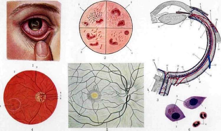
Figure 1. According to Nussbaum, fig. 1 - according to Kölliker; 2 - according to v. Kölliker; 3 - according to Mauthner-Axenfeld; 4-6. according to Nussbaum. Fig. 3. Schematic representation in the horizontal plane of the fundus of the right eye: 1 - optic papilla; 2 - central artery of the retina; 3 - central vein of the retina; 4 - papilla of the optic nerve; 5 - scleral ring; 6 - a. and v. centralis retinal; 7 and 8 - a. and v. ciliares ant.; 9 - choroidea; 10 - m. rectus med.; 11 and 12 - a. and v. ciliares post.; 13 - v. ophthalmica; 14 - sclera; 15 - choroidea; 16 - a. and v. ciliares breves; 17 - m. rectus lat. and med.; 18 - a. and v. centrales retinal; 19 - a. and v. ciliares longae; 20 - lens; 21 - vitreous body. Fig. 4. Normal fundus of the eye: 1 - yellow spot; 2 - central artery of the retina; 3 - central vein of the retina; 4 - papilla of the optic nerve; 5 - scleral ring; 6 - choroidal vessels. Fig. 5. Fundus oculi in ophthalmoscopy and red-free light. Figure 6 - 1 - rod cells; 2 - nucleus; 3 - body of Prota-Kolliker's cells; 4 - dendrites. From the article. Eye. Fundus oculi.
Related articles
Mentioned in
Cite this page
“Fundus Oculi.” Soviet Medical Encyclopedia. English translation of Bolshaya Meditsinskaya Entsiklopediya, 1st ed. (Moscow, 1928–1936), ed. N. A. Semashko. https://sovietmedicalencyclopedia.pages.dev/article/fundus-oculi/