Ophthalmoscope
Historical document, translated for reference. It reflects medical knowledge of the 1920s–30s and is not medical advice.
Summary
The ophthalmoscope is an instrument for examining the fundus of the eye, invented by Helmholtz in 1851. It operates either directly or indirectly, with various techniques to correct for refractive errors and eliminate corneal reflection.
Encyclopedia article (1928–1936)
OPHTHALMOSCOPE, OPHTHALMOSCOPY. Ophthalmoscopy is a method of examination that makes it possible to view the fundus of the eye. It is performed using an eye mirror (ophthalmoscope) and biconvex lenses with a refractive power of 10 to 20 D, or else with only an eye mirror. Ophthalmoscopy was first proposed by Helmholtz in 1851 based on the following considerations: if in a dark room a lamp is placed in front of and to the side of the patient, the pupil appears to us as a black opening. This occurs because the rays of light entering the eye from outside and illuminating the fundus, upon exiting the eye, again turn toward that light source from which they originated. In order for rays originating from the fundus of the eye being examined to enter the eye, it would be necessary to look along the beam of light falling into the patient's eye, that is, to place one's head between the light source and the eye being examined. But then the head blocks the light source, and the pupil remains black. Therefore, examination of the fundus is possible only if the eye is illuminated not directly by the light source, but with the aid of a mirror. Ophthalmoscopy is performed in a dark room. The lamp is positioned to the left and behind the patient. The doctor sits in front of the patient, holds the ophthalmoscope before his right eye, and with it illuminates the eye being examined. The eye mirror, first proposed by Helmholtz, consisted of 3-4 plane-parallel plates placed one on top of another. The light rays from the lamp are reflected by the mirror into the patient's eye, reach its fundus, and then, upon exiting the eye, pass through the transparent glass plates of the mirror and enter the doctor's eye. However, Helmholtz's ophthalmoscope proved inconvenient, because its glass plates 1) were heavy, 2) reflected only part of the rays falling on them from the light source, while the other part passed through the plates. Due to this loss of part of the rays, the illumination of the fundus was very weak. To avoid this, it was proposed to use a concave mirror with a small hole in the middle, which type of ophthalmoscope is now generally accepted. Ophthalmoscopy is performed in two main ways: direct and indirect. In direct ophthalmoscopy, examination is performed using only the eye mirror, and the examiner sees a direct image of the fundus. In this case, the fundus is viewed directly through the refractive media, which in this case act as a magnifying glass. In direct ophthalmoscopy, one must get very close to the patient's eye. In order to see the fundus clearly, the doctor must first correct his own refractive anomaly, if he has one, that is, make his eye emmetropic. The rays originating from the patient's fundus will have different directions depending on what his refraction will be. In emmetropia these will be parallel rays, in myopia-converging rays, and in hypermetropia-diverging rays (see Refraction). An emmetropic eye can focus only parallel rays on its retina without accommodative effort. Therefore, the doctor will be able to clearly see the patient's fundus in direct ophthalmoscopy without any auxiliary aids only if the eye being examined is emmetropic. Otherwise, it is first necessary to give the rays originating from the patient's fundus a parallel direction, that is, to correct his refractive anomaly. For this purpose, behind the ophthalmoscope mirror there is either a frame into which lenses can be inserted, or, what is even more convenient, a small rotating disk attached behind the mirror containing a variety of lenses. In indirect ophthalmoscopy, in addition to the eye mirror, a biconvex lens of 10 to 20 D is necessary, it is most convenient to use a 13 D lens. In indirect ophthalmoscopy, the mirror is held at a distance of about 50 cm, and the lens at a distance of 7 cm from the patient's eye. In this case, in front of the lens, between it and the doctor's eye, an inverted image of the fundus is obtained. Each of these two methods of ophthalmoscopy has its own disadvantages and advantages. In direct ophthalmoscopy, the fundus is visible at greater magnification than in examination by the indirect method, but in the latter case the field of view is larger than in the direct method. Thus, on average, in direct ophthalmoscopy the fundus is visible at 16 times magnification, while in examination by the indirect method-4 times. The degree of magnification depends on the refraction of the eye being examined. Indirect ophthalmoscopy is used for general examination of the fundus, while direct ophthalmoscopy is used for more detailed study of its individual parts. In ordinary ophthalmoscopy, there is one circumstance which in many cases hinders the examination of details of the fundus-this is the corneal reflex. When the eye is illuminated by a concave mirror, part of the rays are reflected from the anterior surface of the cornea. Since this surface represents a small convex mirror, a bright, diminished image of the light source is formed behind the cornea. With proper skill, it is usually possible to shift this reflex to the side by slightly turning the mirror, so that it does not interfere with the examination. But for beginners and for doctors who only occasionally perform ophthalmoscopy, this reflex represents
one of the main difficulties. With the help of special
constructions in recent years special apparatus for reflection-free
ophthalmoscopy has been created. These include ophthalmoscopes
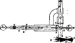
Figure 1
by Wolf, Thorner, Gullstrand. Of these, the large reflection-free ophthalmoscope by Gullstrand has become most widespread. The corneal reflex is caused by the fact that the illuminating beam of light is in no way separated from the beam of rays coming from the fundus of the eye being examined; the rays reflected from the cornea to a large extent follow the same path as the rays that give the image of the fundus. Obviously, the reflex can be completely avoided if the illuminating beam and the beam giving the image of the fundus are separated from each other, that is, if the 'entrance pupil of light' into the eye being examined and the 'exit pupil of light' from it are separated. In Gullstrand's ophthalmoscope this separation of the exit and entrance pupils is achieved in the following way (fig. 1, 2 and 3). The light source here is a very bright incandescent filament L. With the help of a condenser lens K, an image of this filament is obtained in the slit S. Lens Ah throws the image of the slit by means of a glass wedge G onto the pupil P of the eye being examined, and the entire area through which light enters the eye being examined represents a very narrow, elongated strip. The image of the illuminated fundus of the eye
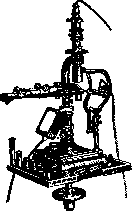
Figure 2. N is thrown by means of lens A in the direction of the objective H of the microscope, through which the observer's eye views the aerial image. With the help of the slides visible in fig. 2, the illumination system can be moved perpendicular to the plane of fig. 2 and thereby change the place where the narrow illuminating beam enters the eye. The optical system consisting of the microscope OH and the ophthalmoscope lens A has a very small entrance pupil, which is the exit pupil from the eye being examined. Consequently, with the help of the aforementioned movement of the illuminator, it is not difficult to separate the exit pupil of light from the eye and the illuminating image of the slit S so that there is sufficient distance between them, as shown in fig. 3. This eliminates the corneal reflex.-To the ophthalmoscope is attached a set of eyepieces, which provide
Fig. 3 allows for examination of the fundus of the eye with magnification from 5 to 40 times. In addition, eyepieces for binocular, stereoscopic ophthalmoscopy are also provided. The latter circumstance is especially valuable in the diagnosis of early stages of papilledema, where it is important to establish the presence of even slight protrusion of the optic disc into the vitreous body. The hand electric ophthalmoscope by Turner is also very convenient. In it, the corneal reflex is also eliminated, but it provides only small magnification. Recently, the firm of Zeiss has released a very convenient apparatus by Nordenson for photographing the fundus of the eye. Essentially, it represents the same construction as the large ophthalmoscope by Gullstrand, but only the observer's eye is replaced by a photographic camera. The same firm has released for educational purposes the polyophthalmoscope by Wes-sely, which allows eight observers to simultaneously see the fundus of the eye. The apparatus is equipped with an eyepiece with a pointer, by means of which the teacher can accurately point out to the students the place on the fundus of the eye to which attention should be paid. There are also special ophthalmoscopes with which one can examine the fundus of one's own eye—auto-ophthalmoscopy. The most rational form of these is the auto-ophthalmoscope by Wes-sely, the principle of construction of which is visible from the schematic fig. 4. Rays of light from the lamp fall on the ophthalmoscope mirror, located for example in front of the right eye. From here, by means of reflection from two mirrors inclined to each other at an angle of 85°, they are directed into the pupil of the left eye. When this pupil is illuminated, a biconvex lens is placed in front of it, by means of which the inverted image of the fundus of the eye is obtained.
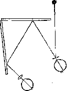
Figure 4.
A valuable addition to ordinary ophthalmoscopic examination is the method of ophthalmoscopy in red-free light, proposed by Vogt. The theoretical foundations of this research method consist of the following. The rays of light coming from the fundus of the eye in ordinary ophthalmoscopic examination consist of two components. 1. Rays of light reflected by the retina. This light beam, little changed in its composition, Vogt designates as "retinal light" (Netzhaut-licht). 2. Rays of light reflected from the pigment epithelium, choroid, and sclera. This light differs sharply in its spectral composition from the composition of the incident light, because when light passes through tissues rich in blood vessels and pigment, it loses short-wave rays and takes on a red color. This greatly altered light beam Vogt designates as "choroidal light" (Aderhautlichit). This choroidal light, mixing with the light rays coming from the retina, contributes to the fact that the details of the retina become poorly visible, as if the details of more deeply lying tissues are superimposed on them. If in ophthalmoscopy one uses light devoid of red rays, then in the light beam coming from the fundus of the eye, the "choroidal light" should be completely absent, and then many details in the structure of the retina, previously invisible, appear quite clearly. (The appearance of the fundus of the eye in this case and the significance of this method—see Fundus of the eye.). In the examination of the fundus of the eye in red-free light, direct ophthalmoscopy is used. The source of light is an electric arc. As a light filter, special glasses colored with copper sulfate are used, on which a small solid layer containing erioviolacin is applied. The combination of copper sulfate with erioviolacin absorbs all red rays.
Related articles
Mentioned in
Cite this page
“Ophthalmoscope.” Soviet Medical Encyclopedia. English translation of Bolshaya Meditsinskaya Entsiklopediya, 1st ed. (Moscow, 1928–1936), ed. N. A. Semashko. https://sovietmedicalencyclopedia.pages.dev/article/ophthalmoscope/