Eye Accommodation
Historical document, translated for reference. It reflects medical knowledge of the 1920s–30s and is not medical advice.
Summary
Eye accommodation is the ability of the eye to adjust to different distances when viewing objects. This process involves changes in the curvature of the lens, controlled by the ciliary muscle, to maintain clear focus on objects at varying distances.
Encyclopedia article (1928–1936)
Eye Accommodation, the ability of the eye to adapt to different distances when viewing objects. When two objects are located at different distances in front of us, either one or the other can be seen clearly, but both cannot be seen distinctly at the same time. This is because the optical system of our eye produces a clear image on the retina only with respect to one particular distance, specifically the distance at which the object being viewed is located at that moment; objects closer or farther than the point of fixation produce blurry images on the retina and therefore appear indistinct. To shift from viewing objects at one distance to another, we must accommodate: during this process, the conditions of refraction in the eye change in such a way that each time a clear image of the object being viewed is formed on the retina. The entire mechanism of A. was clarified by Helmholtz as early as the mid-19th century; his explanation is still considered fundamentally correct to this day. First, he established that in the eye during A. for a near object, the anterior chamber of the eye flattens due to the fact that the iris moves forward slightly, which in turn depends on the lens becoming more convex without changing its position. Changes in the curvature of the lens during A. can be studied by means of experiments with the reflection of luminous objects from the surfaces of the lens. In a dark room with proper positioning of the light source (e.g., a candle), the observer, and the eye being observed, in addition to the image from the cornea, one can also observe (a) the direct, virtual image from the anterior surface of the lens, and (b) the inverted, real image from its posterior surface. During A. for a near distance (a) moves forward and becomes smaller, (b) changes very little. From this it must be concluded that during A. for a near distance the anterior surface of the lens becomes more convex and the radius of its curvature becomes smaller. With the help of his ophthalmometer (see), Helmholtz determined the magnitude of the radius of curvature of the anterior surface of the lens during A. of the eye for distant and near objects. By calculating the data obtained in this way, it was proved that during A. for a near object, the change in the curvature of the lens leads to exactly such an increase in the refractive power of the eye as is necessary to obtain a clear image on the retina under the given conditions. As for the mechanism of changes in the lens during A., Helmholtz pointed out that the natural conditions for the attachment of the lens in the eye by means of the zonular fibers hold the lens in a stretched, »8 flattened state. When the eye is accommodated for infinity, the tension of the zonular fibers and, consequently, the flattening of the lens is maximal. As the object approaches, the zonular fibers relax due to the contraction of the ciliary muscle, and the lens, due to its elasticity, becomes more convex. The ciliary muscle (m. ciliaris), consisting of smooth muscle fibers, is innervated by the third cranial nerve (n. oculomotorius). With the strongest tension of A., the zonular fibers relax to such an extent that the lens drops down due to gravity. A. should be interpreted as refractive power and expressed in diopters. To determine precisely the tension of A. or its width, amplitude, volume, means to know by how many diopters the refractive power of the eye increased when looking at the nearest point compared to the refractive power when looking into the distance. Let us denote the refractive power when looking at the closest, clearly visible point by the letter P and at the farthest by the letter R, then P = V + A (accommodation). From this equation it follows that A = P - R, that is, in order to know the volume of A., it is necessary to subtract the refractive power needed to view the farther point from the refractive power needed to view the nearest point. Suppose we are dealing with a myope whose far point of clear vision lies at 10 cm in front of the eye, while the nearest point of clear vision is at a distance of 5 cm. The refractive power of this eye at rest will be 10.0 D; the refractive power needed to view the nearest point will be 20.0 D. The volume of A. in this case will be 20.0 D - 10.0 D, i.e., 10.0 D. In an emmetrope whose far point lies at infinity, but the nearest point, let us say, at 10 cm in front of the eye, A = 10.0 D - ∞ = 10.0 D. From this it is evident that the volume of A. is the same in both cases. As for the range of A., i.e., the space within which A. is possible, it is different. In the first case the range is 5 cm, in the second the space within which accommodation is possible lies between infinity and 10 cm. Thus, in the study of accommodation, two concepts must arise: first, the volume, width, amplitude of accommodation, expressed in diopters, and the range, space, length of A., expressed in units of length. When measuring the increase in the curvature of the lens and calculating on this basis the increase in refraction, i.e., accommodation, one never obtains the volume of it that is observed in life. Obviously, in the increase of refraction not only the increase in the curvature of the lens plays a role, but also changes in some other conditions, possibly the refractive index of the substance of the lens. Its refractive index is different at the periphery and in the center. Therefore, when the equatorial diameter decreases, which occurs during the act of A., the peripheral parts of the lens with a higher refractive index move toward the center and thereby increase the refractive index of the part of the lens through which the rays pass, which leads to an increase in refraction (Gullstrand). So far we have been speaking of one eye. The width of A., measured for each eye separately, is called absolute. A. when looking with both eyes, i.e., with a certain convergence, is called relative. For viewing closely located objects with both eyes, i.e., for clear binocular vision, it is necessary 1) that the visual lines of both eyes intersect on the object being viewed and 2) that A. be adjusted to this distance. Due to exercise, a close connection develops between the tension of the internal rectus muscles and accommodation. This connection is quite possible, since the nerve fibers for both muscles go in the trunk of the n. oculomotorii, and the nuclei lie at the bottom of the third ventricle and the Sylvian aqueduct very close to each other. Previously it was thought that convergence and A. coincide, e.g., if the eyes are directed at a point lying at 33 cm, i.e., they converge at three meter-angles, then they also accommodate to this distance and are unable to make any change in the degree of A. Donders in 1846 proved the incorrectness of this view. Between the convergence of the visual lines and A. there is some connection, but not as inextricable as was thought before. This can be easily verified by applying to the eyes, converging to a certain degree, various convex and concave lenses. Let us imagine that the subject being examined fixes some object located at 33 cm in front of the eye. In this case he converges at three meter-angles and accommodates to a certain number of diopters depending on the refraction he has. By applying convex lenses to the eye, one finally makes him relax all the amount of A. expended, replacing it with convex lenses. If, on the contrary, concave lenses are applied, they make the subject tense his A., i.e., involve his reserve of it. Reading at a distance of 33 cm will be possible as long as the entire reserve of A. is not used. The part of A. expended when reading at 33 cm and determined by the convex lens is called the negative part of relative A. That part of relative A. which is revealed by means of a concave lens and which is the reserve is called the positive part of relative A. Together they constitute the volume of relative A., i.e., the A. to which the eye is capable at a given convergence. The correct ratio of both parts of A. has great practical importance. Work at close distance is not accompanied by fatigue if the positive part of relative A. is sufficiently large, but if it is small, rapid fatigue occurs during work. Activities at close distance without special fatigue can occur when at least V» of A. remains in reserve. The question of the impulse to A. in people with different refractions in both eyes (in anisometropes) deserves attention. The different strength of A. of each eye could equalize the difference in refraction at least to a certain distance. But in binocular vision, according to Hering and Schmidt-Rimpler, both eyes accommodate equally. This can be verified by applying a concave lens to one eye and alternately closing first one eye and then the other. For clear vision, it turns out that some time is needed for the eye in front of which the lens is placed; consequently, its A. is not greater than that of the eye in front of which the lens is not placed. The pupillary reaction observed during A. is associated with the convergence of the axes, not with the act of A. The strength of A. undergoes certain fluctuations, and first place should be given to its dependence on age.
Familiarization with the changes in accommodation depending on age makes it possible to determine whether one is dealing with a pathological decrease in accommodation or whether this decrease is a physiological phenomenon. The width of accommodation gradually decreases with age, so that by the age of 40, the nearest point of clear vision in an emmetrope is located 22 cm in front of the eye, and work at close distances becomes difficult. Donders' table shows the width of accommodation at various ages. Age Width of accommodation Age Width of accommodation 10 15 20 25 30 35 40 14.0 12.0 10.0 5.5 4.5 45 50 55 60 65 70 75 3.5 2.5 1.75 1.0 0.75 0.75 The cause of the weakening of accommodation with age lies in the gradual decrease in the elasticity of the lens; the activity of the ciliary muscle remains sufficient in this case, while the impulse to accommodation is intensified. Accommodation pareses, which occur at all periods of life and pass unnoticed in youth, become noticeable in old age.
N. Pletneva, A. Samoilov.
Paralysis of accommodation. In addition to the decrease in the width of accommodation with age, i.e., a purely physiological phenomenon, there are also pathological changes in the act of accommodation, namely, paralysis, which can be complete or partial. In the emmetropic eye, paralysis of accommodation is characterized by a complete loss of the ability to distinguish small print at close distances; in paresis, this ability is only weakened; vision at a distance in an emmetrope, as not requiring accommodation, does not suffer damage. The causes of paralysis of accommodation are as follows: 1. Local, affecting the neuromuscular apparatus of accommodation; these include a) toxic paralysis from medicinal substances (atropine, scopolamine) and b) traumatic. 2. Various processes in the orbit affecting the ganglion ciliare or the trunk of the n. oculomotorii. 3. Basal processes: diseases of the brain, meninges or bones of the base of the skull involving the n. oculomotorius. 4. Central brain processes: a) lesions of the fibers of the oculomotor nerve on its path in the area of the cerebral peduncle (rare); b) lesions of the nuclei of the n. oculomotorii themselves (nuclear paralyses), the most common; c) cortical paralyses (rare). Of the acute infectious diseases affecting accommodation, epidemic encephalitis (Nonne's symptom), diphtheria should be noted; of intoxications - ptomaine poisoning and scorpion sting.
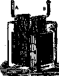
Accumulator: each of the electrodes (A and B) consists of several lead plates.
In addition to paralysis, spasm of accommodation can also be observed, which occurs more often in youth and gives the impression of nearsightedness.
Related articles
Mentioned in
Cite this page
“Eye Accommodation.” Soviet Medical Encyclopedia. English translation of Bolshaya Meditsinskaya Entsiklopediya, 1st ed. (Moscow, 1928–1936), ed. N. A. Semashko. https://sovietmedicalencyclopedia.pages.dev/article/eye-accommodation/