Oculomotorius Nervus
Historical document, translated for reference. It reflects medical knowledge of the 1920s–30s and is not medical advice.
Summary
The oculomotor nerve is the third cranial nerve that innervates most of the eye muscles. This article describes its nuclei location, pathway through the brain, branches in the orbit, and the precise localization of centers for individual eye muscles within its nuclei.
Encyclopedia article (1928–1936)
OCULOMOTORIUS NERVUS, oculomotor nerve, III pair of cranial nerves, innervates the majority of the eye muscles. Its nuclei (nucl. n. oculomotorii) are located in the mesencephalon in the cerebral peduncle, beneath the Sylvian aqueduct; they consist of the main or lateral nucleus, the unpaired central [or median nucleus (nucleus Perlia), and the lateral nucleus of Westphal-Edinger (see Westphal-Edinger nucleus). The main nucleus is 5 mm in length and, like the central nucleus, is composed of large cells, while the nucleus of Westphal consists of small cells. Fibers originating from the lower parts of the nuclei cross between both nuclei (fig. 1), while others, without crossing, run arch-shaped, cross the nucleus ruber, and emerge with numerous roots at the base of the brain, at the inner edge of the cerebral peduncle, near the substantia perforata posterior. Surrounded by the soft meninges, the oculomotor nerve passes between the a. cerebri posterior and a. cerebelli superior, directing outward and forward toward the sinus cavernosus, where it first enters its upper wall, and then the outer wall. In the sinus near the oculomotor nerve lies the a. carotis interna, nn. IV and VI; upon exiting the sinus, the nerve divides into two branches and through the fissura orbitalis superior enters the orbit cavity; in the fiss. orbitalis it lies in the inner angle, and to the outside of it is placed the n. IV. In the orbit, the oculomotor nerve innervates the eye muscles: the upper oculomotorius nervus branch, thinner, innervates the m. rectus superior and m. levator palpebrae superioris, while the lower branch innervates the mm. rectus internus, inferior and obliquus inf. (fig. 2). Fibers going from the nucleus of Westphal-Edinger are thinner in caliber; they terminate in the ganglion ciliare, located on the outer side of the n. opticus; from this ganglion begin the nn. ciliares breves, which penetrate the eyeball and innervate its smooth muscles m. ciliaris and m. sphincter pupillae (see Pupillary fibers, reflexes, centers). On the basis of experimental data and anat.-clin. observations, a more or less precise localization was established in the nuclei of the oculomotor nerve for individual eye muscles: the nucleus of Westphal-Edinger has a relation to the smooth muscles of the eye (mm. ciliaris et sphincter pupillae); in the main nucleus, the centers for various eye muscles are arranged in the following order
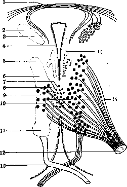
Figure 1. Diagram of the location of nuclei of III and IV pairs along the Sylvian aqueduct. On the left side in the main (lateral) nucleus of the III pair, the approximate localization of centers for external eye muscles is indicated: 1-commissura posterior; 2-nucleus of Darkshevich; 3-posterior longitudinal bundle; 4-aquaeductus Sylvii; 5-m. levator palpebrae; 6-m. rectus superior; 7-nucleus medialis; 8-m. rectus internus; 9-m. obliquus inferior; 10-m. rectus inferior; 11-nucleus trochlearis; 12-n. oculomotorius; 13-n. trochlearis; 14-nucleus lateralis; 15-nucleus of Westphal-Edinger.
(fig. 3): at its very upper end is the center for m. levator palpebrae superioris, then for muscles turning the eye upward (mm. rectus superior and obliquus inferior), and more inward for the adductor muscle of the eye (m. rectus internus); fibers originating from this middle part of the nuclei partially cross; at the lower end of the main nucleus is located the center innervating the muscle turning the eye downward (m. rectus inferior); fibers originating from this center completely cross. For combined movements
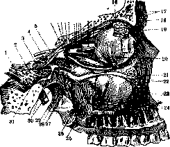
Figure 2. Nervus oculomotorius in the orbit cavity, its division into branches, termination in the oculomotor muscles, and relation to other structures: 1-n. trigeminus; 2-ganglion Gasseri; 3-n. ophthalmicus; 4-n. trochlearis; 5-n. oculomotorius; 6-its ramus inferior; 7-radix brevis n. oculomotorii; 8-radix sympath. n. oculomotorii; 9-radix longus n. oculomotorii; 10-ganglion ciliare; 11-ramus inferior n. oculomotorii; 12-m. levator palpebrae superior; 13-nn. ciliares breves; 14-n. opticus; 15-m. rectus superior; 16-n. supraorbitalis; 17-ramus frontalis; 18-n. supratrochlearis; 19-m. obliquus inferior; 20-m. rectus inferior; 21 and 30-n. infraorbitalis; 22-ramus alveolaris superior anterior; 23-plexus dentatus superior; 24-rami gingivales; 25-ramus alveolaris superior medius; 26-ramus alveolaris superior posterior; 27-nn. sphenopalatini; 28 and 29-ganglion sphenopalatinum; 31-n. mandibularis.
these centers are united by axons of internuncial neurons, as well as by the fasciculus longitudinalis posterior and fasciculus tecto-spinalis. Through the first bundle, the oculomotorius nervus connects with the nuclei of the abducentis nervus, and also fibers from the vestibular system pass through it - fibrae vestibulo-oculomotoriae (the presence of these fibers explains the nystagmus in diseases of the vestibular apparatus). These nuclei connect with the cerebellum through 1) vestibular nuclei and fibrae vestibulo-oculomotoriae; 2) nucleus oculo-pontinus, located in the substantia reticularis of the Varolian bridge and connected with the cerebellum, and through o-xi 0&% *~Щ)a±1 Oo o Сг-4 Figure 3. Diagram of oculomotor centers (according to Stulp'y): 1 and 2 - groups of Westphal-Edinger; 3 - unpaired median nucleus; 4 - trochlearis; 5 - rectus inferior; 6 - obliquus inferior; 7 - rectus internus; 8 - rectus superior; 9 - sphincter iridis; 10 - levator palpebrae superioris; 11 - accommodatio. emerging fibers (tractus pontis ascen-dens) - with the nucleus of the oculomotorius nervus; 3) directly through the brachium conjunctivum, from which fibers to the nuclei of the oculomotorius nervus separate after the decussation (fibers of Klimov-Wallenberg). The pathway connecting the nuclei of the oculomotorius nervus with the cortex of the cerebral hemisphere - oculogyre (levogyre et dextrogyre) - is still insufficiently studied. As stated above, the oculomotorius nervus supplies motor fibers to almost all external eye muscles, excluding mm. rectus externus et obliquus superior; in addition, it innervates the m. levator palpebrae superioris; parasympathetic fibers for the sphincter pupillae and ciliaris muscles pass through it. When the oculomotorius nervus is irritated, strong pupil constriction occurs, and when it is paralyzed - pupil dilation and paralysis of accommodation: the eye sees distant objects clearly only. Pathology of the oculomotorius nervus. The nerve can be affected by various processes at the base of the brain and in the orbit cavity: tumors, aneurysms, meningeal exudates, hemorrhages, changes in bones, inflammation of tissue in the orbit; all these processes can compress the nerve up to its complete destruction; in injuries to the skull or orbit, the oculomotorius nervus can also be affected. Inflammations of the oculomotorius nervus are possible on the basis of infection, intoxication, and auto-intoxication. One of the most frequent infections causing neuritis of the oculomotorius nervus is syphilis (paralysis of the oculomotorius nervus in tabes, progressive paralysis, in lues cerebro-spinalis) in both early and late stages; next comes diphtheria (several weeks after the onset of the disease); selective damage to the oculomotorius nervus is more often observed, namely paralysis of accommodation; among other infections, cerebro-spinal meningitis, typhoid fever, measles, influenza, malaria, etc. can be noted. Toxic substances that cause neuritis of the oculomotorius nervus include alcohol, lead, botulism toxin. Diabetes mellitus can also cause changes in the oculomotorius nervus. Neuritis of the oculomotorius nervus can be isolated or be part of multiple neuritis, both general and exclusively of cranial nerves (in combination with nn. IV, VI, VII, etc.). Congenital and hereditary paralyses of the oculomotorius nervus have been described. Clinically, the disease of the oculomotorius nervus manifests as paralysis of the muscles innervated by it. In complete paralysis, ptosis, deviation of the eyeball outward and slightly downward, pupil dilation, absence of pupillary reaction to light and accommodation, exophthalmus, diplopia, nystagmus are observed. Since the nerve can also be partially affected, the clinical picture varies depending on which fibers and for which muscles are affected (see Ophthalmoplegia); the damage can be unilateral or bilateral; the intensity of the process also varies. Paralysis of the oculomotorius nervus in processes in the cerebral peduncles is part of the picture of alternating syndromes (see). In congenital and hereditary forms (congenital paralysis), paralysis of the oculomotorius nervus can be in various combinations, often combined with spasms in the mm. levator palpebrae superioris, sphincter iridis, and m. ciliaris. The course of paralyses is very diverse; they can develop in several hours or very slowly, over several days; after reaching the peak of development, the paralysis phenomena may decrease and completely disappear, but in some cases the paralysis may not respond to treatment and remains forever. - TREATMENT - etiological: anti-syphilitic, improvement of the environment or removal of the patient from a situation threatening him with any (e.g. lead) poisoning, abstinence from alcohol, tobacco smoking, etc. Among symptomatic measures, electrisation of the paralyzed eye muscles is applied. - One form of paralysis of the oculomotorius nervus is recurrent or periodic paralysis, a very rare disease with not fully clarified etiology and pathogenesis; apparently it has a connection with migraine (see). The disease is characterized by paralyses in the area of the oculomotorius nervus, appearing periodically at significant intervals; precursors of the disease - headache resembling migraine pain, vomiting. The regression of paralysis varies in time; ophthalmoplegia interna remains the longest. In a prolonged disease and in the intervals between attacks, oculomotor disorders do not disappear, but only decrease in intensity. - TREATMENT of periodic paralysis is symptomatic, as in migraine.
Related articles
Mentioned in
Cite this page
“Oculomotorius Nervus.” Soviet Medical Encyclopedia. English translation of Bolshaya Meditsinskaya Entsiklopediya, 1st ed. (Moscow, 1928–1936), ed. N. A. Semashko. https://sovietmedicalencyclopedia.pages.dev/article/oculomotorius-nervus/