Vestibular Nerve
Historical document, translated for reference. It reflects medical knowledge of the 1920s–30s and is not medical advice.
Summary
This article from the 1928–1936 Soviet medical encyclopedia provides a detailed anatomical description of the vestibular nerve, its nuclei, and its central connections. It explains the nerve's origin, structure, and pathways to the cerebellum and spinal cord, and outlines its critical role in maintaining balance and posture.
Encyclopedia article (1928–1936)
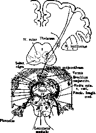
VESTIBULAR NERVE, VESTIBULAR SYSTEM. The vestibular nerve is one of the roots of the auditory nerve, belonging to the eighth pair of cranial nerves. It originates in the Scarpa ganglion, located deep in the internal auditory canal; the peripheral processes of the cells direct themselves toward the semicircular canals, while the central processes form the actual vestibular nerve, which in the auditory canal is connected with the cochlear nerve into one common trunk—the auditory nerve. At the boundary between the medulla oblongata and the pons, the fibers of the vestibular nerve penetrate into the stem part of the brain, medially and anterior to the cochlear nerve, and direct themselves toward the lateral angle of the floor of the fourth ventricle, between the restiform body and the descending root of the trigeminal nerve; before reaching the floor of the fourth ventricle, in the internal parts of the restiform body it divides into ascending and descending branches; the descending fibers (descending root of the auditory nerve of Roller) are surrounded by the nucleus of the descending root (nucleus of Roller's root); descending, they reach the external nucleus of the cochlear nerve; part of them also terminate in the dorsal vestibular nucleus or triangular nucleus (the posterior nucleus of the auditory nerve) and in Deiters' nucleus; the ascending ones terminate in the Bechterew nucleus. The dorsal vestibular nucleus is composed of small cells, reaches its maximum size in the upper parts of the medulla oblongata, where it occupies the entire floor of the fourth ventricle, reaching the middle parts of the medulla oblongata at its lower end; in the pons it rapidly decreases and disappears. Deiters' nucleus, or medial vestibular nucleus, is composed of large cells, appears in the upper parts of the medulla oblongata, between the bundles of the internal part of the peduncle of the cerebellum to the medulla oblongata, and reaches the lower parts of the pons. Bechterew's nucleus, or angular nucleus, lies in the pons, in the outer angle of the fourth ventricle, and is composed of not very large cells. Part of the ascending fibers of the vestibular nerve terminates in the fastigial nucleus of the cerebellum; from the same nucleus a significant part of the axial cylinders from the nuclei described above goes through the internal part of the cerebellar peduncles to the medulla oblongata to the fastigial nucleus; Nucleus fastigii; Nucleus emboliformis; Nucleus globosus; Nucleus dentatus; Cerebellovestibular fibers; Nucleus Bechterew; Nucleus Deiters; Nucleus of the vestibular nerve (triangular) (Schwalbe).
The reticular substance; Vestibulospinal fasciculus; Rubrospinal fasciculus (Monakow); Spinal fasciculus; Corticospinal and corticobulbar tracts. Vestibular system: vestibular nerve, its nuclei, and central connections. Deiters' nucleus gives rise to the vestibulospinal fasciculus (or Lowenthal's fasciculus), which, descending downward between the inferior olive and the ambiguous nucleus, reaches the spinal cord, is located on the periphery of the anterior column, between the anterior roots, and terminates in the cells of the anterior horns. From the same nucleus go fibers to the posterior longitudinal fasciculus, where they take a descending direction and reach the spinal cord. Fibers from the dorsal vestibular nucleus and the descending nucleus of Roller also go into these bundles. In Bechterew's nucleus begin the vestibulo-oculomotor fibers; they go in an ascending direction in the posterior longitudinal fasciculus and terminate in the oculomotor nucleus. From the cortex of the cerebellum (cerebellovestibular fibers), from the fastigial nucleus and embolus, fibers also go to the terminal nuclei of the vestibular nerve described above; part of the fibers passes directly from the cerebellum into the vestibulospinal fasciculus. The paths connecting the vestibular system with the cerebral cortex are unknown: it is possible that they do not go directly, but through the cerebellum. The connection existing between the vestibular apparatus, the labyrinth, the cerebellum, and the spinal cord indicates the function of the vestibular system. The vestibular apparatus has the leading significance in the sense of static equilibrium, which serves for determining the position of the body and its parts in space and for maintaining the correctness of actions in connection with the movement of the body. Centripetal impulses arising upon excitation of the vestibular apparatus exert a tonic and regulating influence on the muscular centers, reflexively causing the contraction of certain muscles of the neck and trunk and a series of involuntary movements necessary for the preservation of equilibrium. Through the posterior longitudinal fasciculus the same impulses influence the eye muscles, reflexively producing the corresponding conjugate deviation of the eyes, thanks to which the direction of the gaze does not change with changes in the position of the head. Through the fibers connecting the vestibular nuclei with the cerebellum, irritation from the labyrinth is transmitted to the cerebellar vermis and stimulates the activity of the centers located there that manage the position of the trunk and the whole body in space. The cerebellum, through the cerebellovestibular fasciculus, keeps the function of the vestibular nuclei in dependence, which, together with the vestibulospinal fasciculus, form important systems of centrifugal cerebellar paths. Disease of the vestibular system is expressed by a number of subjective and objective symptoms (dizziness, unsteady gait, nausea, vomiting, disturbance of equilibrium). The totality of these symptoms has been isolated as Meniere's syndrome (see Meniere's syndrome). Each of these symptoms has its characteristic peculiarity, allowing one to differentiate disease of the vestibular apparatus from other diseases in which similar symptoms occur. Moreover, the vestibular character of these symptoms is revealed by the application of caloric, mechanical, and galvanic irritations; a known reaction to these irritations speaks for their vestibular nature (see Bastian's law, Vestibular methods of investigation, Internal ear).
A. Dementiev.
E. Kononova.
Related articles
Mentioned in
Cite this page
“Vestibular Nerve.” Soviet Medical Encyclopedia. English translation of Bolshaya Meditsinskaya Entsiklopediya, 1st ed. (Moscow, 1928–1936), ed. N. A. Semashko. https://sovietmedicalencyclopedia.pages.dev/article/vestibular-nerve/