INNER EAR
Historical document, translated for reference. It reflects medical knowledge of the 1920s–30s and is not medical advice.
Summary
The article describes the development, anatomy, and physiology of the inner ear, including its bony and membranous labyrinth, cochlear and vestibular apparatuses, and the associated nerve pathways.
Encyclopedia article (1928–1936)
INNER EAR. Development of the inner ear. On both sides of the hindbrain, above the first gill cleft, a thickening of the ectoderm forms, which separates from the corresponding surface in the form of a vesicle. This rudiment of the inner ear subsequently connects with nerve fibers to the brain and then, descending into the mesoderm of the skull, divides into two parts. From the upper part, the semicircular canals and the recessus ellipticus develop, in which the utriculus is located; from the lower part, the cochlea and the rec. sphaericus with sacculus. Subsequently, processes of consolidation and finally ossification occur here. Anatomy. The inner ear consists of several cavities that communicate with each other and have a common bone capsule surrounding them (see Figure 2). In the inner ear, a bony labyrinth and a membranous labyrinth contained within it are distinguished. The former consists of compact bone, is located in the pyramid of the temporal bone, and is surrounded by a layer of spongy bone and air cells. The bony labyrinth (see Figure 1), and consequently the cavities contained within it, is divided into three parts: the vestibule (vestibulum), the semicircular canals (canales semicirculares), and the cochlea (cochlea), with the cochlea being the anterior part, the semicircular canals the posterior part, and the vestibule located in the middle. From the anatomophysiological point of view, another division is also adopted: 1) the cochlear apparatus (cochlea) and 2) the vestibular apparatus (semicircular canals and vestibule). The entire labyrinth contains labyrinthine fluid, which in the membranous labyrinth is called endolymph, and in the space between it and the bone walls is perilymph. The vestibule is a small cavity, the outer wall of which is largely occupied by the oval window and faces the middle ear. On the inner wall of the vestibule, there are two depressions—recessus sphaericus (anteriorly) and recessus ellipticus (posteriorly)—which contain the vestibular sacs: the first so-called sacculus, and the second—utriculus. Both of these sacs are lined with bridge-like epithelium, which in the area of the maculae (the site of nerve endings of the otolithic apparatus, see below) is replaced by cylindrical epithelium, which in turn continues into typical sensory epithelium. This epithelium is covered by an otolithic membrane, which envelops the so-called otoliths—crystals of calcium carbonate, which together with the aforementioned membrane and macula constitute an essential part of the otolithic apparatus. There are three semicircular canals (see Figure 5): the lateral, or horizontal, the superior (vertical), and the posterior (vertical). Each canal has one ampullary arm (at its exit from the vestibule) and one simple arm (at its entrance into the vestibule); both simple arms of the posterior and superior canals unite into one common crus (crus commune). The membranous canals contained within the bony ones emerge from the utriculus and empty back into it. The corresponding planes of the canals, in which the latter are located, are perpendicular to each other. The cochlea has a spirally coiled canal 21/\ times (in humans), ending in the anterior and inner part of the vestibule. The axis around which the coils are wound (see Figure 4) is called the modiolus. Toward the apex of the cochlea, the bony canal, narrowing, ends blindly in the so-called cupula. From the modiolus to the outer wall, partly bony and partly membranous, a partition (lamina spiralis ossea, resp. membranacea) divides the cochlear canal into two parts: the part facing the apex—scala vestibuli—and the part facing the base—scala tympani. From the first, a limited space, the so-called ductus cochlearis (cochlear duct), branches off, formed by the lamina spiralis membranacea, the outer wall of the cochlea, and the so-called membrana Reissneri (extending from the surface of the lamina spiralis ossea to the outer wall). The cochlear duct ends blindly at the apex of the cochlea, and both scalae connect through the so-called helicotrema; at the base of the cochlea, no connection between the two scalae occurs. The cochlear duct connects via a canal (ductus reuniens) with the sacculus, scala vestibuli with the perilymph of the vestibule, and scala tympani (through the aquaeductus cochleae) with the subarachnoid space. With the middle ear, the cochlea (resp. scala tympani) communicates on a bone specimen through the round window, which on a fresh specimen is closed by the secondary tympanic membrane. The peripheral end apparatus of the cochlear nerve is located in the cochlea and is called the Corti organ. The sensory and supporting cells that make up it are located on the lamina spiralis membranacea, which is also called the basilar membrane (membrana basilaris) and consists of fibers (resp. 'strings') of different lengths. The entire Corti organ is covered by the tectorial membrane (membrana tectoria Corti). The peripheral end of the vestibular nerve enters the vestibular sacs, into their maculae, and in the area of the ampullary ends of the semicircular canals—into the so-called crista ampullaris (see Figure 3). The ganglion of the vestibular nerve is located in the internal auditory canal (ganglion vestibulare s. Scarpae). Its peripheral fibers go to the aforementioned end apparatuses, while the central ones go to the medulla oblongata to the corresponding nuclear groups that give rise to the second neuron. The peripheral neuron of the cochlear nerve consists of fibers going from the ganglion spirale located within the cochlea to the Corti organ. Another part of the fibers from this node goes centrally as part of the auditory nerve trunk and ends in the so-called tuberculum acusticum and in the nucleus ventralis n. cochleae, from which the second neurons already originate, which form part of the central cochlear pathway.
I. Aleksandrov. Physiology of the Inner Ear. Should be divided into two departments: physiology of the cochlear (snail) apparatus and vestibular apparatus. The purpose of the snail, according to the theory, resp. hypothesis, held until the present time, by Helmholtz,-is to take together with the central links of the organ of hearing an active part in analyzing the excitations transmitted through the organs of hearing. The vestibular apparatus, from a physiol. point of view, represents part of a very complex mechanism that manages the orientation of our consciousness relative to the position and movement of the body in space and the maintenance of the equilibrium of the body and its parts. Among the system of centripetal nerves supplying various (visual, auditory, kinesthetic, tactile) signals to the coordination center, the vestibular nerve is a specific analyzer of the spatial relations of our body both in a state of rest and during movements. Being a special organ of static sense, the vestibular apparatus manifests its functions in two directions: it sends signals to consciousness that prompt it to voluntary movements for the purpose of maintaining and restoring equilibrium, and, at the same time, it activates the reflex apparatus which achieves the same goal, without the participation of consciousness, by means of corrective movements. By means of physiol. experiment and clin. observations, it has been possible to detail our knowledge of the physiology of the vestibular apparatus. The entire complex symptom-complex of the reflex activity of the labyrinth, united by the general idea of maintaining the equilibrium of the body, has two receptive groups and, corresponding to them, two groups of motor effects. The signals of the first category, arising from the mechanical effects of statoconia on the auditory hairs (in maculae acusticae), are analyzers of position (see Perception, perception of position), which activate the automatic apparatus for maintaining and equalizing the normal relationship between the axis of the head and the vertical line of gravity. This group can be called statoconic reflexes. The other group of labyrinthine reflexes includes the reflexes of the semicircular canals and, in essence of motor effects, can be characterized as movement reflexes. The perceiving apparatus here is the cupula terminalis on the elevation of the sensory epithelium in the ampullae of the semicircular canals; irritation of the cupula is produced by currents of endolymph arising from various rotations of the head. If, from the aspect of the given grouping, we consider the reactive movements, we can form an idea of the origin of their individual forms. Eye movements are observed in two forms: compensatory movements and oscillatory movements; the first form appears as a reaction to changes in the position of the head and is evaluated as a tendency to maintain the eyes in the original position, convenient and customary for orientation in the surroundings; when the head is tilted forward or backward, the eyes remain in the position fixing the horizontal; when the head is tilted to the side, the eyes rotate around their optical axis in the direction opposite to the tilting of the head; these phenomena are not stimulated by irritations from the retina, as they are observed in the dark and even in the blind; these movements, following every change in the position of the head, depend on the displacement of statoconia.-The second form, oscillatory movements, or nystagmus of the eyes, as well as rotational nystagmus-appears when an animal or person is rotated with moderate speed around a vertical axis; the mechanism of the appearance of nystagmus during rotation is as follows: to compensateate Inner Ear 1 Ampulla of the superior semicircular canal Superior semicircular canal Endolymphatic sac Endolymphatic duct Dura mater Perilymphatic space of the superior semicircular canal Utricle Posterior semicircular canal Perilymphatic space of the posterior semicircular canal Ampulla of the posterior semicircular canal
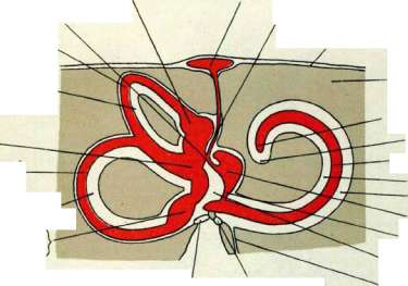
Oval window (Fenestra vestibuli) Round window (Fenestra cochleae) Bone Cupula Helicotrema Cochlear duct Scala vestibuli and scala tympani Scala vestibuli Scala tympani Reissner's membrane Ductus reuniens Perilymphatic space of vestibule Perilymphatic duct Fig. 1. Diagram of the bony and membranous labyrinth of the right side (after Spalleholz). Superior semicircular canal Common crus Posterior semicircular canal External (horizontal) semicircular canal Oval window
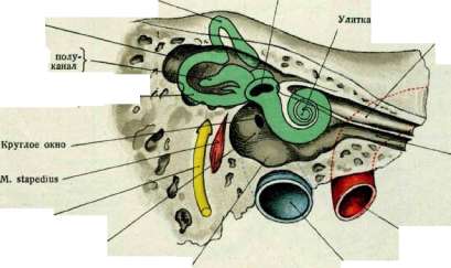
Semicircular canal m. Perilymphatic space of the tympanic cavity N. facialis Medial wall of the tympanic cavity Projection of the bulb of v. jugularis Eustachian tube A. carotis V. jugularis Figure 2. Projection of the labyrinth on the medial wall of the tympanic cavity (after Denker-Kahter). Inner Ear II Cupula Cubical epithelium of the inner surface of the ampulla N. vestibularis
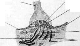
Supporting cells
\ stria «Hair cells / ^pullaris Membrana basilaris Connective tissue cristae N. vestibularis Figure 3. Crista ampullaris of the superior semicircular canal (original drawing from a histological preparation from the collection of A. Ivanov). .-*-, ^ Superior turn Capsule of the labyrinth Modiolus, Membrana Reisneri Membrana basilaris and Organ of Corti
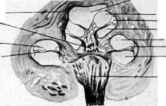
Scala vestibuli Ductus cochlearis Scala tympani Scala vestibuli Ductus cochlearis Scala tympani N. acusticus 2nd turn 1st turn Figure 4. Section through the cochlea along its axis (original drawing from a histological preparation by the author). Capsule of the superior semicircular canal Membranous superior semicircular canal Crus commune, Posterior semicircular canal' External (horizontal) semicircular canal Ductus reuniens' Ampulla of the posterior semicircular canal
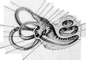
Ampulla of the superior semicircular canal Crista ampullaris and ampulla Ampulla of the lateral semicircular canal Utriculus Macula utriculi Sacculus Membrana Reisneri Scala vestibuli Ductus cochlearis Scala tympani - Lamina spiralis ossea Ductus cochlearis Figure 5. Right bony labyrinth and the membranous labyrinth contained within it; enlarged 4:1 (original figure, combined from casts of the museum of the Clinic of Diseases of the Ear, Nose and Throat of 1st Moscow State University). movement, the eyes lag behind the rotation, returning in jerks to the original position; in some animals (pigeons) nystagmus of the eyes during rotation is accompanied by nystagmus of the head, i.e., pendulum-like swinging of the head in the horizontal plane. After extirpation of both labyrinths, nystagmus is not observed, but does not disappear after turning off the statocysts; in such an experimental setup, the role of the semicircular canals as a source of oscillatory eye movements is revealed. The theoretical justification for the rotational reaction was given by Mach-Breuer: during rotation of the animal with the head in a normal position, the horizontal canal is predominantly irritated; this results in a flow of endolymph in the canal of the ear toward which the rotation is directed, while in the canal of the other ear, the flow of endolymph is away from the ampulla; both flows reinforce each other, causing nystagmus in the direction of rotation. Experiments with caloric irritation (see Barany's method of investigation) and irritation with galvanic current (see Voltaic reaction) also confirm the dependence of nystagmus of the eyes and head on signals from the semicircular canals. In the musculature of the limbs and trunk, the following complexes of reactive movements dependent on the semicircular canals have been experimentally revealed; these experiments are of particular interest as they establish the reaction of the semicircular canals not only to rotation but also to linear accelerated movement of the body. The experiment was conducted by lifting and lowering the animal; in the first case, the animal's legs bend, in the second they extend, straightening (the elevator reflex, according to Magnus and Kleine). In the experiment with jumping (Sprungbereitschaft), the animal is lowered while holding it by the pelvic region; at this time, the forelimbs begin to extend forward, the body curves upward, i.e., a muscular posture for the horizontal plane appears. Thus, the system of semicircular canals serves to regulate the position of the body when there is a danger of loss of balance due to passive changes in position in any plane. The reactive movements of the musculature of the neck, trunk, and limbs, studied in experiments, are of deep interest in that they make it possible to establish the dependence on the labyrinth of the state of muscle tone of the entire body, as well as the mutual influence of different groups in the distribution of the toning action of the labyrinth. The significance of the labyrinth in this respect is guiding, but not absolutely direct, but extending primarily to the tone of the neck muscles and to a lesser degree to the muscles of the trunk. Experiments were conducted on decerebrated (transection of the medulla oblongata through the anterior part) dogs; a dependence of the tone of certain muscle groups on turns of the head (without the participation of neck muscles) was established: in a dog placed on the ground, a sharp increase in tone (Sherrington's decerebral rigidity) of the extensors of the limbs and hypotonia of their antagonists was observed; with passive lowering of the head of the experimental animal, the state of tone showed the opposite arrangement. Being a typical example of a position reflex, this experiment cannot be performed on an animal with destroyed statocysts. It must be borne in mind that experiments on decerebrated animals have clearly demonstrated that the distribution of tone to different muscle groups depends on two sensory apparatuses: the labyrinth and the sensory nerves of the muscles, tendons, ligaments of the neck. In the complex physiology of the vestibular apparatus, not everything has yet been clarified, but even the material that is available gives valuable methodological indications for the clinical analysis of diseases in which there are symptoms of disturbance of the functions of the apparatus governing the equilibrium of the body; patients with symptoms of dizziness (see) especially require in-depth research, as this symptom should always arouse suspicion of disease of the labyrinth as the main component of the static apparatus. For differential and topological diagnosis, all methods of investigation of the vestibular apparatus indicated above serve. d. Surov. Pathology of the inner ear is extremely diverse. Timpanogenic labyrinthitis (see), occurring when the process spreads from the middle ear to the labyrinth, occurs 1) either as a result of infection of the labyrinth through the vessels of its lateral wall, common to the middle ear and the labyrinth, 2) or from traumatic damage to the walls of the labyrinth (damage to the base of the skull with a concomitant disease of the middle ear, surgical opening of the labyrinth when removing granulations from the area of the stapes, cauterization of granulations in the area of the promontory with strong acids, etc.), 3) or from the transition of the inflammatory process from the middle ear to the inner ear. In cerebrospinal meningitis, organic changes often occur in the inner ear in the sensory cells of the organ of Corti and in the nerve trunk; they are often accompanied by phenomena of osteoporosis of the capsule of the cochlea; in the semicircular canals and vestibule, considerable damage can also be found. Investigation of the initial stage of disease of the inner ear in meningitis may be impossible due to the unconscious state of the patient. Later, it is possible to detect shortening of air conduction for high tones, accompanied by shortening of bone and craniotympanic conduction, and then shortening of perception of low tones. In the final stage, one can have: 1) a case of complete deafness, 2) the presence of only individual islands of hearing, and 3) a significant loss of hearing ability in the range. And from the side of the vestibular apparatus, a whole series of disorders is often observed, such as disturbance of gait, dizziness, frequent urges to nausea, etc. Loss of vestibular function is registered objectively by the usual methods of investigation of the vestibular apparatus. It is interesting to note a certain discrepancy between hearing losses and the vestibular apparatus: often significant deafness without disorders of vestibular function is encountered, and vice versa. The question of changes in the inner ear in disorders of the internal secretory glands is extremely interesting. In this respect, one should recall idiopathic, congenital or acquired, diseases of the thyroid gland. It is interesting that different groups of patients in this case give a different picture of the disease of the inner ear, but severe disturbances of hearing function as a rule are not encountered. The latter especially applies to acquired myxedema (cachexia strumipriva); in idiopathic forms, certain lesions of hearing ability are noted. It must be borne in mind that in these cases, not only from the side of the bony parts of the labyrinth were there certain histological changes (osteosclerosis of the labyrinth capsule, etc.), but such changes could also be established in other parts of the skeleton. Closely connected with the question of diseases of the inner ear in general skeletal diseases is its condition in the disease osteogenesis imperfecta Vrolik. Functional investigation of hearing in most cases reveals a picture of otosclerosis, but individual cases of labyrinthine deafness have also been observed. Histological investigation of two such cases also revealed a picture of otosclerosis. Among diseases of the inner ear in disturbed metabolism, one of the first places should be given to the lesion of the inner ear in diabetes. Here one can observe two forms of lesion of the inner ear: 1) suppurative labyrinthitis and 2) non-suppurative diseases of the inner ear. Suppurative labyrinthitis as a complication of the suppurative process of the middle ear are not more common than suppurative labyrinthitis of other etiology. Nevertheless, in operations on the mastoid process in diabetes, in cases of large bone destruction, serious attention should be paid to the condition of the bone around the destroyed tissue, as labyrinthitis often creep up completely unnoticed, sometimes even with very good postoperative course. The study of non-suppurative diabetic lesions of the inner ear is owed to modern otology to Edgar. The latter in almost a quarter of cases of investigation of the inner ear in 52 diabetics found a lesion of the labyrinth and the trunk of the cochlear nerve, which he connects with the influence on the inner ear of toxins circulating in the blood in diabetes. But sometimes the lesion can be explained by the very often accompanying diabetic exhaustion of arteriosclerosis (see below). The pathological manifestations develop slowly, and patients consult a physician when there is already a significant loss of hearing. In gout, too, disease of the inner ear can often be noted. A number of authors attribute them to the simultaneously observed arteriosclerosis. Gouty diseases of the inner ear have been thoroughly investigated by Scheube.
The latter describes the two most frequently occurring forms in this condition: 1) one acutely manifests as tinnitus and dizziness, accompanied by loss of low tones, is unilateral, and does not present with severe deafness; 2) in the second form, a sharp decline in hearing ability should be noted as the primary feature, without accompanying dizziness; in this form, a characteristic sharp deafness in noisy rooms is very prominent, while in silence patients can still hear satisfactorily. Hearing examination reveals narrowing of the upper hearing limit with a normal lower limit. In the first form, apparently, there is deposition of uric acid salts in the internal auditory canal, which causes loss of low tones in these cases (lesion of the retro-labyrinthine portion). Detailed histological studies of both forms are lacking. Among diseases of the inner ear in diseases of the respiratory organs, hemorrhages in the labyrinth in whooping cough should be noted. Authors mention that deafness sometimes occurs in whooping cough, which is apparently connected with the aforementioned etiological factor. - From the group of diseases of the circulatory system, arteriosclerosis very often causes a series of changes in the inner ear, consisting mainly atrophy of the organ of Corti and the trunk of the n. cochleae. These changes obviously early begin to cause subjective tinnitus and decreased hearing ability. Functional examination reveals narrowing of the upper limit and shortening of air conduction for high tones, with simultaneous shortening of bone conduction. Symptoms from the vestibular apparatus are much rarer. The observed dizziness is more likely of cerebral rather than vestibular origin. - In general vasomotor disorders, certain phenomena are also observed from the inner ear. Angioneurotic paralysis of the n. acustici is still mentioned by Politzer (Politzer). Recent authors also point to sudden attacks of nausea or dizziness, noise in the ears, and decreased hearing, which they associate with vasomotor influences. Sometimes forms of sudden anemia of the labyrinth may be observed; in this case, damage to either the cochlear or vestibular apparatus can sometimes be confirmed (hemiplegia cochlearis, hemiplegia vestibularis). - In kidney diseases, attention has long been paid to the frequently associated decrease in hearing, which could not be attributed to the middle ear. Interestingly, damage to the upper limit in kidney diseases serves as a poor prognostic symptom; such cases lead either to death or do not heal completely. Hearing examination, therefore, should always be undertaken in kidney diseases for a more accurate prognosis. Disorders of the vestibular apparatus compared to cochlear ones occur much less frequently in kidney diseases. - Diseases of the blood (leukemia, anemia, hemophilia) also often affect the condition of the inner ear. Regarding the localization of these lesions, authors differ: some attribute them to changes in the organ of Corti, others put central connections in the foreground. Infectious diseases of the body also often leave traces for the inner ear. Among them, epidemic parotitis should be mentioned first. The pathological anatomy of the inner ear in this disease has not yet been fully elucidated. Its course is characterized by the speed, "apoplectic" nature of the occurring phenomena: ear noises, ringing, whistling in the ears, accompanied by hearing loss, sometimes up to complete deafness. After the noises disappear, hearing either recovers, improves, or the hearing impairment remains stationary. Disorders of equilibrium also often occur. - Diseases of the inner ear observed in influenza, scarlet fever, and measles belong to the type of tympanogenic labyrinthitis already described above. In some cases, especially with influenza and measles, hematogenous spread of infection can be confirmed; very rarely forms of meningogenic labyrinthitis are observed. In measles, forms of deafness are observed that depend on pathological changes not only in the peripheral auditory apparatus but also in the nerve trunk. These central lesions can also be confirmed in diseases of the inner ear during diphtheria. The vestibular apparatus to a greater or lesser degree can be involved in the suffering in all these infections. - Typhus and typhoid fever are often accompanied by lesions of the inner ear. Most recent researchers believe that the main site of localization of the disease is in the retro-labyrinthine portion of the n. VIII, up to its entry into the medulla oblongata. Hearing disorders occur in both diseases during the 2-4th weeks. The phenomena observed from the n. vestibularis disappear faster than cochlear ones and are expressed significantly less intensely. Both diseases do not always lead to hearing loss, but sometimes complete deafness can remain forever. - Syphilis often affects the inner ear. Congenital and acquired syphilis of the inner ear are distinguished. In both, lesions can affect both the cochlear and vestibular apparatus. From the vestibular apparatus, a characteristic feature is the inconsistency of vestibular reaction results with each other, as well as of the same reaction with respect to both ears. In cases of congenital syphilis, so-called pressor nystagmus (or fistular symptom without fistula; see Vestibular examination methods) is more often encountered than in acquired syphilis. From the diseases of the nervous system that are accompanied by lesions of the inner ear, multiple sclerosis should be mentioned. Just as in other nerves, one can observe either normal nerve function or its loss, so with respect to the n. cochlearis and n. vestibularis, the same alternation of phenomena can often be noted. From this group of diseases, congenital-degenerative forms of deafness should be mentioned. This lesion is localized in the peripheral nuclei of the n. VIII, in the nerve trunk, or in the organ of Corti. Functional examination reveals disorders in the area of the upper (sometimes also lower) limit and shortening of bone conduction. - From other diseases of the labyrinth, cancer of the inner ear should be noted, which differs from other malignant tumors by a slower growth of the tumor and insignificant cachexia. - Age-related changes of the inner ear (presbyacusis) consist in increasing rigidity of the basilar membrane, especially sharply expressed at the base of the cochlea. This corresponds to the picture of hearing: narrowing of the upper limit and shortening of bone conduction. Sometimes in this condition, lesions of central conductors and a combination with arteriosclerosis are also encountered. - Lesions of the inner ear observed in various poisonings and professional hazards give a varied picture both in terms of pathological anatomy and symptomatology. - Therapy of diseases of the inner ear is partly causal (tympanogenic labyrinthitis, endemic cretinism, syphilitic diseases, etc.), directed against the general underlying disease (arteriosclerosis, kidney disease, diseases of the circulatory system, metabolic disorders), or symptomatic (pilocarpine, iodine and bromine preparations, vaccineurin, quinine, galvanization, protein- and osmotherapy, Otosclerol, Panitrin, Natr. nitrosum, etc.) in diseases of the inner ear in typhus, epidemic parotitis, epidemic cerebrospinal meningitis, etc.
i. Aleksandrov.
Related articles
Mentioned in
Cite this page
“INNER EAR.” Soviet Medical Encyclopedia. English translation of Bolshaya Meditsinskaya Entsiklopediya, 1st ed. (Moscow, 1928–1936), ed. N. A. Semashko. https://sovietmedicalencyclopedia.pages.dev/article/inner-ear/