Ear
Historical document, translated for reference. It reflects medical knowledge of the 1920s–30s and is not medical advice.
Summary
This article describes the anatomy and embryological development of the outer ear, including the auricle and external auditory canal, explaining its formation from embryonic tissue and its relationship to the tympanic membrane and middle ear.
Encyclopedia article (1928–1936)
Ear (external) consists of the auricle (auricula) and the external auditory canal (meatus auditorius externus); it develops from a ridge surrounding the embryonic tympanic membrane, which is initially located at the level of the skin. In this formation, the cartilaginous framework of the external ear arises, with its deeper part forming the ossifying tympanic ring (annulus tympanicus) on the third month of intrauterine life, which serves as a frame for the tympanic membrane. The rudiment of the auricle first appears as a thickening on the posterior edge of the aforementioned ridge. Subsequently, all these parts grow: the cartilaginous rudiments give rise to the cartilaginous sheath of the external auditory canal and the convolutions of the auricle; the tympanic ring in the first years of a child's life transforms into the bony sheath of the internal, deeper parts of the external auditory canal. The lumen of the canal, which has the shape of a slit in a newborn, gradually expands to the size of a canal with a round or oval cross-section, separated from the tympanic cavity by the tympanic membrane, which embryologically should be considered as an outpouching of the end of the Eustachian tube, protruding between the external ear and the labyrinthine formations, i.e., the descended 'auditory pits' and 'auditory vesicles'.
The lumen of the canal, which has the shape of a slit in a newborn, gradually expands to the size of a canal with a round or oval cross-section, separated from the tympanic cavity by the tympanic membrane, which embryologically should be considered as an outpouching of the end of the Eustachian tube, protruding between the external ear and the labyrinthine formations, i.e., the descended 'auditory pits' and 'auditory vesicles'.
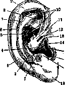
In its fully developed state, the auricle (fig. 1) is a fantastically curved cartilaginous plate, serving as a continuation of the cartilaginous sheath of the external auditory meatus, covered with skin, which below forms its antitragus (lobule); the cartilaginous framework is attached to the surface of the skull by a ligamentous apparatus, while one distinguishes the upper (supporting), posterior and anterior ligaments (lig. Valsalvae). The central depression on the lateral surface of the auricle is called cavitas conchae auriculae, behind which is located the semicircular projection-the antihelix (anthelix), which below passes into the tubercle-antitragus (tuberculum antitragicum); still further to the periphery on the very edge of the auricle is located the helix (helix), forming in its anterior part a knee (crus), an upper tubercle (tuberculum helicis) and a posterior-inferior tail (cauda). The central depression leads into the entrance of the external auditory meatus (porus acusticus externus), and on the anterior edge of this entrance is located the projection-tragus (tuberculum tragicum). The tubercle of the helix is otherwise called Darwin's (tuberculum Darwini). It is considered a rudiment, analogous to the tip of the ear of mammals. In the cartilage of the external auditory meatus there are transverse slits-incisions of Santorini (incisurae Santorini). The rudimentary muscular apparatus of the auricle consists of several superficial weak bundles, located partly on the lateral surface of the cartilaginous framework, e.g., the tragic muscle (m. tragicus) and its superficial bundle (fasciculus accessorius, s. superficialis), or on the posterior, e.g., the oblique muscle (m. obliquus), and from more pronounced muscular formations connecting the skeleton of the auricle with the aponeurotic helmet of the skull (galea aponeurotica cranii); the latter bear the names of the anterior, superior and posterior auricular muscles (mm. auriculares anterior, superior Henle, posterior, s. pyramidalis Jungi). The skin covering of the external ear in general corresponds in its structure to other areas of the skin, but in the depth of the external auditory meatus it is greatly thinned; of the skin appendages, hairs are often strongly developed on the tragus and in the opening of the external auditory meatus; the glands (ceruminous) resemble in structure the sweat and sebaceous glands, but the secretion they secrete (earwax-cerumen, sordes aurium) differs in its color (from yellowish to brown), viscosity and bitter taste, which makes it unlike the usual skin secretions.-The blood and lymphatic system of the external ear and its innervation. Arteries are branches of the temporal and maxillary arteries (e.g., deep artery of the auricle-a. auricularis profunda). Veins flow into the external jugular and mastoid, temporal and maxillary. Lymph. vessels flow into the retroauricular, submandibular and parotid lymph. nodes. Motor innervation comes from the facial nerve, sensory-from the trigeminal and vagus (Arnold's nerve). The ear is examined by inspection, palpation, probing, measurement with a caliper (meatometer), radioscopy and some other methods, the main one being inspection with the help of illumination by a mirror (reflector) and otoscopy (ear endoscopy). In most cases, otoscopy is performed without the help of auxiliary instruments by simply pulling the tragus and auricle in opposite directions with the fingers to expand this opening and look into it more easily; but if the passage is very curved, slit-like, or there are many hairs in it, then otoscopy is performed with the help of ear funnels inserted into it, i.e., small metal or ebonite tubes of various calibers. Of pathological processes, one most often has to deal with malformations and developmental anomalies, to which belong excessive or too small size of the auricle (macrotia, microtia), complete absence of it (anotia), excessive number of auricles (polyotia); partial excessive growth or partial underdevelopment of some parts of the auricle, e.g., 'satyr's ear', 'monkey's ear', absence of the lobe, additional cartilages (tragus), protruding ears, etc. The external auditory meatus may also be absent (congenital atresia). In the area of the external ear, fistula openings are encountered-the result of an unfused first branchial cleft. The listed anomalies may be combined with defects of deeper parts of the ear, e.g., the labyrinth, and give rise to their surgical correction (cosmetic operations-otoplasty; operations to improve hearing). The psychiatrist Morel (in the 1830s) pointed out anomalies of the external ear (e.g., such as underdevelopment or excessive development of its individual parts, unusual attachment of the auricle, etc.) as one of the external signs of psychogenic degenerative diseases ('Morel's ear'). Since the auricle and external auditory meatus are covered with skin, they are the site of manifestation of ordinary skin diseases; certain forms have a certain specificity, such as the hematoma of wrestlers, perichondritis, frostbite of the auricle (due to its easy vulnerability, thinness of the organ, susceptibility of the cartilage to purulent infection); in turn, for the external auditory meatus, due to its narrowness and curvature, are characteristic: blockage with wax, epidermoid masses ('external cholesteatoma'), foreign bodies; all this is often combined with eczema, otomycosis (fungal disease of the ear and its special case-aspergillosis of the ear), strictures of the auditory meatus of various origins-from trauma, collapse of the cartilaginous walls after operations, after ulcerative processes, etc. The external auditory meatus is a favorite place for parasites, e.g., larvae of insects and 'false' parasites (N. A. Khodkovsky), i.e., accidental finding of living beings; in purulent otitis, otorrhea, i.e., ear discharge, is bait for flies, cockroaches and other domestic insects. Living foreign bodies often cause the so-called objective noises in the ear, i.e., subjective auditory sensations depending on an objective sound phenomenon (just as e.g., when clicking of tendons and muscles of the tympanic cavity).-Acute and chronic infectious diseases also often localize their manifestations in the external ear (erysipelas, smallpox, tbc, lupus, syphilis and rarer-scleroma, Pinday's ulcer, etc.). Of neoplasms, cysts (atheromas), angiomas, keloids, exostoses are more common, of malignant-cancers, more rarely-sarcomas. Of metabolic diseases and diatheses, gout is related to the external ear, in which painful thickenings (tophi arthritici) are observed on the auricle and in the opening of the external auditory meatus. Irradiation of pain to the external ear can accompany processes in neighboring organs (rheumatism of muscles, caries of teeth, etc.); occasionally the term 'otodynia' or 'otodynia' is used. Prevention consists mainly in observing the following rules: nothing should be inserted into the external auditory meatus, even for the purpose of cleaning it, nothing should be scratched, wiped or liquid admitted into it, especially soapy water. During bathing, water getting into the ear is undesirable, and in persons with a damaged eardrum it is harmful. Such people should at least protect the external auditory meatus with pieces of vaseline-soaked cotton. The treatment of the listed diseases is for the most part based on the general principles of dermatology and surgery, including plastic surgery (otoplasty). Special features are those techniques for which the topography of the external ear is important, for example extraction of foreign bodies, epidermoid and wax plugs, fighting strictures, removal of EXOSTOSES AND SOME OTHERS.
V. Vojacek. The ear of vertebrates consists actually of two inseparably connected sense organs-the organ of hearing and the organ of equilibrium. The ear is located in the auditory capsule of the skull ('inner' ear), but in terrestrial vertebrates it is supplemented to a greater or lesser extent with complex adaptations for the transmission of sound vibrations ('middle' ear) and for their capture ('outer' ear). Embryonically, the ear is laid as a thickening of the ectoderm on the sides of the head (the so-called dorso-lateral placodes, from which also develop the lateral line organs of fish). The rudiment of the ear sinks in the form of a pit, which then forms the 'auditory vesicle', initially connected with the ectoderm by a hollow stalk, and later usually completely separating from it. The auditory vesicle differentiates into two parts, separated by a small constriction: upper and lower. In the upper part, three pocket-like folds are formed in mutually perpendicular planes-the rudiments of the three semicircular canals (i.e., the most important part of the organ of equilibrium). The remaining part of the upper part, on which these canals develop, receives the name of the oval sac (utriculus). In the lower
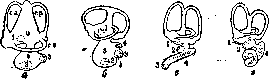
Figure 2. The membranous labyrinth of a bony fish (a), frog (b), bird (c), and mammal (d): 1-macula utriculi; 2-macula sacculi; 3-macula lagenae; 4-papilla basilaris, or Corti's organ; ca-canal semicircularis, anterior; ce-canal semicircularis externus; cp-canal semicircularis posterior; I-lagena; s-sacculus; u-utriculus. (After Hesse.)
In the lower part, a projection (lagena) is formed, which in mammals is elongated into a spirally coiled canal, the cochlear canal (canalis cochlearis). The remaining part of the lower section forms a sac of the labyrinth (sacculus). Both sacs together are called the vestibule (vestibulum) (Figure 2). The canal, which initially connected the cavity of the auditory vesicle and then the vestibule with the external environment-the endolymphatic duct (ductus endolymphaticus, s. aquaeductus vestibuli)-opens in some cartilaginous fish even in the adult state with a small opening on the dorsal side of the head. In all other vertebrates, it becomes separated from the ectoderm of the integument and closes at its end (often forming a terminal swelling). Thus, from a simple ectodermal rudiment, a rather complex structure develops-the membranous labyrinth. The history of its development, structure, and connections with the nervous system prove its origin from the system of the lateral line organs of fish. Around the membranous labyrinth, a cartilaginous and then a bony auditory capsule develops, the inner wall of which in higher vertebrates repeats the complex shape of the inner ear, forming the so-called outer or bony labyrinth. The cavity of the membranous labyrinth is filled with fluid-endolymph. Between the membranous labyrinth and the bony one, there is a slit-like space, also filled with fluid-perilymph. In higher vertebrates, this cavity communicates with the lymphatic cavities of the head by means of the perilymphatic duct (ductus perilymphaticus). In lower vertebrates, the membranous labyrinth consists of both sacs, separated by a constriction, and three semicircular canals-the external horizontal and two vertical (anterior and posterior). At one lower end of each canal, there is a swelling-the ampulla-in which a strip of sensory epithelium (the auditory crest, crista acustica) is located. The ampullae of the external and anterior canals lie next to each other at the anterior end, the ampulla of the posterior canal-at the posterior end. The internal limbs of both vertical canals merge with each other. The sensory epithelium consists of short pear-shaped sensory cells, provided with cilia and surrounded by terminal branches of the fibers of the auditory nerve, and cylindrical supporting cells. This sensory epithelium forms in the ampullae protruding into their cavity 'auditory crests' (cristae acusticae), the cells of which are provided with long cilia, and in the vestibule-'auditory spots' (maculae acusticae). The latter are located in the oval sac (macula acustica utriculi and a small macula neglecta) and in the round sac (macula acustica sacculi). In the endolymph, numerous small otoliths (statoliths) are suspended, giving it a milky appearance, and in addition, separate larger stones, consisting mainly of calcareous carbonate (see Otolith apparatus). Any change in the position of the body causes displacement of the endolymph and statoliths, which in this act on the cilia of the auditory crests and spots. Thus, the ear of lower vertebrates, and especially its semicircular canals, are primarily an organ of the sense of equilibrium. However, oscillatory movements of the statoliths must also act on the cilia of the auditory cells. In most fish, a small projection (lagena) is observed on the round sac, and in this projection there is also a separate auditory papilla (papilla acustica lagenae), which becomes separated from the auditory spot of the sac. At the same time, the corresponding branch of the auditory nerve (n. lagenae) also becomes separated. This projection is actually the rudimentary organ of hearing. However, only in terrestrial vertebrates does it receive more significant development. In amphibians, part of the lagena is strengthened on the skeletal frame and forms a small 'basilar membrane' (membrana basilaris), on which a separate part of the auditory papilla (papilla acustica basilaris) is located. In reptiles, this part develops more strongly and in crocodiles it is elongated into a rather long, slightly curved canal (canalis cochlearis). At the same time, the basilar membrane, strengthened on the skeletal frame, and the main papilla located on it are elongated in length, and to it now approaches the separated branch of the auditory nerve (ramus basilaris). Since the basilar membrane with its edges is attached to the wall of the skeletal labyrinth, the perilymphatic space around the canal is thus divided into two parts-the upper (scala vestibuli) and the lower (scala tympani). The auditory apparatus of birds is constructed in a very similar way. In mammals, the inner ear (see) reaches the highest degree of development. The cochlear canal is further elongated in length and at the same time curves in a spiral, forming from 1½ to 5 turns. The cochlear canal borders with its basilar membrane on the lower perilymphatic space (scala tympani) and with its upper wall, the Reissner's membrane-on the vestibular staircase (scala vestibuli) (Figures 3 and 4). Both perilymphatic canals pass into each other at the apex of the cochlea. The membranous cochlea connects with the rest of the labyrinth through a narrow canal. The constriction between both sacs also has the character of a narrow canal, from which the endolymphatic duct departs, ending in a blind swelling in the meninges. In connection with the differentiation of the inner ear, the auditory nerve of mammals has divided into two portions-the nerve of the cochlea and the nerve of the vestibule. The auditory crests and auditory spots are innervated by branches of the latter nerve, which also has a separate ganglion. The main papilla of the cochlea has been transformed into a complexly constructed Corti's organ. The basilar membrane, i.e., the lower wall of the cochlear canal, is attached with its inner edge to the spiral bony projection of the bony cochlea, along which the nerve of the cochlea (n. cochlearis) passes to the Corti's organ, which also has its own separate ganglion (ganglion spirale). The outer edge of the basilar membrane is attached by means of a ligament (ligamentum spirale) to the outer wall of the bony cochlea. The basilar membrane contains numerous connective tissue fibers stretched across between the bony spiral projection and the spiral ligament. According to Helmholtz's theory, these fibers, the length of which gradually decreases toward the apex of the cochlea, are set into vibration by the sound waves reaching them, similar to strings. On the basilar membrane, as stated, lies the perceiving auditory apparatus-the Corti's organ, consisting of supporting and sensory cells arranged in a specific way. From the supporting cells, two special rows of cells-pillars-stand out, inclined toward each other and limiting a special channel in the middle of the organ (the Corti's channel). The sensory cells are arranged in one row on the inner side of the pillar cells and in several rows on their outer side.
Figure 3. Cross section of the cochlea of a bat:
1-ductus cochlearis; 2-membrane tectoria; 3-Corti's organ; 4-scala tympani; e-ganglion spirale; 5-scala vestibuli. (After Hesse.)
Scheme of the auditory organ of a mammal; the perilymphatic space is marked in black: 1-incus; 2-t. tensor tympani; 3-stapes; 4-ductus endolymphaticus; 5-scala vestibuli; 6-cochlea; 7-tuba Eustachii; 8-ductus perilymphaticus; 9-tympanic membrane; 10-membrana rotunda; 11-cavum tympani; 12-malleus; 13-outer ear. (After Weber.)
To the sensory cells from above there is applied a special covering cuticular membrane (membrana tectoria), secreted by the epithelial cells of the inner edge of the canal. For terrestrial vertebrates and especially for mammals, there is thus a progressive development of the actual auditory apparatus. This is expressed not only in the deep differentiation of the parts of the inner ear and in the development of the cochlea with its Corti's organ, but also in the development of various additional parts serving for the transmission of sound vibrations of the air medium and constituting the middle, and then in higher vertebrates, the outer ear. The upper part of the hyoid arch, which in fish is the suspensorium for the lower jaw (hyomandibulare), loses this function in terrestrial vertebrates and is transformed into the auditory columella-the columella of the ear (columella). The cavity lying under the suspensorium of the first visceral cleft of fish ('gill')
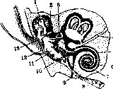
cartilage') grows around the columella in terrestrial vertebrates and is thereby transformed into an air-filled tympanic cavity (cavum tympani), located between the auditory capsule on one side and the integument on the other. The integument in this area thins out and forms an elastic tympanic membrane. Similarly, a portion of the wall of the auditory capsule thins out, forming the membranous 'oval' window (fenestra ovalis), to which the inner end of the columella is connected. The latter thereby gains greater mobility and serves as a transmitter of vibrations from the tympanic membrane through the oval window to the perilymphatic space of the inner Ear. The primary connection of the tympanic cavity with the posterior part of the oral cavity is preserved in the form of a canal—the Eustachian tube. In reptiles and birds, a second membranous 'round' window (fenestra rotunda) appears in the wall of the auditory capsule, separating the perilymphatic space from the tympanic cavity. The tympanic membrane is set somewhat below the general level of the skin, forming a rudimentary external auditory canal, along the edge of which there is sometimes a small skin fold—a rudimentary auricle. The middle Ear (see) of mammals differs mainly in the greater complexity of its transmission apparatus. Of the three auditory ossicles of mammals, only the innermost ossicle, which closes the oval window, the 'stirrup' (stapes), corresponds to the columella of amphibians, reptiles, and birds. The other two ossicles are the result of the transformation of the posterior parts of the jaw arch. The middle ossicle—the 'incus' (anvil), connected to the stirrup by its long process—represents the quadrate bone of lower vertebrates, while the outer ossicle—the 'malleus' (hammer), attached by its handle to the tympanic membrane (see)—represents the articular bone of the lower jaw of lower vertebrates. The transmission of vibrations from the tympanic membrane therefore occurs in mammals through a chain of three ossicles, of which the middle one, i.e., the incus, is attached by its short process to the wall of the tympanic cavity via a ligament (see Middle Ear). A special muscle, attached to the handle of the malleus (tensor tympani), can tense the tympanic membrane, while another muscle, the stapedius muscle, tenses the membrane of the oval window, regulating the amplitude of their vibrations with sounds of different intensity. In higher vertebrates, the tympanic cavity forms air-containing protrusions in adjacent bones. In mammals, the external auditory canal reaches significant development, and additionally, at its posterior edge, a projection—the auricle (concha)—develops, which has a cartilaginous framework and is moved by its own special musculature.
I. Shmal'gauzen. Professional diseases of the ear. Diseases of the inner ear. Etiology and pathogenesis. Under modern production conditions, there are a number of factors that have a harmful effect on the inner ear. In the general sum of factors, it is necessary to distinguish two main groups: adequate and inadequate. The first group includes all those irritations to which the ear is specifically adapted (sounds, noise, angular accelerations), for which it is an organ of perception. The action of these factors is direct. The second group includes factors that are not capable of causing a corresponding irritation of the organ of hearing. Their action is indirect—through the blood or nervous system (intoxication by industrial poisons). In addition to these two groups, there are certain factors that, while not being adequate stimuli, can nevertheless, upon reaching a certain intensity, cause direct irritation of the organ of hearing (fluctuations in atmospheric pressure). Adequate stimuli (noise and concussion) undoubtedly play a very significant role in the etiology of industrial lesions of the organ of hearing. They cause diseases of the inner ear incomparably more often than inadequate stimuli. With inadequate stimuli, the auditory nerve is usually involved in the process secondarily or along with lesions of other nerves or the body as a whole; with adequate stimuli, the lesion of the auditory nerve is primary, usually isolated. Thus, for example, in acute poisoning, the phenomena from the organ of hearing recede into the background against the symptoms of general body intoxication; with acute acoustic trauma, the phenomena from the organ of hearing come to the forefront, prevail, and are often not accompanied by other symptoms at all. Lesions of the inner ear are very common in noisy groups. According to Zacher, in experienced groups of boilermakers, the frequency of lesions reaches 100%. Pathological anatomy.—The basis of professional lesions of the organ of hearing lies in a number of pathological-anatomical changes in individual parts of the inner ear. Histological studies of the temporal bones of people with professional diseases of the inner ear and experimental animals subjected to prolonged exposure to individual harmful factors have revealed the following changes. In deafness caused by noise and concussion, a complete absence of the organ of Corti has been found in all turns of the cochlea; in its place there is a flat border consisting of cubic cells. Reissner's membrane is completely fused with this border. Nerve fibers preserved in the upper turn of the cochlea are partially or completely absent in the middle and main turns. Accordingly, in the main turn, the spiral ganglion completely atrophies; only individual ganglion cells are preserved in places. The trunk of the auditory nerve is not pathologically altered. There are also no changes in the vestibular nerve. The pathological-anatomical picture presented corresponds to a severe professional hearing impairment. The dynamics of the lesions, revealed in experimental studies, are as follows. In the initial stages of the lesion under the influence of airborne noise, the changes are localized in the main turn of the cochlea, mainly involving the sensory cells of the organ of Corti. Subsequently, the lesion spreads along the cochlear ladder; at the same time, it deepens and involves the nerve fibers and ganglion cells. The more intense the source of sound, the faster the process spreads. With simultaneous exposure to noise and concussion, the changes described are joined by a process in the upper turn of the cochlea, which is localized in the nerve cells and fibers; in this case, the organ of Corti is not affected. In this case, the process, arising at both poles of the cochlea (upper and lower turns), develops concentrically. In intoxications of the organ of hearing (lead, arsenic), pathological changes are localized in the ganglion cells. The organ of Corti is usually preserved.—Changes resulting from gas embolism (see) during rapid decompression (see Caisson work) or due to rapid fluctuations in pressure are manifested mainly in hemorrhages and small extravasations. The vessels of the labyrinth are engorged with blood; sometimes they are completely empty while the lumen is preserved. Diagnosis. Recognition of lesions of the inner ear does not present particular difficulties. In addition to otoscopy, which makes it possible to exclude a disease of the middle ear, the main importance is the study of the functional state of the cochlear and vestibular apparatus. Qualitative examination of hearing can give the following results: a) lowering of the upper limit of sound perception, positive Rinne test; normal perception of low sounds and reduced perception of high sounds; b) raising of the lower limit, positive Rinne test, shortening of bone and air conduction for low sounds; c) raising of the lower limit and lowering of the upper limit, reduced perception of low and high tuning forks, shortening of bone conduction, perception of whisper is reduced in all cases. The three types presented generally reflect the diversity of the clinical picture, depending on the peculiarities of the mechanism of action of individual harmful factors. Examination of the vestibular apparatus by means of rotational and caloric tests indicates, in cases where the labyrinth is involved in the process, an increase or decrease in its excitability. Sometimes, however, very rarely, there are also spontaneous symptoms of labyrinthine lesion in the form of nystagmus, disturbances of statics and dynamics, accompanied by a number of subjective complaints of dizziness, unsteadiness when walking, etc. It is much more difficult to diagnose the professional nature of the lesion. A carefully collected general and professional anamnesis, familiarity with the nature and degree of exposure to the corresponding professional factors on the organ of hearing, provides substantial help. Professional harmful factors are often not the only cause; in these cases, they represent a factor that promotes the development of a pathological process that arose due to another etiology. Course and prognosis. Professional diseases of the inner ear develop slowly and gradually. The rate of their progression depends on the nature of the noise and its intensity, on individual peculiarities of the organism, on the duration of daily exposure, on socio-domestic conditions, and on the general cultural level of the workers. According to pre-revolutionary statistical data, these diseases were extremely common; in particular, in noisy groups, almost all workers suffered from hearing impairment, which in experienced groups reached a degree bordering on complete deafness. The reduction in the length of the working day, the system of labor protection and preventive measures, regular vacations—all this has already resulted in a significant decrease in morbidity even in the group with adequate stimuli (noisy); in other groups, diseases of the inner ear have become a rarity. Among subjective complaints, the most common is the complaint of noise in the ears, which is very disturbing, especially in the first period. The lesion usually affects both ears. In young workers, complaints of dizziness are common. Hearing impairment, developing slowly, can remain unnoticed for a long time. It attracts attention only when, having reached a significant degree, it becomes an obstacle in social intercourse. In the initial stages, the perception of high sounds is reduced, i.e., the process is clearly limited. Subsequently, along with the continuing deterioration of the perception of high sounds, the perception of lower sounds also decreases. In pronounced cases of professional hearing impairment, the perception of all sounds is reduced, and at the same time, the perception of whispered speech is also reduced. At the same time, the maximum degree of reduction for high sounds is preserved, gradually decreasing in the direction of low sounds. The picture described is typical, but in addition to it, other variants are also observed. Thus, with simultaneous exposure to noise and concussion, the curve of the auditory relief has a different direction: reduction at both poles—high and low sounds—with normally preserved perception of medium sounds. In intoxications, a form of hearing impairment is observed with maximum reduction in the perception of low sounds, decreasing in the direction of high sounds. The types presented naturally do not exhaust all the diversity of professional hearing impairment, which is determined by the interaction of the external environment and the reactive peculiarities of the organism. The state of the ear before entering production and the processes occurring in it already in production play a great role in determining the nature and degree of lesion of the organ of hearing. In this respect, chronic suppurative otitis plays the greatest role. At present, it can be considered established that a disease of the middle ear is often a factor predisposing to a more rapid lesion of the inner ear. The picture of the lesion in these cases has some characteristic features.—Disturbances from the vestibular apparatus are less common; they are observed mainly in acute intoxications and acute diseases of the inner ear as a result of violation of decompression rules. Along with subjective complaints, disturbances of statics and dynamics, vomiting, and spontaneous nystagmus come to the forefront. The prognosis of these diseases is usually favorable.
Within 1-2 weeks the symptoms subside and working capacity is restored. In slowly developing diseases of the vestibular apparatus, especially if the process simultaneously involves both labyrinths, symptoms of balance disturbance may be completely absent, the loss of labyrinthine functions being compensated for by other organs that regulate the balance of our body. The loss or impairment of labyrinthine functions can only be established by means of special vestibular methods of investigation (see).-The prognosis for professional diseases of the inner ear is favorable. If the effect of the harmful factor ceases in the early stages of the disease, restoration of functions can be achieved; in far-advanced cases, restoration of functions is impossible, but further deterioration is arrested. An exception is represented by certain toxic neuritis (lead), which sometimes continues to progress even after the worker leaves this production. The arsenal of methods for treating professional diseases of the inner E., as well as for these diseases in general, is very limited. The therapeutic effect of the methods proposed to date is insufficient. Diseases of the outer and middle ear.-Eczema and furunculosis of the external auditory canal are observed in workers of a number of dusty workshops, especially in those where chemically active substances are present. The occurrence of eczema and furunculosis is explained by the traumatization of the external auditory canal by the dust itself, as well as by the fingers of the hands when attempting to remove it, if the dust causes skin irritation and itching. Constant scratching and irritation lead not only to deep violations of the integrity of the skin cover, but also facilitate skin infection. In most cases, staphylococcal infection is present. Sometimes the process is not limited to the skin alone and extends to the cartilage, causing perichondritis. Eczema and furunculosis occur comparatively rarely.-More commonly observed are the so-called plugs of the external auditory canal. Dust settles on the walls of the external.. auditory canal and clogs the ducts of the glands. The epidermis quickly atrophies and desquamates. The unevenness of the walls, in places devoid of epidermis, hinders the removal of accumulations from the external auditory canal. All these factors create the possibility of the formation of epidermoidal cerumen plugs and dust concretions. Diseases of the middle ear. The eardrum is also often changed due to the effect of dust on it. The epidermis covering it from the outside becomes less transparent and the eardrum becomes cloudy. Under the influence of noise, prolonged oscillations of the eardrum occur, its significant excursions, and reflex contractions of the muscles of the middle ear. As a result, a number of degenerative changes can develop in the middle ear. These changes can be recognized during otoscopic examination: the eardrum is atrophic, thin, retracted; sometimes their nature can be judged by the character of hearing loss - impairment of sound conduction. With sharp fluctuations in atmospheric pressure, ruptures of the eardrum can occur with subsequent infection and suppuration of the middle ear cavity. Professional selection. Clinical and experimental studies have established that the reaction of the cochlea to prolonged irritation by noise is not the same in different individuals. Particularly vulnerable in this respect are persons who, before entering this production, already suffered from diseases of the inner E. or have a hereditary predisposition to deafness. Such persons should not be admitted to noisy industries. The admission of persons suffering from chronic purulent inflammations of the middle ear, accompanied by symptoms of damage to the inner E., is also absolutely contraindicated. As for all other forms of chronic diseases of the middle E., the question of admitting persons suffering from them should be individualized depending on the nature of the process in the middle ear, functional examination, and the professional hazards of the given production. Persons with chronic catarrhs of the nasal and nasopharyngeal mucosa, with impaired patency of the Eustachian tube, should not be admitted to work under conditions of fluctuations in atmospheric pressure. - Measures to combat NOISE-SEE Noise.
Related articles
Mentioned in
Cite this page
“Ear.” Soviet Medical Encyclopedia. English translation of Bolshaya Meditsinskaya Entsiklopediya, 1st ed. (Moscow, 1928–1936), ed. N. A. Semashko. https://sovietmedicalencyclopedia.pages.dev/article/ear/