Deafness
Historical document, translated for reference. It reflects medical knowledge of the 1920s–30s and is not medical advice.
Summary
Deafness is defined as the loss or reduction of hearing ability, with various classifications based on degree, quality, origin, and other characteristics. The article details the causes of deafness, including defects in the outer, middle, and inner ear, as well as systemic diseases and environmental factors.
Encyclopedia article (1928–1936)
431 Deafness (surditas) - loss or reduction of hearing ability, i.e., the ability to perceive sounds. In the first case, they speak of complete deafness, in the second - of partial deafness. Partial deafness can have different degrees; the degree of hearing loss is considered weak if the subject can distinguish whispered words at a distance of 1 m or more; a strong degree is considered the inability to understand whispered speech even directly next to the ear; intermediate degrees are considered moderate. Qualitatively, deafness can also be different; if the ability to perceive low sounds is lost, this deficiency is designated as 'bass' deafness; if higher sounds corresponding to soprano or treble are excluded from perception, they speak of 'treble' deafness. Furthermore, depending on its origin or other characteristics, deafness can be congenital, acquired, unilateral and bilateral, peripheral or central, progressive or persistent, true or feigned, etc. Causes of deafness. All the listed forms of reduced hearing depend on defects of the hearing apparatus, which in humans and related animal species consists of three main parts: 1) a special neuroepithelial (Corti) organ, possessing specific sensitivity to sounds (peripheral receptor), 2) auxiliary sound-conducting parts (tympanic cavity, external auditory canal, auricle) and 3) brain auditory centers, connected to each other and to the Corti organ by conducting auditory pathways. Violation of integrity, underdevelopment and pathological processes in each of the listed parts are accompanied by more or less pronounced degrees of deafness. With defects of the outer parts of the ear (auricle), noticeable hearing disorders are usually not observed, since the physiological significance of this organ is apparently small. A certain role is attributed to it in auditory orientation in space, i.e., determining the place from which a given sound originates. Only in cases where an anomaly of the auricle is combined with an anomaly of deeper parts of the ear, the tympanic cavity, or even more so, the inner ear, is deafness noticeable and usually quite significant. Diseases and abnormalities of the external auditory canal also comparatively little impair hearing, since the external auditory canal serves mainly only as a passive conductor of sounds and also has the peculiarity that it loses this property only with hermetic sealing. The role of the eardrum is significantly greater. When an opening forms in it from any injury, even if small, the victim immediately feels stunned to a greater or lesser degree, but this deafness is still very far from complete and has a partial character ('bass' deafness), and subsequently in most cases, ruptures of the eardrum occurring in acute purulent inflammations of the tympanic cavity tend to heal and give in general a favorable prognosis. Also considered safe in terms of hearing are surgical punctures of the eardrum ('paracentesis'), so frequently used now as a therapeutic measure in middle ear otitis. The nature of deafness from dysfunction of the middle ear (tympanic cavity) varies depending on which part of its mechanism is excluded from action. The cavity can become blocked by transudate or exudate, the ossicles can be immobilized by infiltration of the mucous membrane, ankylosis of the joints, from the formation of scar bridges during the organization of inflammatory products, from ventilation abnormalities of the cavity with blockage of the Eustachian tube, from congenital defects of the middle ear and from other causes. Deafness in these processes also has the character of bass deafness, but in degree it is often significantly stronger than in diseases of the outer ear, and if ankylosis of the stapes in the oval window occurs, then according to some authors, complete absence of hearing on the corresponding side can even be observed. But in the majority of cases, with pure diseases of the middle ear, without involvement of the inner ear, deafness does not reach a very strong degree, for example, patients freely distinguish sounds of conversational and even whispered speech at a not distant distance; moreover, their deafness is capable of fluctuations both independently and depending on therapeutic procedures. In some forms, when dysfunction depends mainly on mechanical causes (from accumulation of transudate, retraction of the eardrum), sometimes one successful inflation of the Eustachian tube is sufficient for hearing to immediately improve to normal. Persistent perforations of the eardrum, usually accompanied by other defects in the mechanism of the middle ear (e.g., greater or lesser fixation of the joints of the auditory ossicles, thickening of the mucous membrane, rigidity of the muscles of the tympanic cavity), are not so amenable to therapeutic procedures that help in catarrh of the middle ear and Eustachian tube, but with such perforations the degree of deafness changes from other factors, e.g., often in connection with the moisture or dryness of the mucous membrane (with increased purulent discharge, hearing improves, with its cessation - worsens); further, factors such as an artificial eardrum and some other purely mechanical circumstances affect the acuity of hearing. The phenomenon of 'paracusis Willisii' observed in some cases of immobility of the auditory ossicles consists in the fact that deafness paradoxically decreases in a noisy environment, e.g., on the streets with the rumble of carriages. This phenomenon is thought to be explained by the fact that from the vibrations experienced by the ear, the joints of the auditory ossicles lose their rigidity and function better. The Corti organ (see separate plate, figs. 1 and 2) and the spiral nerve ganglion located in close proximity to it (see separate plate, figs. 3 and 4) are important parts of the hearing apparatus in the sense that only their death absolutely leads to complete deafness; but if these parts function to any extent, one can always count on some residual hearing. On the other hand, these organs show extreme sensitivity to the most diverse harmful influences. Very often, hereditary and congenital processes (otospongiosis, hereditary labyrinthine deafness, underdevelopment of one or another parts), inflammations extending from the tympanic cavity or from the meninges, degenerations on the basis of various intoxications (by bacterial poisons, chemical and medicinal substances); they also suffer from abnormalities in the chemistry of the body in endocrine anomalies and finally from the action of prolonged or excessively strong noises ('professional' deafness in certain works and occupations in 'noisy' industries, in artillerymen, from gunshot and other contusions, etc.). Careful research shows that the described form of deafness, so-called 'labyrinthine,' i.e., depending on processes in the inner ear, occurs much more frequently than was thought before. In any chronic purulent inflammation of the tympanic cavity, continuing for not less than several years, one can always expect some involvement of the inner ear; it reacts either also with purulent or more often with a milder form, e.g., serous inflammation, which some call induced, i.e., resembling the usual edema occurring in parts neighboring the inflamed nest. Besides such influence from a closely located infectious focus, we very often encounter the action of bacterial poisons entering the labyrinth through the bloodstream; at this time, the infection itself, though brief and relatively mild in general, can prove fatal for hearing, causing complete deafness in the affected ear (for example, in mumps, mild forms of measles and flu). It is not surprising that the inner ear often suffers in diphtheria, scarlet fever, smallpox, various forms of typhus, malaria, tuberculosis, syphilis and other acute and chronic infections. The connection with some of them is complicated also by the fact that some medicines used in the listed diseases and as it were obligatory in their treatment can themselves cause deafness (e.g., salvarsan and quinine). From the numerous list of non-bacterial poisons causing labyrinthine deafness, first place should be given to substances whose widespread use in modern society and apart from this constitutes a social problem (namely, alcohol and nicotine). Harmful influence is also attributed to some chemical substances, main or by-products in certain industries (chlorine, lead, arsenic, aniline), as a result of which deafness can also be considered professional in these cases. (For details on deafness as a professional disease - see Hearing, pathology.) Diseases of the circulatory system, blood, endocrine glands and various diatheses can also be accompanied by a reaction from the inner ear in the form of trophic changes or vasomotor disorders, as a result of which more or less persistent deafness occurs (arteriosclerosis, gout, leukemia, Basedow's disease, diabetes, etc.). New growths can destroy the inner ear and the auditory nerve either directly, by being located in them or nearby, or else, in tumor cachexia, poisoning these organs with products of their life activity.
Hemorrhages from various causes (trauma, contusions, the action of combat toxic substances) add to what is already a long list of factors causing processes in the inner ear. For the 'labyrinthine' Deafness under consideration, it is not the degree of it, which can be any, from barely noticeable Deafness to complete loss of hearing, but its qualitative characteristics, namely, the perverted type of it, i.e., the predominant 'treble' form of Deafness, and poor perception of tuning forks through bone—impaired 'bone conduction.' If the pathological process is even more central, i.e., it affects the conducting auditory pathways or the auditory centers in the brain itself, then, as surprising as it may seem, Deafness in such cases usually does not manifest as obviously as in the previous forms. This is explained by the fact that in the brain stem, the right and left pathways and centers lie very close to each other and to other vital centers, while in the cerebral hemispheres and cortex they separate, and often innervation turns out to be bilateral. Thanks to this, a significant part of the brain processes generally does not affect the state of hearing, and another, also significant part, which could cause Deafness, is accompanied by such severe general phenomena that it makes accurate hearing research impossible. To date, only a very limited number of observations are known where central Deafness was unequivocally confirmed. A characteristic feature of this form was proposed to be particularly poor perception of sounds of intermediate pitch, i.e., not bass or treble, but medium. However, this cannot be considered strictly mandatory. Another difference can be seen in that with damage to the centers, the analysis of sounds perceived by the Corti organ should be disrupted, and from this, for example, a disproportion arises between the acuity of hearing for words and the acuity of hearing for tuning forks. A third difference is specific Deafness (e.g., the inability to understand the meaning of spoken words, even if individual sounds are perceptible): such Deafness makes a person as if forget their native language and corresponds to damage to certain parts of the cerebral cortex in the temporal lobe gyri (cortical auditory centers—the cortical sound analyzer). This form of Deafness is remarkable also in that for most right-handed people, this center exists only in the left hemisphere. In functional disorders of the brain, for example, in hysteria, the perceiving part of the auditory apparatus can also be paralyzed, and 'nervous' Deafness arises, differing in the peculiarity of its characteristics, as well as the fact that it often appears and passes suddenly, either spontaneously or under the influence of such methods as suggestion or accidental affect (fright), etc. From what has been said so far, it is clear that more severe forms of Deafness depend on organic changes in the tympanic cavity, the cochlear part of the inner ear, or in the internal auditory canal (the trunk of the auditory nerve). Pathological anatomy. The main pathological-anatomical and pathological-histological changes found in Deafness in the organs of hearing consist of the following: parts of the tympanic cavity undergo a series of deformations under the influence of developmental anomalies, trauma, or inflammatory processes. With developmental anomalies, the tympanic cavity can narrow, become empty, and generally be distorted in its structure in the most bizarre ways. In ear trauma, the tympanic membrane is very often affected, namely, a perforation forms in it, distinguished by an angular shape, often with hemorrhages at the edges (see separate table, fig. 5 A). With prolonged catarrhs and persistent strictures of the Eustachian tube, the prolonged retraction of the membrane leads to immobility of the ossicles and distortion of their shape. Adhesive catarrhs are characterized by the formation of adhesions in various places in the tympanic cavity, the attachment of the membrane to the promontory, the ankylosis of the ossicles (see separate table, fig. 5 C). In dry forms, the mucous membrane and areas of the tympanic membrane appear atrophied, the lumen of the Eustachian tube may gape, resulting in abnormally free communication between the tympanic and nasopharyngeal cavities and the flapping of the tympanic membrane. The muscles of the tympanic cavity undergo degeneration and atrophy. In purulent processes, destruction is more pronounced: the membrane is perforated, and sometimes completely destroyed. If later the hole in it grows over, this occurs due to scar-like pliable tissue that easily bulges inward and flaps. The mucous membrane can thicken, ulcerate, form adhesions, keloid masses, granulations, and polyps (see separate table, fig. 5 B). The bony walls often undergo caries and, as they destroy, form defects of various sizes; the auditory ossicles also often necrose and disappear. In the cavities of the middle ear, epidermoid masses accumulate—the so-called cholesteatoma. Deafness is accompanied by specific changes in the inner ear. With developmental anomalies, bizarre distortions of various parts of the auditory labyrinth are encountered: atrophy and, conversely, improper growth of certain bony areas, for example, the cochlear pillar, hydroptic changes in its membranous canals, aplasia of the Corti organ, etc. Inflammatory processes in the inner ear ultimately also lead to the degeneration of the delicate structures of the cochlea, i.e., mainly the Corti arches, hair and supporting cells, or the cells of the spiral nerve ganglion and the fibers emanating from it. To trace these details, very fine histological processing of preparations of the human labyrinth or appropriate experimental research on animals is necessary. To avoid supravital changes, the method of intravital fixation by Witmak is used.
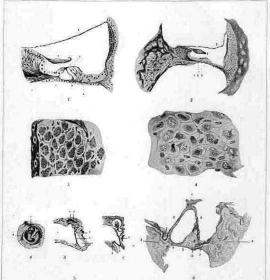
Pitt, I. Anatomical structure of the ear: 7 - Tympanic membrane; 2 - Malleus; 3 - Incus; 4 - Stapes; 5 - Oval window; 6 - Round window; 7 - Tympanic cavity; 8 - Eustachian tube; 9 - Mastoid cells; 10 - Mastoid process; 11 - External auditory canal; 12 - Auricle; 13 - Tensor tympani muscle; 14 - Stapedius muscle; 15 - Internal auditory canal; 16 - Cochlea; 17 - Vestibule; 18 - Semicircular canals; 19 - Cochlear nerve; 20 - Vestibular nerve. Fig. 2. Cross-section of the temporal bone with the internal ear structures: 1 - Auricle; 2 - External auditory canal; 3 - Tympanic membrane; 4 - Malleus; 5 - Incus; 6 - Stapes; 7 - Oval window; 8 - Round window; 9 - Tympanic cavity; 10 - Eustachian tube; 11 - Mastoid cells; 12 - Mastoid process; 13 - Internal auditory canal; 14 - Cochlea; 15 - Vestibule; 16 - Semicircular canals; 17 - Cochlear nerve; 18 - Vestibular nerve. Fig. 3. Various sections of the cochlea: 1 - Basilar membrane; 2 - Vestibular membrane; 3 - Spiral ligament; 4 - Spiral ganglion; 5 - Ductus cochlearis; 6 - Scala vestibuli; 7 - Scala tympani; 8 - Modiolus; 9 - Osseous spiral lamina; 10 - Foramina nervosa; 11 - Spiral vessels; 12 - Spiral organ of Corti. Fig. 4. Structure of the spiral organ of Corti: 1 - Hair cells; 2 - Supporting cells; 3 - Tectorial membrane; 4 - Nerve fibers; 5 - Basilar membrane; 6 - Tunnel of Corti; 7 - Cells of Claudius; 8 - Cells of Hensen; 9 - Phalangeal cells; 10 - Deiters' cells. Fig. 5. Vestibular apparatus: 1 - Utricle; 2 - Saccule; 3 - Ampullae of semicircular canals; 4 - Semicircular canals; 5 - Nerves of the vestibular apparatus. Fig. 6. Pathological changes in the inner ear: 1 - Otosclerotic foci in the region of the oval window; 2 - Otosclerotic foci in the region of the round window; 3 - Otosclerotic foci in the cochlea; 4 - Atrophy of the spiral ganglion; 5 - Degeneration of the nerve fibers; 6 - Thickening of the basilar membrane; 7 - Atrophy of the spiral organ of Corti. 12 Wittmaack. After Wittmaack improved the histological research technique of the labyrinth and through his experiments made it obvious the extreme sensitivity of the auditory nerve to various medicinal substances, further research proved the harmful effect of a number of other medicinal and non-medicinal substances, for example, alcohol, tobacco (M. F. Tsytovich), salicylates (N. S. Orembovsky). One of the most disfiguring diseases of the inner ear is otosclerosis, or otospongiosis, in which various areas of the compact bone labyrinth capsule undergo spongiosis and increase in volume, barricading the openings and canals of the labyrinth (see separate table, fig. 6). If the bone growths occupy the area of the oval window, then ankylosis of the stapes occurs; if such exostoses arise in the cochlea, those or other parts of the organ of Corti may be affected. Both of these lead to progressive weakening of hearing, sometimes reaching complete D.; similarly destructive for hearing is another process-spontaneous degeneration of the cochlear branch of the auditory nerve, causing clinically a picture very similar to otosclerosis. Both mentioned diseases are hereditary, but usually do not manifest themselves before the period of maturity, in contrast to those forms of underdevelopment and deformation of the inner ear, which are congenital and cause complete or almost complete D. from the very earliest age; these latter forms are discussed in detail in the description of deaf-mutism, since, existing from childhood, they prevent the child from learning to speak in a timely manner. There are still other, rarer and less studied forms of distortion of the normal structure of the inner ear, similar in type to those just listed, namely: rachitic form, hereditary multiple exostoses, Paget's disease, etc. Some of these conditions are associated with greater or lesser certainty with anomalies of internal secretion. Clinical picture of D. consists of its quantitative and qualitative features, as well as of accompanying phenomena. Already from the degree of hearing loss, one can often make an orientation, albeit rough, in the localization of the process. For example, if the subject does not hear ordinary conversation right next to the ear, and even more so if he is completely deaf, this should be attributed to processes in the labyrinth or generally in the nervous hearing apparatus, because only such kinds of conditions can bring the loss of function to such a strong degree. Progressive hearing loss is characteristic of the hereditary forms of dystrophy of the inner ear mentioned above or to多年 purulent inflammations of the tympanic cavity, in which secondary degenerative processes can develop in the labyrinth. Sudden deterioration of hearing in women, synchronous with each new pregnancy or childbirth, is considered characteristic of otosclerosis. On the contrary, sharp fluctuations in the acuity of hearing and, especially, a sudden return to normal under the influence of certain therapeutic procedures (for example, irrigation of the external auditory canal, inflation of the Eustachian tube) or else suggestion are characteristic of cases of mechanical obstacles to hearing (ear plugs, narrowing of the Eustachian tube, functional nerve deafness, partly traumatic neuroses). Qualitative features of D. are also very diverse. This group includes uneven sharpness of hearing for sounds of different character, for example for bass and treble areas. If bass sounds are relatively poorly perceived, one can assume a disorder of the sound-conducting function, and this sign is very pronounced in pure forms of otosclerosis, in its initial stages; treble deafness is characteristic of lesions of the sound-perceiving parts; so-called gaps in the 'ladder of tones' are observed in the most severe forms of labyrinthine D., including in deaf-mutes with remnants of hearing ability. Discrepancy between air and bone conduction is also characteristic of various diseases; for example, shortening of bone conduction is characteristic of processes in the inner ear and generally in the nervous hearing apparatus; unusually strong shortening of it (up to complete absence) is often observed in syphilitic lesions, and often the simultaneous contrast between it and good hearing with air conduction is striking (Wanner, V. E. Perekalin). The symptom paracusis Willisii is a companion of those processes in which rigidity of the auditory ossicles can be assumed (for example, otosclerosis and ankylosing forms of dry catarrh of the ear). The symptom diplacusis consists in that certain tones are heard in one ear at a normal height, while in the other they seem lower or higher; if the tone splits in height, this is denoted by the term diplacusis dysharmonica; if the tone splits in time, then-diplacusis echotica. The latter forms, as well as more complex hearing disorders (of the kind of verbal deafness), indicate processes in the sound-perceiving apparatus and hearing centers. Accompanying symptoms provide very substantial help in recognizing the form of deafness. In practice, all cases of D. are divided into 2 groups-with visible changes in the ear and without objective data. If during otoscopy signs of any pathological process in the external auditory canal, on the tympanic membrane or in the tympanic cavity are determined, it is very natural to attribute the cause of the existing D. precisely to these changes; but very often in the subject suffering from D. to one degree or another, the visible parts of the ear, as well as the Eustachian tube, appear completely normal ('negative objective data'). This is considered the rule in non-purulent diseases of the labyrinth and central lesions. In otosclerosis, it is sometimes possible to observe the so-called Schwarz sign - pink translucence of the tympanic membrane. In labyrinthitis, a supplementary sign can be disorders of the part of the inner ear adjacent to the cochlea, i.e., the vestibular apparatus. The diagnosis is, of course, reinforced by all other methods of research, data from the anamnesis, accompanying general phenomena in the organism, as well as
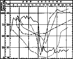
air conduction acuity of hearing whispering speech
bone conduction temperature Figure 7. and subjective sensations - noises, which also characterize certain forms of Deafness: for example, very distressing noises occur in otosclerotic processes, they are less pronounced in other diseases of the labyrinth and are not so noticeable in middle and external otitis. An important diagnostic tool is trial treatment (insufflation, lavage), the role of which was indicated above, as well as the course of the process, for which purpose it was proposed to compile for each patient a 'hearing curve' similar to a temperature curve (see Figure 7). Detailed determination of hearing function is carried out according to the rules of audiometry, for which sets of tuning forks, organ pipes and other sound sources (in the very recent time radio generators) and human speech are used. To expose 'feigned' Deafness, special methods are used, partly of a purely scientific nature, for example special experiments (Stenger's, Govseev's), comparison of data on different days, establishment of a conditioned reflex to sound, or else not strictly scientific, of the type for example using the factor of surprise or the sleepy state of the suspected malingerer. It is much simpler to determine 'concealed' Deafness; here ordinary audiometric methods are used, but necessarily with those precautions that the subject cannot use for guessing other sense organs (for example so that his vision is completely excluded, etc.). For details see below (Deafness in a medico-legal aspect). Prevention of Deafness. Improvement of preventive measures against Deafness is one of the main tasks of modern otiatry, especially because a significant number of 'deaf' people with the current state of science should be recognized not so much as patients amenable to one or another therapy, but as hearing disabled persons, for which type of persistent loss of function there are as yet no such mitigating devices as for example glasses for the eyes or crutches for the legs. That Deafness which gives a relatively good prognosis (for example in acute otitis, catarrhs of the Eustachian tube, certain forms of labyrinthine processes) passes along with the main disease, and no special measures specifically against Deafness are taken. But there is another category of diseases, very numerous, which not only does not give spontaneous recovery, but also resists all treatment methods; in it one especially feels helpless, because modern devices for artificial improvement of hearing often turn out to be ineffective. All preventive measures against diseases that impair hearing can be divided into measures of a general nature, of the type of anti-epidemic measures (with the understanding that if for example scarlet fever disappears from the population, then there will be no scarlet fever ear complications), and into specifically ear prevention, having a narrower task - to prevent the possibility of ear diseases as such. To special ear prevention belongs the fight against all the immediate causes of Deafness, which can be divided into accidental and constitutional. As an example of an accidental cause one can cite the habit of children sticking various objects into their ear out of mischief, which leads to injuries of the ear, eardrum and even the labyrinth. Raising the sanitary literacy of the population, intensified supervision of children, in connection with the development of measures for the protection of childhood, should prevent these cases. Purulent and catarrhal otitis often arise on the basis of hypertrophy of the pharyngeal tonsils. Their removal in a child with normal constitution in all other respects can prevent these processes. Certain forms of chronic purulent otitis often cause intracranial complications (meningitis, brain abscesses). Timely surgery can prevent these complications and is in this sense a preventive operation. Abstinence from smoking, alcoholism, immoderate or irrational use of certain drugs (quinine, salvarsan) is a prevention against alcoholic, nicotine and salvarsan lesions of the auditory nerve. Wearing earplugs during work associated with loud noise protects against professional Deafness. The question of prevention of constitutional factors is much more complex. In the reception rooms for ear diseases, patients with abnormal constitution prevail, not with normal constitution, but with anomalies of it of both a general nature (lymphatics, asthenics) and with specific local forms, which are usually designated by the term dystrophies and are divided into categories. Anomalies of the constitution of the mucous membrane of the tympanic cavity in connection with the constitution of the nose and throat are best designated as dystrophy with a shade of gigantism (hypertrophic type) and nanism (atrophic), however without a sharp difference between both forms and with frequent fluctuations and transitions of one into the other. A more obvious sign of abnormal local constitution should be considered the multiplicity of lesions (examples: simultaneous hypertrophy of various tonsils; simultaneous homogeneous process in both ears; simultaneous involvement of several organs of the same system, for example rhinitis and dacryocystitis; maxillary sinusitis and otitis; otitis, rhinitis and laryngitis, etc.). Characteristic of constitutional diseases is the persistence of the process, expressed in apparently causeless recurrences (for example in nasal polyposis or its accessory sinuses), and special resistance (chronicity) of processes, opposing without visible cause therapeutic measures, with the characteristic sign being the ineffectiveness of the same surgical methods, the action of which in normal constitution is considered quite reliable. Constitutional Deafness very often shows signs of hereditary suffering, being transmitted most often apparently recessively, but often also by the type of dominance. Factors of heredity are connected with factors of sex, which is manifested for example by a marked predominance of certain forms of Deafness (otosclerosis) in one sex - in this case in women. Prevention of Deafness in cases of constitutional anomalies cannot be limited to individual measures alone, because the factors of constitution are too powerful to yield to such a weak influence. Some benefit is obtained from the application of general measures (nutrition, strengthening regimen, change of climate). Treatment of Deafness Therapeutic measures, i.e. measures to combat existing deafness, can be divided into two groups: the first will include measures directed against the disease which causes loss of hearing, with the aim to cure it or weaken its action. Through these measures they strive however not only to restore hearing, but also to free the patient from other possible consequences of the disease; therefore here therapy does not yet have a specific character. To the second group belong measures directed specifically against Deafness. An example of them are the so-called 'acoustic' operations: artificial perforation of the eardrum, division of adhesions of the tympanic cavity, tenotomy in case of contracture of its muscles, mobilization of the stapes, plastic restoration of the external auditory canal in its atresia, the latest operations on the labyrinth with the help of an operating microscope (Holmgren) and others. Another example are bloodless methods - the use of artificial eardrums and hearing-improving devices. As 'artificial eardrums' are used either materials resembling a real eardrum (gutta-percha circles, egg membrane, thin plaster) or simply wads of cotton, dry or moistened with diluted glycerin, with which they cover the hole in the eardrum or fill the tympanic cavity through a defect in the eardrum. This method in some patients with partial or complete absence of the eardrum gives an increase in acuity of hearing, often quite noticeable. The cause of such a change in hearing is not yet clear; probably here more favorable conditions are created for the transmission of sounds to the labyrinth due to the pressure of the artificial eardrum on the labyrinthine windows and thus the regulation of intralabyrinthine pressure. Hearing-improving devices have the form of hearing tubes, which are applied to the ear with the narrow end and catch sounds with the bell (see Figure 8 and 9) or else are based on the action of mic Figure 8.

Figure 9.
of the telephone and consist of a set that includes a battery of dry cells (see figure 4, g), a receiver for sounds in the form of a diaphragm and a telephone plate with an amplifier (see figure 10, 1 and 2). There are various systems of such devices, but their common property is the distortion of sound images, which often nullifies other advantages of the device. It is impossible to predict in advance that such a device will help a particular patient. For some, it helps to the extent that it restores their ability to work and allows them to engage in a profession requiring hearing; for others, only temporarily, and soon patients abandon it. In cases where hearing loss can be assumed to be due to inactivity or congenital underdevelopment, it is logical to attempt to sharpen hearing through appropriate acoustic exercises ('activation') with the help of various devices (Urbančich's harmonica, Dr. Maurice's 'kinésiphone', Professor Skritsky's 'activator'). The last-mentioned device appears to be particularly practical; moreover, it allows teachers of the deaf to spare their own voice when working with students, thanks to which, in a certain number of cases, with certain forms of incomplete deafness, it was possible to somewhat increase the acuity of hearing. Further experiments are certainly necessary to determine how much activation will enrich our medical arsenal. In the event of failure of the listed means, the hearing-impaired are subject to appropriate social measures for education and care. The care for deaf children coincides with or follows the principles of education for the deaf and mute
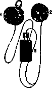
Figure 10. (see Deaf-mutism, deaf-mutes) or, in less severe forms, are limited only to measures that separate them from the general composition of a given class or group of students. Such children are seated in the first desk, separate lessons are arranged for them, etc. For those who have lost their hearing, there remains the resource (in terms of communication with other people) to sharpen their normal ability to guess speech by the speaker's facial expressions (lip reading). Corresponding courses can train the vision of the deaf and hard of hearing to a considerable extent. Finally, in the distribution of work, the hearing-impaired should be used for such labor where they could freely compete with the hearing on the basis of its nature. Deafness in a forensic medical sense. The question of hearing loss can be the subject of a forensic medical investigation if Deafness is the result of some professional injury, accident, or intentionally inflicted trauma. The task of the examination is to establish a causal relationship between the state of the person's hearing ability and the circumstances to which its weakening is attributed (e.g., if the plaintiff claims that he became deaf from a bruise received in a catastrophe that occurred at such and such a time and place, the examination must show whether the hearing loss of the person examined could have occurred from the injury he sustained, and also whether there was actually an injury to the hearing apparatus). The second task is to determine the actual degree of deafness or to establish the very fact of hearing loss, since the interested party may give intentionally false testimony. To determine the cause of Deafness in a forensic medical examination, generally the same methods are used as are usually applied in the examination of ear patients; they inquire about all the details of what happened, what the person examined feels at the present time; then they examine the ear, determine (with certain special precautions) its function, as well as other signs that have significance here (e.g., symptoms from neighboring, and sometimes distant organs), and assess how probable the connection is between the given hearing defect and its supposed cause. Objective signs of injury to the ear or other pathological process in it greatly facilitate the examination. For example, if fresh bruises, abrasions, or tears are found on the external auditory canal or eardrum upon examination, then it becomes plausible that partial hearing loss is due to the inflicted trauma. Conversely, if the person examined shows signs of a long-standing disease that in itself causes a certain degree of Deafness (e.g., chronic suppurative otitis), and there are no signs of injury, then in this case it is more possible to exclude traumatic etiology. Severe forms of Deafness, for example, complete loss of hearing in one or both ears, are always associated with processes in the inner ear or auditory centers; therefore, with corresponding complaints, trust in them increases if at the same time it is possible to establish a severe form of injury (e.g., skull fractures, often extending to the petrous part of the temporal bone, symptoms of cerebral hemorrhage, general shock, etc.). Intentionally inflicted injuries and artificially induced diseases often have distinctive features by which they can be recognized. For example, when corrosive liquids are poured into the ear (e.g., carbolic acid), traces of chemical burns often remain on the skin of the cheek; the interior of the external auditory canal may also be ulcerated and even necrotic, which almost never occurs in natural ear diseases. Determination of the acuity of hearing in cases where insincerity is suspected from the person examined is conducted according to special rules, which aim 1) to discover concealed Deafness or 2) to expose the person examined in feigned deafness. Concealed Deafness, as mentioned above, is recognized relatively simply, because if hearing is examined with precautions that exclude guessing words by the speaker's facial expressions, then the decrease in hearing ability immediately reveals itself in that the person examined does not repeat the words said to him. The second task is more complicated - exposing the simulation of deafness. Here different cases are possible. Sometimes complete, absolute, bilateral Deafness is simulated. The methods for ascertaining the truth in this form of pretense are divided into strictly scientific and more general. The first group includes attempts to use some reflex in which the auditory pathways participate and which is determined objectively. Some suggest using unconditional reflexes, for example the auriculo-palpebral (blinking of the eyelids with sudden strong sound stimulation). Others prefer conditioned reflexes; this method is more troublesome, as it is necessary to produce a series of combinations of two irritants (e.g., sound and faradic) and develop a conditioned reflex on a certain muscle group (e.g., finger extensors) that manifests itself, for example, in the withdrawal of the fingers with sound stimulation (electric bell). If the subject withdraws his fingers only at the bell, without the accompanying faradic shock, it is concluded that he hears this sound. The second more or less scientifically substantiated method is the so-called Govseev's experiment ('brush experiment'). Its technique is as follows: the simulator is placed with his back to the examiner, who simultaneously runs the palm of one hand along the subject's back, and with the other hand, armed with a clothes brush, along his own chest (or vice versa); the experiment is repeated many times, and the subject must each time guess what was used to stroke his back - a hand or a brush. Truly deaf people in both ears usually guess the hand and brush either every time or at least in a very high percentage of all cases; among simulators, the vast majority make mistakes (people with normal hearing also mostly cannot distinguish between the hand and brush under the conditions of this experiment). Finally, the methods of the third order are based on the psychological peculiarities of human nature and have little in common with otology in essence. Most of them aim to act by surprise, to catch the subject unawares; for example, to call out to him during sleep: a truly deaf person will not wake up from this alone, but a feigningly deaf person may wake up and thus betray himself. There are many other similar tricks, which however are only effective when they are varied and improvised in each individual case; if already used methods are employed, there is no guarantee that the subject does not know them and is not on guard against them. In the simulation of complete unilateral Deafness, the methods just mentioned cannot be applicable, because our science does not have methods that absolutely guarantee separate examination of the right and left ear, and therefore the subject who reveals his hearing can always say that he heard with the other, healthy ear. But in such simulators, methods based on this very property of mutual compensation or generally on the influence of the function of one ear on the other become valuable; for example, such an experiment is used: to the subject who claims he is deaf in one ear, two identical tuning forks are brought close to the ears, the sound of which he must listen to. It is known that if sound from two identical sources is directed into both ears, only the ear to which the source is closer is heard (because then the sound of the corresponding pitch is felt stronger in this ear and masks the sensation of the second ear). The same phenomenon can be reproduced in the just mentioned method - for this it is necessary to place the tuning forks at different distances from both ears: with normal hearing, only the ear to which the corresponding tuning fork is closer is heard. In truly deaf people in one ear, this phenomenon does not occur; if it is accurately determined at what distance their healthy ear hears the corresponding tuning fork, this distance will remain constant, even if the second tuning fork is brought closer to the other ear. In simulators, however, with such an experimental setup, the sensation in the 'healthy' ear will be drowned out by the sensation in the supposedly deaf ear, and he is forced either to admit that he no longer hears with the 'healthy' ear, or that he hears with the 'deaf' one. Exposing simulation in cases of incomplete Deafness in one or both ears requires considerable skill. The main method here is repeated measurement of the acuity of existing hearing; non-simulators give more constant data in such experiments than simulators. However, all the listed methods suffer from the common defect that they mostly do not directly determine the acuity of hearing in the subject, but only the fact of whether he is giving testimony sincerely or not. Thus, the methods used to expose simulation only fulfill part of their purpose. Moreover, the results of these methods mostly speak only of a certain probability of simulation, and do not establish it with such obviousness as would be convincing to the uneducated in medicine persons present at the examination.
Related articles
Mentioned in
Cite this page
“Deafness.” Soviet Medical Encyclopedia. English translation of Bolshaya Meditsinskaya Entsiklopediya, 1st ed. (Moscow, 1928–1936), ed. N. A. Semashko. https://sovietmedicalencyclopedia.pages.dev/article/deafness/