Hutchinson Triad
Historical document, translated for reference. It reflects medical knowledge of the 1920s–30s and is not medical advice.
Summary
The Hutchinson Triad is a set of three clinical signs—dental deformities, parenchymatous keratitis, and nerve deafness—that are highly characteristic of congenital syphilis. This article details each component and discusses the diagnostic significance of their combination.
Encyclopedia article (1928–1936)
HUTCHINSON TRIAD (Hutchinson), a name assigned to a specific combination of stigmata or signs of late congenital syphilis, the enormous diagnostic importance of which was first noted by the famous English dermatologist Hutchinson (in the 1860s). It includes: 1) dental deformity, 2) parenchymatous keratitis, and 3) nerve deafness. Each of these signs, taken separately in its clinically typical form, is already highly suspicious of congenital syphilis, but the combination of even two and especially three of them (which, however, is not very frequent) indicates the presence of late congenital syphilis with almost absolute probability. Only during the first 11/*-2 decades after the publication of Hutchinson's main works (1859-63) did some clinicians (Mooren, Giraldes, Panas, etc.) attempt to dispute the indisputable belonging of the indicated signs to congenital syphilis. However, in these cases, the discussion concerned rather the significance of each sign individually than their triple coexistence, known by the name of the Hutchinson Triad; this name was first given to the group of these signs by Fournier. Let us examine in more detail each of the three components constituting the Hutchinson Triad: I. Dental deformity, or so-called Hutchinson's incisors. Here we are speaking of the upper central incisors of the second eruption (permanent). They are characterized by: 1) a crescent-shaped, even elegantly curved notch located at the free edge of the tooth [see Figure 1 and 2; Figure 1 shows typical Hutchinson's teeth: a solid notch on the upper incisors, the teeth are spaced apart; Figure 2 shows a typical Hutchinsonian right incisor (the left one is artificial)]; 2) the configuration of the tooth, resembling a screwdriver due to its widening at the neck level and narrowing at the free edge; 3) the indirect concave direction of the upper central incisors with the crescent-shaped notch. As is known, a crescent-shaped notch on the free edge can also be found on other teeth (incisors and canines), however, the symptomatic value of Hutchinson is attached only to the crescent-shaped notch affecting the upper central incisors of the second eruption (Figure 3 shows pseudo-Hutchinsonian teeth, often mistaken for Hutchinsonian: a notch only on the front surface, correct tooth position). In later times, to this basic dystrophy were added a whole series of other changes of the dental system occurring in late congenital syphilis, often in combination with the other components of the Hutchinson Triad, such as: slight fragility of this system, microdontism, amorphosis, widely spaced incisors (so-called diastema dentium, or Gaucher's sign), prognathism, and finally, the Gothic palate, etc. II. Parenchymatous keratitis is, in Hutchinson's opinion, one of the most important signs of congenital syphilis. Later, with the help of the Wassermann reaction, which is positive in almost 100% of cases with this symptom (Igersheimer), it was possible to fully and objectively confirm the correctness of Hutchinson's views. Parenchymatous keratitis most often appears between the 7th and 14th years (after the 20th year its frequency noticeably decreases, and after the 25th year it becomes rare); however, due to the opacities remaining after it, it retains diagnostic significance even at a later age. Typical parenchymatous keratitis always affects both eyes. Usually inflammation of the second eye follows the first after 6-12 months. Often this interval can be significantly shortened or, on the contrary, lengthened to several years. Clinically, parenchymatous keratitis in its developed state is expressed by a uniform or partial milky-colored opacity of the cornea, the surface of which is dull and almost always penetrated by vessels. Sometimes, on the contrary, the cornea has a spotted appearance, and the spread of vessels is limited to individual areas (see the color plate to the article Keratitis). Subjective symptoms include irritation, photophobia, lacrimation, etc. These phenomena come to the fore especially in the presence of severe complications, for example, inflammation of the iris, etc. Purulent processes are never observed in parenchymatous keratitis. Its course is usually chronic—months and even years. Complete restitutio ad integrum never occurs, although vision can recover quite well, more often, however, b. or m. serious disorders of vision remain, and even almost complete blindness (in 5-6%). III. Nerve deafness, usually beginning with deafness, which, occurring almost always suddenly (within a few days or even hours), affects both ears simultaneously, progresses relentlessly, and in the end can end in complete deafness. Sometimes the development of deafness may stop at any stage, so that impairment of hearing in one ear may be combined with complete deafness in the other. Subjective disorders (noise in the ears, dizziness, etc.) may be more or less pronounced or even completely absent. The matter is based on a disease of the labyrinth, and this process is very often incurable. In cases where deafness is combined with a long-standing and even completed parenchymatous keratitis, the latter, often with the onset of ear involvement or even shortly before it, may flare up again. If, between the 7th and 13th years, with signs of closure of the Eustachian tube, deafness suddenly develops and rapidly increases in combination with noise in the ears, one should always, first of all, think of congenital syphilis. The diagnosis becomes almost absolutely certain if, in addition, there is parenchymatous keratitis. To the three symptoms described, constituting the classic Hutchinson Triad, Heubner adds syphilitic gonorrhea, often accompanying the triad. All these signs usually appear and reach their full development, approximately, in the 3rd-5th-13th years. The Hutchinson Triad is therefore very characteristic precisely of late congenital syphilis, but one must not forget that even the absence of all four signs does not exclude the presence of this form of the disease. Hochsinger, in contrast to the Hutchinson Triad, which he recommends calling the "Late Triad" (Spat Trias), proposes his "Early Triad" (Fran Trias), the signs of which are characteristic of the first year of life of a syphilitic infant. The Hochsinger Triad includes: 1) skeletal deformity (caput natiforme, Olympic forehead, saddle nose), 2) scars radially located from the lips and corners of the mouth, and 3) involvement of the cubital glands.
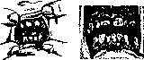
РИС. 1. Рисунок 2. Рисунок 3
Related articles
Mentioned in
Cite this page
“Hutchinson Triad.” Soviet Medical Encyclopedia. English translation of Bolshaya Meditsinskaya Entsiklopediya, 1st ed. (Moscow, 1928–1936), ed. N. A. Semashko. https://sovietmedicalencyclopedia.pages.dev/article/hutchinson-triad/