Syphilis
Historical document, translated for reference. It reflects medical knowledge of the 1920s–30s and is not medical advice.
Summary
Syphilis is a chronic infectious disease caused by the pale spirochete, primarily transmitted through sexual contact and considered one of the most important venereal diseases. The article explores its controversial origins, historical spread across Europe and beyond, and its social impact during the first major epidemic in the late 15th and early 16th centuries.
Encyclopedia article (1928–1936)
SYPHILIS. Contents: I. History of syphilis...............515 II. Epidemiology.................519 III. Social significance of syphilis........524 IV. Spirochaeta pallida .............,, 527 V. Pathological anatomy...........533 VI. Experimental syphilis.........536 VII. Clinical picture and diagnosis of acquired syphilis ......................54 5 VIII. Visceral syphilis............. 562 IX. Treatment of acquired syphilis......570 X. Personal prevention.............588 XI. Congenital syphilis.............589 Syphilis (syphilis, lues, lues venerea), a chronic infectious disease caused by the pale spirochete; transmitted primarily through sexual contact, it is one of the most important diseases in the so-called group of venereal diseases (see). Supporters of the 'American' origin of S. in Europe (I. Bloch, G. Gaustein) link the emergence of S. to the campaign of Charles VII in Italy (1494-95), whose army, consisting of mercenaries of various nationalities (French, Swiss, Dutch), came into contact with Spanish soldiers who had returned from Barcelona in 1493 after a journey with Columbus. The Spanish physician Diaz de Isla (1462- 1542) states that Columbus's sailors brought S. to Spain from the island of Espanola and quickly disseminated it in the coastal cities of Spain. Charles VII's campaign through Italy, especially his stay in Rome, where, according to Delicado, there were 14,000 Spanish prostitutes at that time, contributed to the spread of S. both in Italy itself and in the countries to which the mercenary armies moved after Charles VII's defeat. In the exile of Charles VII from Italy, Milan, Venice, Germany, as well as Spain, which sent reinforcements by sea from Barcelona to Naples, participated. The victorious troops entered cities whose population had already become victims of S., which had spread among them with extraordinary rapidity, as Charles VII's army, according to historians of that time, indulged in unlimited debauchery. By the beginning of the 16th century, S. had become known almost throughout the European continent. The spread of S. in Europe was facilitated by the social shifts of the emerging capitalist era: the ruin of the peasantry, the growth of cities, the development of trade relations and in particular navigation, the proletarianization of artisans. S. spread rapidly along maritime trade routes and outside Europe. By the beginning of the 16th century, S. becomes known throughout Southern and Southeastern Africa, from where it was soon brought to China and Japan (1512). The medieval way of life, its morals, the almost unlimited sexual freedom of men, the existence of so-called public baths, which were places of open debauchery, and the legalization of brothels, maintained not only by private individuals but also by urban communities and even the clergy, all favored the development of the S. epidemic. For these reasons, S. took on epidemic proportions, characterized during this period not only by massive spread among all classes of the population but also by particularly severe course. The Italian physician of that time Summarina writes: 'No words and no pen can depict the suffering caused by this new disease.' 'Instead of a latent stage lasting several weeks,' writes Boeck (W. Boeck), 'between the appearance of the primary ulcer and the secondary phenomena, the primary period lasts only a few days, accompanied by headaches, tearing pains in the joints, general weakness and insomnia; 8-14 days after infection, the head and then the body become covered with a rash, leaving deep ulcers or rapidly disintegrating tumors, destruction of bones, and often gangrene.' The relatively quickly recognized connection of this severe disease with sexual life gave rise to calling S. 'the sexual plague.' The degree of spread of S. during this first epidemic can be judged by the fact that from 1495 to 1497, as documented by I. Bloch, cases of S. were detected I. History of syphilis. The significant spread of S. throughout the world and the serious consequences it caused to both individuals and society prompted many historians of medicine to study the question of the time of origin of S. in Europe. This study of the history of S. divided historians of medicine into two camps: adherents of its existence among European peoples in the most ancient times and the so-called 'Americanists,' who believed that syphilis appeared in Europe only after the discovery of America. Among other authors, Hensler, Proksch, Sudhoff were defenders of the first assumption. In their opinion, the abscesses and ulcers described by Hippocrates in the mouth, throat, alopecia, eye inflammations, condylomas in the genital area, etc. can be recognized as manifestations of syphilis. Proksch in his 'History of Venereal Diseases' notes as proof that Hippocrates meant precisely S, the sequence in the description of pathological phenomena. The causal connection between nasal lesions and diseases of the genital organs is mentioned by Dioscorides, Galen, Paul of Aegina, Celsus, although the direct causes of nasal defects are not indicated by either Celsus or Susruta. Similarly, as evidence, anatomical findings are presented: the skull of a child found by Parrot, and the famous skeleton Solu-ъгё. However, Virchow asserted that he knew of no case of S. of bones that dated to the period preceding the first and sufficiently well-studied epidemic in Europe at the end of the 15th century. Finally, individual authors (Sudhoff) cite documents on the basis of which they judge the existence of S. in Europe before the end of the 15th century: this includes the edict of King Maximilian I of Germany, allegedly published in 1493, on blasphemy punishable by syphilis; the protocol of a court session in Dijon in 1463 about infection with an infectious disease; and Ordonnances des rois de France of 25/Ш 1493 as evidence of the existence of S. in Paris before Charles VII's campaign in Italy. These documents have been recently studied by G. Gaustein, who in his monograph (Die Fruhgeschichte des Syphilis 1495- 1498) proved that the edict of King Maximilian was actually published only in 1497, i.e. after the return of German soldiers from the campaign in Italy; that in the so-called Dijon protocol, the subject is not S, but apparently a case of epilepsy; that the Ordonnances des rois, to which Sudhoff refers, are in fact a police order of 25/VI 1498 in the following places in Germany: Bamberg, Bayreuth, Breslau, Erfurt, Frankfurt n/M, Hamburg, Hildesheim, Cologne, Munich, Lower Rhine, Oldenburg, Eastphalia, Nordlingen, Nuremberg, Prague, Strasbourg, Vienna, Wurzburg. In 1496-97, S. is already known in the Netherlands, England, Denmark, Scotland. The introduction of the disease from one place to another can also be judged by the popular names of S: thus, along with names of S. according to its clinical manifestations (Blattern, Pustulen, Pok, Bubus, grosse Blattern, pestis inguinalis and others), the French called it mal de Naples, passio italica, morbus hispanicus; in Germany, Italy and Spain, England, Ireland and Switzerland-morbus gallicus, mal francoso, lues aetica; in Poland-deutsche Krankheit (German disease); in Portugal-Castilian disease; in Japan-Portuguese disease, disease from China (T6-Kasa), disease from the Liu-Kiu islands (Liu-Kiu-Kasa), in Persia-Turkish disease; in Russia-Polish or French disease. In these very popular definitions of S, some authors (Proksch) see proof that S. was known to these peoples before the discovery of America, because no people called S. morbus americanus, and the most widespread name among physicians was morbus gallicus. In reality, however, the popular definitions of S. were explained by the fact that medieval medicine knew nothing about this new disease, that, turning to ancient authors, it did not find an exact description of the disease and measures for its treatment, and that the massive spread of S. was obviously connected with military campaigns to new countries, from where the disease was brought. Precisely for this reason, the Spanish physician Diaz de Isola, a contemporary of the first S. epidemic, calls it 'the disease of the island of Espanola (discovered by Columbus); similarly, other Spanish contemporaries, Oviedo and Las Casas, who gave detailed descriptions of the lands discovered by Columbus, call S. 'the West Indian disease.' In their descriptions of S., they note not only the very fact of the 'American' origin of S, but also its milder course among the natives compared to that of Europeans infected with S. Las Casas, a participant in Columbus's second voyage, was in 1498 on the island of Haiti and, questioning the natives about how long S. had existed among them, established that S. had existed here long before the arrival of the Spaniards-'from time immemorial.' As the first and most severe wave of the S. epidemic subsided and the severity of clinical phenomena weakened in the middle of the 16th century under the influence of the widespread use of mercury and Guajak, the popular definitions also disappeared: Bethencourt introduces the name lues venerea, and Fracastoro (Girolamo Fracastoro)-syphilis (after the hero of his work 'The French Disease').
The first epidemic of S., which led to its introduction to all countries of the world, ended with its universal spread, creating a number of endemic foci, from which S. was introduced to new places and, over time, increasingly transformed into a disease inextricably linked to the conditions of capitalist economy. In both the Middle Ages and modern times, the inevitable existence of prostitution under conditions of capitalist exploitation became the main source of the spread of S. (the spread of S. in Western Europe and other countries of the world-see Venereal diseases). A country of particular endemic spread of S. was Russia, where S. first penetrated during the years of its first epidemic. According to Haeser, S. was brought from Italy by some woman to Krakow in 1495, according to Strikovsky-in 1493, and in 1495 and 1496 it was spread by troops throughout Poland, bordering Russia. "One must think that this woman was the founder of S. in Russia," writes Gertsenshtein, pointing out that the appearance of S. to such an extent "alarmed" the then Moscow government that in 1499 the great prince Ivan Vasilievich entrusted his Lithuanian envoy, the nobleman's son Mamonov, on his way to Lithuania to verify in Vyazma the existence of S, namely: "Has anyone arrived with that illness, that sores are wandering? and it is called the French disease and as if it was brought from Vilna." There are no descriptions of S. in Russia before the Peter the Great era, because only under Peter I does the influx of physicians from Western Europe begin, and if individual foreign physicians earlier served only the court, then under Peter I they are intended mainly for the army and fleet he is creating and only later for the rest of the population. Despite the absence of medical descriptions of S. in Russia in the 16th-18th centuries, Gertsenshtein rightly considers that S. "firmly took root" in Russia due to continuous wars, the absolute absence of medical care for the sick, the low cultural level of the masses, the patriarchal way of life in large families, the continuous replenishment of troops with new layers of the population; under such conditions, once introduced, S. turned individual families and villages into foci of its endemic spread.-The significance of the spread of S. can be judged by the fact that the treatment of this disease occupied the largest place among folk healers of all nationalities of the country, as well as by individual later descriptions of the introduction of S. from Russia to other countries. Thus, according to Lancerot, S. was brought by sailors of a Russian ship to Ageresund and Stavanger (Norway) in 1710, where it took endemic spread under the name of "unclean" disease "radesyge" (radesyge), studied in 1758 by Honoratius Bonvi. K. Gron (Gr0n) cites van Swieten, indicating that S. was widely spread in Siberia in 1715-16; he believes that Swedish prisoners of war, interned in Tobolsk after the defeat at Poltava, brought the disease with them from Saxony. The first statistical information about S. in Russia dates to the mid-19th century, and as for its morbidity in the 18th century, one can judge both by publications of special guides, e.g. "A Guidebook on Venereal Diseases for Popular Use" by the chief physician of the St. Petersburg military-land hospital S. A. Venchansky in 1755, and by the "measures of struggle" to which the government was forced to resort. Thus, in 1762, the Senate (quoted from Gertsenshtein) proposed the establishment in cities of special hospitals for the treatment of S: in provincial cities for 100 beds, in district cities for 50, with doctors, pharmacies, baths, etc., indicating "the possibility and necessity of free use by the sick." In the following 1763, in view of the significant spread of "French venereal disease" (S.) among soldiers, the Senate ordered the deportation of all identified sick women to Nerchinsk; in 1783, a secret hospital for sick men and women with S. was opened in Petersburg. As to what extent S. had penetrated the peasant population layers at that time, one can also judge by Gertsenshtein's indication that in the Little Russian provinces when hiring workers, a certain portion of their salary was withheld "for the treatment of French venereal disease" among them. The "measures of struggle" undertaken by the government against S. were naturally powerless: serfdom, poverty and ignorance of the numerous rural population, the absence of systematic medical care-all this could not weaken the endemic spread of S, which when introduced to new areas caused heavy disasters in them. To the same extent as the rural population, and sometimes even more, the population of national districts suffered. Tarnovsky in a note to Lancerot's guide (The Doctrine of S., 1876, SPB) points out that "in 1850 among the Kirghiz, Bashkirs, Meshcheryaks of the Orenburg province S. spread to such an extent that according to reports of local authorities many clans were universally infected, and the disease had an extremely severe character." The emancipation of the peasants and the subsequent development of capitalism in Russia caused significant development of peasant crafts in cities, where they came into contact with regulated prostitution, among which S. was extremely widespread. Similarly, a factor contributing to the introduction of S. into villages was military service with prolonged separation of conscripts from their families. S. brought by migrant workers and soldiers to villages in the second half of the 19th century found in it peculiar ways of spread: the dark life of the village, the absence of primitive hygiene skills, superstition, folk medicine, which had strengthened in the people the view of S. as a miserable and shameful disease-all this contributed not only to sexual but also to extremely strong non-sexual transmission of S. The close connection of S. in tsarist Russia with the life of the rural population determined it as "domestic" S. The presence of endemic foci of "domestic" S. was a factor determining the steady growth of S., about which one can judge from the last quarter of the 19th century, i.e. from the time of the first publications about it in the statistical reports of the medical department of the Ministry of Internal Affairs. II. Epidemiology. The epidemiological feature of S. in prerevolutionary Russia-from its first detailed descriptions, i.e. from the middle and end of the 19th century, until the October Revolution of 1917-was its endemicity, i.e. the identification of new cases of it in certain, sometimes limited areas, cases of all ages, sometimes entire villages or a number of families, with the main route being its "domestic" spread. In endemic foci, the primary source of infection could be sexual infection, however, the further spread as a rule went by non-sexual routes. Descriptions of individual, sometimes quickly passing epidemics of S. are found in the history of almost all European countries both soon after and many decades after the first epidemic (pandemic) of S. described above. Thus, Konrad Meyer-Arens describes an endemic outbreak of S. in the canton of Bern (Switzerland) in 1570, which led to the issuance of special government orders on the isolation and treatment of patients; in 1592 and 1602 these orders were repeatedly confirmed. Among the oldest outbreaks of S. is also the one described by Gilchrist and Benjamin Bell in 1771, then the endemic S. rampant in the 17th and 18th centuries in Scotland, called sibbeus (apparently after the name of one of Cromwell's generals, whose troops introduced S. to Inverness). According to Willemoers, the reason for the significant spread of S, especially among highlanders, was the wide hospitality, shared use of beds with strangers, smoking common pipes, eating from common bowls with common spoons. To the endemic outbreaks of the 1760-70s also belongs the so-called Canadian syphiloid-a disease that gained significant spread in Mai Baie (now Murray Bay), brought, according to Proksch, by the English or Germans (mal anglais, maladie allemande). Under the name morbus dithmarsicus (Dithmarsch disease or Jutland syphiloid) is described an endemic S. in Denmark, the beginning of which dates to 1757; the disease took such formidable proportions that at the government's proposal in 1813 a special investigation of the affected areas was organized. To the endemic outbreaks of S. in Norway, Gron also attributes the so-called radesyge-the above-mentioned infection by Peter I's sailors of the population in Stavanger and Danish sailors in Ageresund (1709-10), which gave the infection long-term existence and universal spread. In 1778, a commission created by royal decree for the investigation and treatment of S. developed measures, among which it is interesting to note the following: in the affected areas, marriages of persons suspected of having S. were prohibited without prior medical examination; priests and other officials had to twice a year report on the number of patients they had; recruits called up had to be examined for the presence of S.; to this time also belong indications of the indisputable non-sexual nature of infection. The greatest severity in both spread and duration of existence is distinguished by the endemic S. in the Balkans. According to descriptions by the Franciscan monk I. F. Jukic (quoted from K.).
Gr0n;y) Syphilis was introduced in 1780 by the Turks into Bosnia, where it received the name skerljevo (in Croatian; or scherlievo in Italian, mal di Fiume, gribbing disease, mal di Fuccine, mal di Radugusa, etc.), and comparatively quickly spread to all Balkan countries. The strength of the endemic syphilis in these countries can be judged by the fact that after the retreat of the French (1814), Emperor Franz I of Austria himself visited in 1816 the areas affected by the disease skerljevo, and by his decree in 1818 a mass examination of the population was carried out in 11 districts under Austrian protectorate; all residents were examined, regardless of property status, sex, and age, with the exception of the aristocracy of the city of Fiume, for which only a certificate from a home physician was required. For the isolation and treatment of patients, hospitals were opened in Fiume and Castel Nuovo with 1,000 beds each. Until 1859, through the hospital in Castel Nuovo, i.e., over 41 years, about 190,000 patients passed. Along with hospitalization, preventive measures were also carried out: some areas were cordoned off by troops for quarantine, the right to marry was restricted, all houses and utensils after the removal of patients were disinfected, with some items even being destroyed. Thanks to the involvement in subsequent years of an increasing number of physicians and professors (e.g. Sieg-mund, Hebra, etc.) in examinations, the organization of hospitals and permanent observation of foci of syphilis, the endemicity of the latter gradually declined, especially in Trieste, Krain and Croatia. The next outbreak occurred in Brenotal in 1860, with infections being transmitted through lymph inoculations from children who died in infancy. In 1880-81, the number of patients in Dalmatia increased so sharply that it again required the implementation of special preventive measures (compulsory hospitalization of patients when necessary). Syphilis spread particularly widely in Bosnia and Herzegovina, as these areas, which until 1878 were under Turkish rule, were almost completely unaware of medical assistance; the epidemic state of these countries was completely unknown. The strength of the endemicity and the degree of spread of Syphilis in these uncultured countries can be judged by the fact that within one decade (until 1889) 11 special hospitals were opened in Dalmatia. The endemic Syphilis in Bosnia and Herzegovina was until the end of the 19th century the subject of special examinations by such large Austrian syphilologists as Auspitz, I. Neumann, M. von Zeissel. However, even at the beginning of the current century, endemic Syphilis in Bosnia and Herzegovina affected entire villages. The prolonged existence of the infection led to the continuous expansion of free medical institutions, both stationary and outpatient, the publication of popular literature in national dialects, distributed by rural teachers, people's judges, etc., as well as systematic house-to-house examinations, sometimes accompanied by treatment on the spot. As a result of these measures, according to the report of Glück (L. Glück), in 1903 a number of small endemic foci were eliminated and tertiary gummatous Syphilis cases became less common. However, periodic observations of Syphilis foci in Bosnia and Herzegovina were carried out until the beginning of the imperialist war in 1914. The colonial policy of European capitalist countries turned the conquered countries into significant foci of endemic Syphilis; thus, Herzenstein cites in his work on Syphilis in Russia (1885) the data of Rohlfs on Syphilis in Morocco: "The local name for this disease mird el kebir - the great disease - indicates the population's view of it. Jackson (Jackson) asserted that in Morocco it is difficult to find a person whose blood would not be infected with this poison. Currently, Syphilis mird frendji (the French disease) is the most widespread. Leo (Leo) says that, 'perhaps only one-tenth of the population is free from it'." Thus, already in the first years of the official registration of Syphilis in Russia, its enormous spread was revealed and its main endemic feature - the significant prevalence of Syphilis among the rural population - was identified. The latter is confirmed both by the registration of Syphilis in medical institutions and by data from house-to-house family examinations. Summarizing the reporting data on Syphilis for 1878, 1879 and 1880, Herzenstein comes to the conclusion that "approximately 2 million of the population of European Russia are afflicted with this scourge". Chistyakov (1880), after examining 25,050 people in Krasnoslobodsky district of Penza province, found among them 4.73% of Syphilis patients even with far from perfect research methods under conditions of house-to-house examination. The number of Syphilis patients among those receiving treatment at medical sites was also significant: D. Zandberg (1893) found among outpatients in Kozlovsky district of Tambov province 20.6% of Syphilis patients; Barsukov (1878) in the same province - 18%; Nikolsky (1886) - 15.7%; Maslovsky (1890) determined the prevalence of the entire province population at 2.5%. - Somewhat later, Dovodchikov, when examining 51,077 peasants in Yaroslavl province, identified 3.2% of Syphilis patients; Lifshits in the former Lithuanian province out of 40,027 examined - 4% of Syphilis patients. - Such a significant prevalence among the rural population was explained by the ease of its non-sexual transmission under rural living conditions. Uvarov reported at a congress on discussing measures to combat Syphilis (1897) that "in Kostroma province, syphilitic ulcers were observed in the mouth in 221 cases, on the lips and cheeks - 141 cases, on other parts - 246 cases; if the latter category includes lesions on genitalia, then infections through everyday contacts must significantly predominate". Kalmanovsky in Lodeinopolsky district of Olonets province (1903) among 1,082 Syphilis patients seeking help found sexual infection in only three cases. On the spread of Syphilis in the USSR see Venereal diseases - spread in the USSR. The implementation of special measures to combat Syphilis in the countryside and the widespread use of salvarsan preparations for its treatment led, along with a decrease in the saturation of Syphilis in individual families, to significant changes in the clinical picture of household Syphilis, which was characterized by the predominance of tertiary gummatous forms of Syphilis over primary and secondary forms according to data from the late 19th - early 20th centuries. According to Herzenstein, the ratio of condylomatous and gummatous forms was 13% and 87%; according to Chistyakov - 32.6% and 67.4%; according to Dovodchikov - 14.0% and 86%; according to Lifshits - 30% and 70%. Primary scleroses were particularly rare: out of 523 cases of syphilis, Zandberg found only 4 primary scleroses (all of non-sexual localization), i.e. 0.8% with 21.7% condylomatous and 72.1% gummatous forms of Syphilis. - A significant change in the ratio of forms of Syphilis was noted by Galperin and Isaev based on the comparative registration data they developed for 1913 and 1924, and they identified a predominance of primary and secondary forms over tertiary even in provinces with the highest registration of Syphilis. Based on an analysis of data from house-to-house family surveys for 1925-26, Galperin stated that Syphilis I and II constituted on average 56.3%, Syphilis III - 43.7% of all detected cases of Syphilis; according to data for 1927-28 - 62% and 38% respectively. In foci with significant spread of syphilis, one of the characteristic features of household syphilis still persisted in 1925, namely the predominant oral localization of syphilids. Thus, Lepukali and Aubrecht among the registered 1640 cases of syphilis found its oral localization in 52%. The possibility of wide detection of manifest forms of Syphilis in its endemic foci facilitated their timely sterilization with salvarsan and thus the gradual extinction of a number of endemic foci. Observations in subsequent years with significantly improved statistics show a significant decrease in the number of Syphilis patients among the rural population: from 1913 to 1931, the decimile registration of Syphilis decreased here from 53.7 to 22.3. Among those examined by ven. teams of the State Ven. Institute of NKZdr. in 1930-31 in various districts of the RSFSR 20,713 people, 2.45% of Syphilis patients were found, compared to 4.9% (according to data from mass surveys of 1927-28). The data from the 1930-1931 survey also revealed the emerging again predominance of tertiary (latent) Syphilis over primary and secondary: in relation to the entire examined population, Syphilis II constituted 0.65% (0.3% active and 0.35% latent forms), and Syphilis III - 1.8%. With the overall decrease in the number of Syphilis patients, this predominance of tertiary forms indicates the complete coverage of patients and the extinction of infectious foci. 'Household' Syphilis disappears in the countryside as patriarchal individual peasant family life is replaced by new socialist forms of collective labor and living. Unlike 'household' Syphilis in the pre-revolutionary countryside, among the urban population Syphilis usually spreads through sexual contact. The sexual route of syphilis infection remains dominant in the spread of the infection among the urban population and in Soviet Russia. With the development of dispensary services, the percentage of non-sexual infections significantly decreases; this is also explained by the development of medical care in rural areas, as a significant number of rural patients began to be registered in the first years of Soviet healthcare in urban venereal institutions.
The percentage of extragenital infections is higher among women than among men; a relatively high percentage of unexplained infections also primarily concerns women; among the latter, the so-called 'unknown' S. (syphilis ignorata) is relatively frequently encountered, when S. is diagnosed as the cause of diseases of internal organs, the central nervous system, etc., unexpectedly for the patients themselves, in women most often in connection with the course and outcome of pregnancy, especially during periodic serological examinations. Veyn and Zilberman in 4,670 cases of S. (latent, gummatous, and of the nervous system), registered in 1926, 1927, and 1928 in two venereal dispensaries in Moscow, identified 24% (1,146 cases) of unknown S, of which 33.5% were women and 18.5% men; according to other authors, the proportion of women with unknown S is even higher: according to Meshchersky it is 45.6% among women, according to Shestakova-67.5%, according to Shoykhet-46.69%, according to Rossiyansky and Gitman-41.6%, according to Levenson and Goldberg-from 25% to 62.1%. Morozova and Yukeles among 6,000 pregnant women who underwent clinical and serological examinations, found 2% suffering from S, among which 10 cases had active manifestations, all the others-latent cases, of which 65-70% constituted unknown S. The predominance of unknown S. in women is consistent with the fact that they seek treatment much less frequently than men for primary S; Levenson and Goldberg in 4,047 cases of S. among women registered S. in 2.8%, Galperin and Isaev in 5,107 cases in 2.2% (among men in 10.8%).-The years of Soviet health care are characterized by the significant involvement of women in treatment in the secondary, i.e., socially more dangerous, period of syphilis: thus, according to data from Batunin and Malkov in the Kazan clinic of skin and venereal diseases from 1889 to 1913, the highest percentage of women under treatment was for S. III (61.9%); for S. I and II only 38.1%; according to the aggregate data of Galperin and Isaev (1924-1925) for S. I and II there were 68.05% of women under treatment, for S. III only 21.95%. In the epidemiological characteristics of S. in the cities of the USSR, it is necessary to note significant changes in the sources of its spread. The main source of S. among the urban population in the capitalist countries of Western Europe and tsarist Russia was prostitution (see Venereal diseases, - social causes of the spread of S.). In the USSR, the role of prostitution in the spread of S. is steadily declining. Along with the main factors that led to a sharp change in the epidemiological curve of S. in the USSR in the direction of its steady decline (increase in the material and cultural level of the working people, widespread development of preventive measures based on the initiative of the working people, creation of a powerful network for free treatment of patients, enormous sanitary-educational work carried out in the fight against S), the sharp decline in prostitution as the main source of S. contributed to the intensive reduction in the incidence of S. among the urban population. Thus, according to data from the State Venereal Institute of the People's Commissariat of Health in Moscow, per 10,000 population there were S. patients: in 1926-84.0; in 1927-57.3; in 1928-46.3; in 1929-44.3; in 1930-38.5; in 1931-31.0; in 1932-21.9; in Leningrad respectively: in 1927-48.1; in 1930-31.1; in 1932-30.2. The intensity of the reduction of S. among the urban population is expressed more strongly than other venereal diseases. Thus, in Moscow per 10,000 population S. decreased from 1926 to 1932 from 84.0 to 21.9, i.e., by 77.4%, gonorrhea-from 77.0 to 37.7, i.e., by 50%; soft chancre-from 7.0 to 3.4, i.e., by 51.4%. In calculation per 10,000 of the total population of the RSFSR only for the 4-year period 1928-1931, S. decreased by 43%-from 42.8 to 24.7. Statistics on S. see also Venereal diseases. III. Social significance of syphilis. The social significance of S. is determined both by the degree of its spread (see above) and by its importance for the patients themselves, their offspring (see below, congenital S.) and the collectives in which these patients live and work. The significance of S. for the patient is determined by the severity and duration of the clinical manifestations of the disease (see below, clinical picture of S), the possibility of both early and late damage to S. of vital organs, leading to temporary or permanent disability and significantly increasing mortality among persons who had S. On the organization of the fight against S. see Venereal diseases. In fact, in the USSR most patients with open forms of S. are temporarily disabled, as for the sake of social prevention of S. they are hospitalized until the danger of them transmitting the infection in the conditions of their work and daily life disappears; thus the Soviet state assumes the hospital maintenance of the said group of S. patients and through social insurance organs pays them temporary disability benefits (for the duration of hospitalization, and in some cases outpatient treatment). Atarov based on materials of the North-Caucasian regional insurance fund for 1927, 1928 and 1929, taking disability for all types of diseases as equal to 100, established a relatively low proportion of S. as a cause of temporary disability-0.18% with 0.29% of disability days; however, the average duration of temporary disability in each individual case of S. is significant; according to the data of Borzov and Larisman the duration of disability in S. averages 20.7 days and ranks second after tuberculosis of the lungs (22.4 days), according to Atarov-14.45 days. It is necessary however to take into account that the data presented refer to fresh forms of S; the significance of S. in the issue of temporary disability increases if one takes into account a number of diseases in the etiology of which S. has a predominant role. S. as a cause of death is determined by studying autopsy material and developing statistical data from insurance societies registering all causes of death among the insured persons. Thus, Runeberg (1898; cit. by Gaushstein) found according to materials of the Finnish insurance society 'Kaleva' among 734 cases of death of insured persons in 11.4% S. as the cause of death; Bruhn-Fahraeus (1902) according to materials of the Swedish society 'Nordstjernan' on 1,569 cases-6.8%, Lennhalm (Lennhalm, 1922) according to materials of the society 'Thule' on 10,554 cases-3%. Kleinschmidt (1906) according to materials of the German society 'Vaterlandische Versicherungs-gesellschaft' on 2,503 cases-6.2%; Blaschko (1904) according to data of the society 'Victoria'-6% and according to materials of two English societies-6 and 10%. According to the data developed by Florshütz of the Gotland insurance society for 44 years (cit. by Oppenheim) it turned out that, if the mortality of all insured is taken as 100, then the mortality of syphilitics increases to 168; thus for all age groups of S. patients there is an excess mortality of 68%; the maximum excess falls on the age from 36 to 50 years (86%).-Matthes (1901) in 160 deceased syphilitics in clinical institutions of Jena found S. as the indisputable cause of death in 15% and probable-in 12%. Gren in 3,729 autopsies in the city of Oslo (for the period 1871-1895) found S. as the indisputable cause of death or 'accidental autopsy finding'-in 96 cases, probable S.-in 47 cases, inaccurately established S.-in 19 cases, in total 4.3%; there Harbitz in 6,529 autopsies for the period 1900-27. found S. as the cause of death in 4.68% (303 cases), and, also taking into account 'accidental findings'-in 5.6%. Gaushtein developed statistics of 11,376 autopsies in Ulevall (Norway), without cases of congenital S, among which there were 465 indisputable and 50 probable cases of S, i.e., in total 515, or 4.53%; S. as the cause of death among these 515 cases was in 247 cases, i.e., 2.17%; comparing the number of new cases of S. with the annual general mortality and the given calculations of mortality from S, Gaushtein believes, 'that 7.06% of those infected with syphilis should die from the syphilis they acquired'.-Along with these data, characterizing syphilis as a cause of death among adults, it is necessary to consider S. as a cause of mortality among newborns. Gaushtein presents the following data, expressing the high proportion of S. in mortality among newborns. Thus, in Switzerland for the decade 1910-1920 there were stillbirths in total: Stillbirths In cantons In cities Absolute number ... 58,271 including: syphilis .... 913(1.56%) macerated fetuses | 2,516(4.32%) 12,791 574(4.48%) 1,061(8.29%) Total (S. and ma-
Among stillbirths, the percentage of syphilis and macerated fetuses is significantly higher: in the cantons as a whole 13.72%; in cities-22.48%. During the indicated decade, for every 10,000 births there were stillbirths from syphilis: from 1901 to 1905-16.9%; from 1906 to 1910-17.2%; from 1911 to 1915-20.4%; from 1916 to 1920-21.5%. While among older ages mortality increases from diseases that are consequences of syphilis, in early age, especially in the first year of life, mortality is a consequence of congenital syphilis. (see table 2), with the highest percentage occurring in the first three months of life; thus, according to Gaushstein, in Berlin among those who died in the first year of life from congenital syphilis, by months: from 0 to 3 months-3.60%; from 3 to 6 months-0.64%; from 6 to 9 months-0.15%; from 9 to 12 months-0. Table 2. Mortality from syphilis and subsequent diseases in Berlin in 1913 (by age). Age groups All deaths Percentage of S. and subsequent diseases 0-1 yr. 1-5 yrs. 5-10 yrs. 10-20 yrs. 20-30 yrs. 30-40 yrs. 40-50 yrs. 50-60 yrs. 60-70 yrs. 70-80 yrs. Over 80 yrs. 120 3 4 10 19 17 14 14 2 5 19 21 12 3 1 7 38 46 34 120 3 4 13 50 98 108 6» 11 1 5 607 1585 648 955 2 041 2 363 2 69Э 3 480 3 998 3 231 1457 2Д ' 0.2 i 0.0 ; <M ! 0.6 ! 2.1 | 3.6 3.1 1.6: 0.3 od ; Total 131 | 74 28 067 1.7 ! According to N. Morozova and I. Yukelis (Moscow), who examined 304 pregnant women suspected of having syphilis, in 50 cases pregnancy ended with the birth of a macerated fetus; in the same maternity ward, out of 788 autopsies of fetuses as a result of pathological labor during a 5-year period of work in the pathology department, syphilis was found in 16.5%; among 108 pregnancies that ended with the birth of a premature fetus, syphilis was found in 19.5% (21 cases); out of 46 full-term stillbirths, syphilis was found in 29% (11 cases).-Regarding the influence of syphilis on offspring, one can judge from the data of Pick and Bandler (Pick, Bandler) (cited by Gaushstein), who followed the fate of 528 children born in syphilitic marriages; they found 37 stillbirths; 95 children died in the first year of life, then shortly after another 21 from syphilis of the brain and nervous system, 28 from paralysis, 23 from tabes, 33 from diseases of the heart and blood vessels, 15 from diseases of the liver and kidneys; thus out of 528, 252 died. The above data indicate the enormous social significance of syphilis, its influence on the fate of the patient, his offspring, on marriage, family and society. According to data from Moscow pathology departments for 1923-27 (cited by Davydovsky), out of 37,899 autopsies, syphilis as the main disease was registered in 3.15% of all cases, as a concomitant disease-in 1-1.5% of other cases.
S. Gelperin. IV. Spirochaeta pallida. The pale spirochete (Spirochaeta pallida), the causative agent of syphilis, was discovered (1905) by F. Schaudinn together with Eric Hoffmann. Due to certain differences from true spirochetes (lack of an undulating membrane), it was later assigned to the genus treponema (Treponema pallidum). The pale spirochete has been found in pathological products of all stages and forms of acquired and congenital human syphilis and experimental syphilis in animals (monkeys, rabbits and others) (in the lymph of scleroses, papules, condylomas, in the organs and blood of hereditarily syphilitic fetuses, in the brain and cerebrospinal fluid of paralytics) (see separate table, figure 6). Schaudinn, like most other researchers, classifies the pale spirochete as protists (presence of an axial filament, lack of a membrane); however, individual authors (Meirovsky) tend to consider it a mold (presence of true branches or lateral buds, lack of a differentiated nucleus). From these data it is seen that it occupies an intermediate position between protozoa and molds. Depending on the conditions of observation, the pale spirochete may show different morphology: in the lymph of syphilitic products in an unprocessed state or in a physiological NaCl solution when observed in the dark field, it has the appearance of a corkscrew of pale gray color on a dark background; its coils (waves) are very delicate, thin, deep, even, their apices are rounded; the number of waves varies from 3 to 25 (on average 8-10); its ends are pointed and sometimes resemble flagella. The anatomical structure has not been finally established: most researchers recognize the presence of an axial filament (Ciarla) connected with the terminal flagellum (Schwanzenende-Oelze), which performs the function of its organ of movement; an undulating membrane has not been proven; the presence of branches, spores or buds is disputed (Sobernheim); a formed nucleus has not been found, but nucleus-like structures of the volutin type (a special nucleoprotein) have been described by some researchers (Zulzer). The pale spirochete possesses mobility characterized by the following features, which distinguish it to some extent from similar saprophytic spirochetes (Sp. refringens, Sp. microdentium, Sp. balanitidis, Sp. calligyra, etc.) and clearly appear in the dark field: 1) slight progressiveness, 2) rotatoriness, 3) pendulum-like motion, 4) a certain rhythmicity of bending movements (in the middle of it). Microkinematic study of the pale spirochete has shown the presence of sharp changes in its morphology during moments of movement, not captured by ordinary methods. Due to the peculiarities of its chemical structure, the pale spirochete is difficult to stain without mordants. Its most characteristic staining is when using the Giemsa method and similar dyes (azure); with this method it acquires a clear pink color, while saprophytic spirochetes get a violet nuance of staining (Sp. refringens, Sp. balanitidis, Sp. Vincenti, etc.) and in general are easily stained by ordinary aniline dyes (gentian violet, fuchsin, etc.). Its morphology is well preserved with this method.-The long-held opinion of exclusively longitudinal division of the pale spirochete (Schaudinn, Provaszek, Nogushi, etc.) is at present shaken by facts that also speak in favor of its transverse division (Hoffmann, Levaditi, Muhlens, etc.); at the place of impending division, a thread-like flattening of the waves (Zwischenfaden, Schellack) is described, and sometimes also nodular points (sporenartiges Körnchen Meirowsky); with transverse division, the disintegration of the mother spirochete into not two, but three and even more daughter individuals is possible (Hoffmann, Zülzer). The question of the cyclicity of its development is still open, since the various forms of so-called cyclicity described by different authors (ring-shaped, spermatozoon-like-Kristalovich and Sedletsky, granule-containing or coccal, described in lymph glands as pre-spirochete stages-Levaditi and S-esari) can more likely be explained as degeneration of the spirochete under the influence of various conditions--artificial medium, immune properties of the host [small short forms in the fluid of the ventricles of paralytics, thread-like in the fluid of the ventricles of rabbit neurosyphilitics (Finkelstein)]. The morphology of the pale spirochete on artificial media shows certain deviations-in liquid and semi-liquid media, small and thread-like forms, disintegrating into 3-4 daughter individuals (Zülzer) with terminal flagella; some specimens resemble 'refringens'; on the surface of blood agar (according to Forster), many long specimens (Oganessian). The morphology of the pale spirochete in the organism of experimental animals (monkeys, rabbits) does not show any characteristic regular differences from that in humans. The current question of the multiplicity of the pale spirochete has been resolved in the negative sense: there is one race of spirochetes, which depending on the conditions of existence (artificial medium, passage, constitution of the host) can be more or less dermato-neurotropic or affect other organs (eyes, blood vessels, liver, etc.). The assertion of the school of Levaditi and some German researchers (Plaut, Mulzer) about a special mutational race that gives changes in the cerebrospinal fluid in rabbits, as based on insufficiently precise experiments, has been refuted both by German researchers (Prigge) and by extensive experiments of the State Venereological Institute (Finkelstein, Bobykina-Strelinskaya, Gurevich) on rabbits and mice.
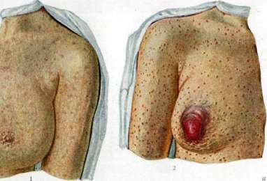
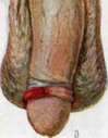
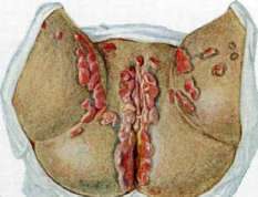
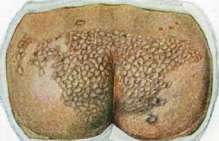
Figure 1. Syphilitic roseola, Figure 2. Hard .....chancre of the nipple and condylomata of the genitalia. Fig. 3 Hard chancre (primary syphilitic chancre). Fig. 4 Chancre of the glans penis. Fig. 5, Ruptions after tubercular syphilide. Fig. 6, Spirochaeta pallida [staining: a-Levaditi; b-unstained field of vision; c-Giemsa]. Fig. 7. Spirochaeta .....culture; d-agar; e-serum; f-fat; g-old; h-blood. The culture of the pale spirochete can be obtained, though with great difficulty and not regularly, on various protein media under conditions of relative anaerobiosis and a certain active acidity (pH = 7.2-7.8, Weiss and Wilkes-Weiss, Krantz); have been proposed: a) liquid media [Kroo-Schultze: rabbit serum and Normosal (1:10):-100 cm3 sterile guinea pig liver to be boiled for 3 days, 6 hours each day on a water bath at 50°C, filter, pH=7.5, pour sterile paraffin]; b) semi-liquid medium of Shereshewsky-see Nutrient media; c) solid media: Forster's blood agar-2% slightly alkaline agar +20% whole rabbit blood. Many other formulas have been proposed, which however do not guarantee success. Of the newest cultivation methods, the method of Forster is interesting in principle-growing on the surface of blood agar in hermetically sealed Petri dishes; anaerobiosis is achieved by the fact that the cultivation is carried out in the vicinity of an oxygen-absorbing microbe (Bac. prodigiosus) on the same agar surface, divided along the diameter by a groove (a strip of agar is cut out along the diameter!); on one half of the agar surface a spirochete is sown, on the other-Bac. prodigiosus; the seam between the covers of the Petri dish is hermetically sealed with plasticine. It has already been stated above (Oganessian) that in this case abundant growth of long spirochetes is obtained, which are not pathogenic for animals; spirochetes, especially in impure culture, may possess pathogenicity for rabbits (Sovade, Tomashevsky, Nogushi and others). In its chemical composition, the pale spirochete contains many lipoid substances, since it does not undergo pepsin-hydrochloric acid digestion, but dissolves in alcohol, saponin, bile (taurocholic acid sodium); in addition, a small amount of protein (lipoprotein) of an alkaline nature has been found in it (Schumacher, Bergel, Tsul'tser) and it contains almost no nucleic acid. The pale spirochete is able to reduce arsenic preparations (especially organic), which explains its salvarsan instability; in addition to arsenic preparations (salvarsan, stovarsol, osar-sol) it is also unstable with respect to antimony (stibosan), related to arsenic, to mercury, iodine, and bismuth. The pale spirochete is little stable in the test tube (in vitro) with respect to various other harmful agents, e.g. to drying; t° about 50° is fatal for it in 1 hour, about 10°-in 20 hours; in tissue pieces in the cold it survives up to 58 days; in cadaveric material, according to Armuzzi, it survived up to 110 days [cases of infection through cadaveric material are known (Hoffman and others)]; in lymph in sealed capillaries, according to Shereshewsky, it survives for several days. Ultraviolet rays are fatal for it, X-rays are ineffective. Nogushi and Bronfenbrenner give the following figures for the death of spirochetes (2 strains) under the influence of minimal solutions of various substances: sublimate-1:200,000-500,000; As2O3-1:20,000-30,000; tricresol-1:500; phenol-1:200; saponin-1:5,000-7,000; taurocholic acid sodium-1:1,000-2,000; NaOH-n/81; HCl-n/3aP; alkaline solution of Neo 1:125 does not kill the spirochete (in vitro) within 12 hours; it is very resistant to a hypertonic solution of NaCl, since it is incapable of plasmolysis. The pale spirochete does not form a true toxin in the liquid medium; the products it produces in the body are of an endotoxic nature; filterable forms for it have not been proven. The pale spirochete possesses antigenic properties with respect to the human and animal organism: in the serum of animals and humans under its influence appear complement-binding properties (Wassermann, Citron, Klopstock and others), precipitating, agglutinating (rather agglomerating), lytic (Gurevich, Timokhina), opsonizing properties. Bacterioscopy. The basic condition for detecting the pale spirochete in pathological materials is the correct taking of material from the main seat of infection-lymphatic paths-syphilitic elements. Method of taking material: 1) the primary chancre or other element is freed from external dirt and coating with the help of a cotton swab (sterile), moistened with physiol. solution of NaCl or better with a hypertonic solution (1g/l-2%) NaCl as enhancing secretion and facilitating the removal of detritus of the coating. 2) Irritation of the chancre to enhance the secretion of lymph containing spirochetes is produced by some sterile instrument-sterile platinum loop or spatula, glass spatula (Elze model), edge of a cover or slide (ground). Bloody material is unsuitable for examination, since in general blood contains few spirochetes. Lymph usually appears 1-2 minutes after the beginning of irritation. 3) A healed epithelialized chancre or a non-ulcerated efflorescence before irritation is carefully scarified with the blade of a sterile scalpel until the lymphatic paths are opened; avoid opening blood vessels. The appearance of lymph can be accelerated by squeezing the chancre from the sides, as well as by applying small Bier cups. 4) Sometimes an important aid is the puncture of regional lymph glands, which is performed as follows: the skin over the corresponding gland (most often inguinal) is sterilized with iodine and punctured with a steeply bevelled sterile needle (preferably platinum), fitted on a cannula of a 5-10 gram syringe (Record) with a tightly fitting piston; after the needle penetrates the underlying subcutaneous tissue, the needle is passed to the gland fixed between the thumb and index finger of the left hand and is injected into the cortical part of the latter; then several (5-6) careful aspirations of the piston are made; after this, usually a few drops of the gland secretion consisting of lymph, lymphocytes, and a small number of red blood cells are found in the needle if the puncture is done correctly. In such material in 60-70% it is possible to find spirochetes in their absence in a healed chancre. Material obtained by the above method from any pathological efflorescence is examined either in the untreated form for the detection of living spirochetes or in the treated form for the detection of them in the dead state. In practice both methods are most often used: 1) one part of the material (1-2 drops) is applied directly to the surface of a well-washed slide (preferably ground) or into a previously applied drop of physiol. solution of NaCl, covered with a clean, ignited cover slip and examined in the dark field (see Microscopic technique). 2) From another part of the material thin smears are prepared on clean slides (preferably ground); the material is smeared with the edge of a cover or ground slide; with a certain skill good preparations are obtained when smearing with a platinum loop. The still wet preparation is fixed with vapors of osmic acid (1%) or pure formaldehyde (in the Gamma apparatus) for 1-2 min. One can also fix with a mixture of alcohol with ether (half and half) for 15 min. or with methyl alcohol for 10 min. Some authors consider alcoholic fixation less acceptable, since due to the extraction of lipoid substances from the spirochetes by alcohol, the latter stain worse and fewer spirochetes are detected in the preparations than with other methods of fixation. About a hundred methods of staining and treating preparations containing spirochetes have been proposed; in general these methods represent 4 types: a) silvering methods (modif. of Levaditi's method-comparatively simple according to Fontana), b) methods of the Giemsa type (see Giemsa, Giemsa staining), c) staining with other dyes (the simplest method-fuchsin according to Shmanin), d) negative methods (with colloidal suspensions of ink, silver, etc.). Of these methods the most practically tested are: 1) Staining according to Giemza (accelerated modification of Shereshewsky); on the preparation fixed with osmic acid or formalin and passed 3 times through the flame, a solution of Giemsa dye is poured (1-2 drops of commercial dye per 1 cm3 of 11/2% glycerin water, prepared ex tempore and boiled until clear; the dye is poured 3 times and washed with dest. water; dried and microscoped. The pale spirochetes in such a preparation are clearly pink; saprophytic spirochetes (Sp. refringens, Sp.
Vincent et al.) with a distinct violet hue. 2) Negative methods of processing specimens with colloidal suspensions of certain substances that create a specific background, on which unstained spirochetes and formed elements are visible: a) processing with ink, according to Burri: a drop of so-called bacteriological, finely ground ink (brand "Pelikan"-Gunter-Wagner) is applied to a clean slide, a drop of lymph from the pathological focus is added, which is mixed with the ink and then spread across the slide with the edge of a coverslip or slide; the preparation should be of medium thickness; the preparation is air-dried and after this, without any fixation, it is ready for microscopy. b) The collargol method (Nitsche's method). A smear is made from tissue fluid; after it air-dries, a thick layer of 2% collargol solution is poured onto it; after 3 minutes, the collargol is drained; after drying, the smear is examined under immersion. Other substances have also been proposed for negative processing (opal blue, cyanohin). When processed with collargol, gray (colorless) spirochetes of quite typical morphology are visible on a reddish-brown background in the preparation.-Among the methods mentioned, the dark-field method should be placed first in terms of practical significance, under which the pale spirochete is studied under conditions closest to natural, where not only its characteristic morphological features but also the peculiarities of its movement are revealed. Second place should go to staining methods like Giemsa, which reveal the characteristic relationship of the spirochete to azure dye, distinguishing the pale spirochete from vulgar spirochetes - the pink color of the pale spirochete and the violet hue of the vulgar spirochetes. Third place should be given to the so-called negative processing methods, among which the most satisfactory results are obtained with the collargol method. Staining of sections and pathological material is best done by the combined silvering method of Levaditi-Manouelian. In peripheral conditions (rural social sectors, expeditions, new construction sites, etc.), the simplest and fastest of the mentioned methods (ink, collargol, fuchsin) will of course find the greatest application. The Giemsa stain is used less often; in general, it is quite labile in quality; it is even more difficult and rare to apply the dark-field method, which requires fairly complex and expensive equipment and electrical energy, which are not always available in peripheral work conditions. Differential diagnosis. Spirochetes are found on both normal and diseased skin and mucous membranes (especially of the genital organs and oral cavity) that have a certain morphological similarity to the pale spirochete and therefore must be taken into account in the differential diagnosis of syphilis. Of these, the so-called Sp. refringens (Schaudin), described by Donne as Sp. lineola, is most often found; its characteristics: it is significantly thicker than the pale spirochete, contains 3-8 uneven flat bends, stains with Giemsa in a reddish-violet color, and also with ordinary methods (solutions of methylene blue, fuchsin, gentian). Other spirochetes can be mentioned: Sp. calligyra (Noguchi) and a whole series of still insufficiently differentiated spirochetes (Tr. balanit., Tr. genital., Tr. urethrae, Tr. vaginal., etc.), found in various materials (in smegma - especially in balanitis, in the oral cavity, on the tonsils, etc.). Their pathological significance has not been established. Most of the mentioned spirochetes are easily distinguished from the pale spirochete both in morphology (coarser, different type of coils) and in their relationship to dyes (easy staining with ordinary solutions of aniline dyes), as well as a different type of mobility in the dark field. Among the oral cavity spirochetes (Tr. microdent. and Tr. macrodent. - Noguchi, Tr. medium, Tr. mucos.), the most attention must be paid in differential diagnosis to the small spirochete of dental plaque (Tr. microdent.). The difference of these spirochetes is as follows: 1) the dental spirochete is shorter and thicker than the pale one; 2) the dental spirochete has sharp, angular zigzag coils, while the pale one has regular arc-shaped coils; 3) the dental one performs a few flexing, arrhythmic movements; 4) the dental spirochete appears brighter, more light-refracting in the dark field than the pale one. To avoid contamination with vulgar spirochetes when examining oral cavity efflorescences, preliminary thorough mechanical cleaning (toothbrush, indifferent rinsing) is necessary to remove surface deposits, with which, in most cases, saprophytic spirochetes are also eliminated. In general, regarding the diagnosis of the pale spirochete, the so-called "Hoffmann's rule" still applies: "in preparations from syphilitic secretions, the pale spirochete is only definitely diagnosed when, in addition to the characteristic spirochetes, there are no spirochetes of a similar morphological type; in such cases, repeated examination with the above precautions must be carried out". Incidentally, for an experienced researcher, only one spirochete - Castellani's - is completely indistinguishable in morphology and staining from the pale spirochete. Cultures of the original material on artificial media for detecting the pale spirochete have no practical significance given the current state of technique for its cultivation, as it is only rarely grown from pathological material. Such cultures currently have only theoretical interest for studying the properties of various strains of the pale spirochete maintained on artificial media, partly for the preparation of so-called luetine, which is occasionally used for skin reactions (in tertiary syphilis). In rare cases, e.g. when deciding on the cure of syphilis after prolonged therapy, animal experiments can be applied according to Mulzer's proposal. For this purpose, the rabbit is used, as is known, to be very susceptible to syphilis; in such cases, the rabbit is injected into the testicles with blood, semen from men, and from women, in addition to blood, secretions from the cervix and vagina. 10. Finkelstein. V. Pathological anatomy. Schematically, the course of S, as indicated above, breaks down into three periods. The most important factor of the primary period is the formation of a hard ulcer (ulcus durum). The development of the ulcer is preceded by the formation of a papule. The ulcer usually has the appearance of a superficial defect or abrasion, its edges and bottom are somewhat firm, parchment-like; sometimes the induration itself is barely noticeable, inflammatory edema and hardening of the surrounding tissue predominate - the so-called primary sclerosus. Relatively rarely, ulcers are multiple. As a rule, the ulcer is accompanied by the development of buboes, i.e., swelling of regional lymph glands, e.g. inguinal (lymphadenitis). The ulcer and the involvement of regional glands can be considered as the primary syphilitic complex, by analogy, for example, with the primary tuberculous complex. Microscopic examination of the hard ulcer reveals pictures of subacute inflammation with a marked predominance of proliferative phenomena from the adventitial and endothelial vascular elements, as a result of which the vessel walls appear loosened, while their lumens are often narrowed. In the infiltrate, an abundant number of lymphoid cells and a few polymorphonuclears are found; later, young fibroblasts, lymphocytes, and plasma cells predominate. The superficial layers of the ulcer usually present a solid cellular infiltrate; deeper, the latter is mainly concentrated along the course of the vessels; often the infiltrate follows the vessels into the limits of the superficial fasciae, e.g. into the area of the cavernous tissue of the penis. Special processing reveals an abundance of spirochetes in the infiltrate. Healing of the ulcer occurs with the development of a delicate, slightly depressed, flat, whitish (depigmented) scar, and microscopic examination does not always reveal smoothing of the papillae, as is the case in vulgar skin scars; the elastic tissue in the papillary layer is also preserved, and apparently neofiber formation occurs. In the lymph nodes, vulgar hyperplastic processes predominate from the follicles and medullary cords with desquamation and proliferation of the sinus endothelium; suppuration as a rule does not occur. The presence of suppuration and necrotic processes indicates the presence of another or mixed infection (soft chancre, inguinal lymphogranuloma, etc.). The next (second) period of the disease, designated as the period of generalization, is expressed not only in a number of usual general infectious clinical phenomena but also in some morphological changes, mainly from the skin, mucous membranes, lymph glands, and spleen. Light roseolar, as well as papular and pustular eruptions on the skin (syphilides), subacute catarrhal processes from the mucous membranes, especially the pharynx, nose, and larynx, characterize the second period. From the histological side, the secondary period syphilides do not present anything specific. Against the background of hyperemia of the vessels of the subpapillary area, loosening of the substrate, perivascular infiltrates (slight in roseolas, pronounced in papules) of lymphoid cells with an admixture of polynuclears are noted; sometimes degenerative changes from the epidermis.
The presence of significant exudative and destructive phenomena serves as a prerequisite for the development of syphilids of the pustule type, pemphigus, which in turn can develop into syphilitic skin ulcers with drying crusts on them, so-called rupia. Particularly acute and diffuse inflammatory phenomena with extensive epidermal detachment are observed in newborn syphilitics. The pustular syphilid, which develops as a result of suppuration of papular, often indicates especially malignant forms of the disease. On the palms and soles, in particular on the heels, papular eruptions are accompanied by powerful development of horny layers resembling calluses (so-called syphilitic psoriasis). The intensity of skin manifestations during the period of generalization extremely varies; in some cases these manifestations are significant, in others extremely limited, and in individual cases apparently completely absent, which however has its analogies in other 'eruptive' infections. The third period of syphilis is designated as gummatous, precisely because it is often accompanied by the development in various organs and systems of so-called gummas (see). In the necrosis itself, one should not mistakenly see a simple nutritional disturbance on the basis of obliterative processes in the vessels of the gumma; necrosis, as well as the subsequent disintegration, constitute an integral stage in the development of the gumma, they are part of the essence of the process itself. Moreover, the necrosis of anatomically intact tissues can be the very beginning of the gumma-forming process, and granulomatosis and scars may only be subsequent moments. On the other hand, the onset of scleroses is a usual prerequisite for the development of new necroses, including the formed scars and vessels. The alternation of phases of necrosis and granulomatosis, which can be repeated several times, and lies at the basis of the growth of the gumma; it also feeds the clinical and anatomical symptomatology of the tertiary period, since the concept of necrosis is associated with the concept of loss of function (e.g. gummas in the conducting system of the heart, in the nervous system, etc.), and the concept of granulomatosis and scarring is associated with the concept of chronic inflammation, irritation, scleroses, etc. The clinical-anatomical significance of gummas also follows from the fact of their tumor-like nature and from the inevitable end of every gumma and gummatous infiltrate—sharp and extensive scar changes in the tissues at the location of the gumma. As a result of these changes, various deformities and disfigurements of organs (e.g. saddle nose), development of pathological lobulation of them (hepar lobatum, pulmo lobatus, etc.), strictures of canals, e.g. trachea, larynx, stomach, etc., often occur. The disintegration of the gumma itself, insofar as it affects the structural elements of the organ, can lead to the development of ulcers, perforations, e.g. of the palate, nasal septum, etc. Gummas are observed in various tissues and organs (meninges, liver, muscles, skin, etc.). Rarely however, at autopsy, gummatous S. of many organs is found; rather, on the contrary, the development of gummas in any organ to a certain degree as it were guarantees against their development in other places of the body. The next feature of the III period is the development of so-called diffuse syphilitic scleroses (or cirrhoses) on the basis of chronic specific inflammation; sometimes such scleroses are accompanied by the development of gummatous infiltrates in some places. Cirrhoses are most often observed in the liver (of both adults and newborns), in the pancreas, adrenal glands, lungs, testicles. It must however be admitted that the specification of such cirrhoses requires considerable caution, and in all relevant cases it is necessary to pay special attention to differential diagnosis. When developing syphilitic cirrhoses in children, especially with congenital S., the phenomena of proper cirrhosis are often combined with significant defects in the general development of the organ.-A very important feature of the same period should be considered the development of syphilitic vasculitides, mainly along the arterial system [see separate table (p. 591 - 592), fig. 2, 4 and 5]. Finally, as a fourth type of pathological processes in the late periods of S. (so-called parasyphilis or metasyphilis), one should point to diffuse and predominantly degenerative processes from the nervous system. These include in the first place tabes and progressive paralysis. In general, it must be emphasized that S., like any other infection, can along with extremely characteristic pictures and even instead of them give atypical and quite vulgar and rather acute degenerative-inflammatory processes. Among such, along with catarrhs of the II period, one can name e.g. fibrinous-purulent peritonitis in newborn syphilitics, diffuse leukocytic infiltrates of organs in the same cases, osteoplastic periostitis, etc.-S. as a chronic infection is often accompanied by deep and progressive metabolic disorders. Disorders of protein metabolism can manifest as amyloidosis of internal organs (spleen, kidneys, liver, intestines). As a manifestation of lipid metabolism disturbance, one can consider the lipid (often chronic) nephrosis of syphilitics; sometimes a mixed 'amyloid-lipid' nephrosis is observed. Disorders of both protein and lipid metabolism are especially common in young syphilitics, in particular in lues congenita tarda. Special pathological anatomy of S.-see corresponding Organs.
I.
Davydovsky. VI. Experimental Syphilis. The first reports on the possibility of infecting monkeys with S. appeared at the end of the 19th century: Klebs in 1871, and in 1886-88 the St. Petersburg physician Shperk and other authors report on infecting monkeys with S. material from sick humans and on the development of syphilitic ulcers (chancres) and rashes in monkeys. However, these isolated successful experiments were lost among the mass of negative results of S. vaccinations on animals and were not properly appreciated by contemporaries, just as the undoubtedly successful infection of S. in rabbits' eyes by Hansel in 1881. In December 1903, Metchnikoff and Roux published the results of their famous experiments on 2 chimpanzees: they inoculated a female in the fold of the praeputii clitoridis with material taken from a syphilitic. After this, works by a whole series of researchers (Neisser, Finger and Landsteiner, Nicolle and others) appeared, not only confirming the infectivity of S. for monkeys but also clarifying a whole range of questions related to this phenomenon. It turned out that with appropriate technique, infection succeeds in all cases without exception. Monkeys contract S. not only different species of higher anthropoid monkeys (chimpanzees, orangutans, gibbons) but also lower ones (Macacus, Cercopithecus, Cynocephalus). In the latter, however, susceptibility is somewhat weaker than in higher animals, as shown by the need to use material with a large number of spirochetes for their infection; in syphilitic chancres in them, spirochetes appear in very small quantities, and infection itself is only possible on certain areas of the skin. The most reliable results of monkey infection are obtained by skin inoculation through intensive rubbing into scarified skin. Through intact epithelium, infection does not succeed. Neisser did not obtain infection even when rubbing virulent material into the nasal mucosa, tonsil surfaces, or conjunctivas. According to most authors, subcutaneous infection does not succeed (Metchnikoff, Roux, Neisser and others). The same applies to intravenous and intraperitoneal infection and infection through the cornea. However, Salmon reports positive results of infection through the cornea, and Tomashewsky succeeded in infecting monkeys with S. by introducing material under the skin of the eyelid. Infection by inoculation into the testicle was obtained by Hoffmann, Lobe, Mülzer and some other authors. Passages of the virus from lower monkeys often give worse results, but Neisser, Finger and Landsteiner and others believe that this depends not on weakening of the infectious principle (virus) in lower monkeys, but on the smaller amount of spirochetes in the material, which primarily determines the strength and speed of manifestation of the infectious process. Infection succeeds best when inoculating material from fresh primary sclerosis or glands or from manifestations of secondary S. (papules, condylomas). Products of tertiary S. do not always give infection; Neisser, Hoffmann and others described both negative and positive results. Experiments of infection with blood from syphilitics also succeeded only in a small percentage of cases, apparently due to the small content of spirochetes in the blood. Hoffmann succeeded in infecting a monkey with the cerebrospinal fluid of a syphilitic. S. was successfully inoculated to monkeys by introducing blood and pulp from organs of congenital syphilitics. The manifestations of the disease in monkeys are in general similar to what is observed in humans. Scarifications at the site of inoculation first heal, and only after a more or less prolonged period of incubation do specific changes appear, even in the case where the site of inoculation was excised several hours after infection. The duration of the incubation period is 3-4-5 weeks (from 15 to 65 days according to Neisser, Baermann and Halberstadter), on average 30 days (Metchnikoff and Roux). The primary lesions have a diverse appearance: sometimes a firm dark blue infiltrate, sometimes ulcers with disintegration, sometimes they have a papular character or completely resemble the primary sclerosis of humans; the character of the primary lesion does not depend on the species of animal. Secondary S. phenomena were observed in all species of monkeys. In higher monkeys, especially chimpanzees and gibbons, secondary phenomena in the form of cutaneous papular rashes are usually clearly expressed. In orangutans and lower monkeys they occur much less frequently and are completely denied by many authors. But other authors (Zabolotny, Hoffmann, Lobe and others) describe in lower monkeys spotted and papular rashes on the skin, papules on mucous membranes. The internal organs of monkeys (spinal cord, liver, lungs, muscles and adrenal glands) according to Neisser's research proved non-infectious when transplanted to other monkeys, but the spleen, bone marrow, lymph glands and testicles sometimes caused infection. The presence of spirochetes in the spleen and bone marrow of lower monkeys despite the absence of secondary phenomena indicates generalization of the virus in these animals. Worms in monkeys (Rhesus) infected with S. but not showing external manifestations of the disease proved the presence of infection by transplanting inguinal glands to rabbits after 6½ months with positive results and thus established the presence of asymptomatic infection in monkeys (see below). According to histological structure, syphilitic lesions in monkeys are analogous to the corresponding lesions in humans. Spirochetes in S.-infected monkeys were found in smaller quantities than in cases of S. in humans. In higher monkeys, syphilitic lesions usually contained significantly more spirochetes than in lower species; in the latter, the presence of spirochetes can always be demonstrated in lymph glands. In 1906, Bertarelli repeated Hansel's experiments (1881) and proved that S. can also infect rabbits. By inoculating syphilitic material on the cornea or introducing it into the anterior chamber of the eye, he after a rather long incubation period obtained syphilitic inflammation of the cornea of a characteristic nature, which was proven by the histological picture and the presence of spirochetes. Other authors confirmed Bertarelli's data; both Bertarelli and other authors proved the transmissibility of S. from rabbit to rabbit and to monkeys. Infection is produced by introducing small pieces of tissue containing spirochetes into the anterior chamber of the eye or by rubbing the material into the scratched cornea. Schellack, Brown and Pearce obtained infection by simply implanting* material containing spirochetes into the conjunctival sac. Syphilitic keratitis in rabbits obtained by inoculation of syphilitic adrenal glands, juice from lung tissue and products of tertiary S. have been described. The incubation period lasts 4-6 weeks, however, a later infection has also been described - after 2-3 months and even after 150 days after infection. Usually the following phenomena are observed: first, at the site of inoculation, inflammation develops with clouding of the cornea and injection of vessels, sometimes with suppuration; after 5-10 days complete healing occurs with clearing of the cornea, and after a more or less prolonged incubation period, phenomena of parenchymatous keratitis of varying strength develop - from clouding to suppuration and disintegration. Papular and gummatous formations on the cornea have been described. The iris is also involved in the process (Tomashewsky, Finkelstein). Syphilitic keratitis in rabbits usually heal after some time; recurrences are quite frequent (Grigoriev, Yarysheva, Batrachenko, Kashinsky and others). Uhlenhuth and Mülzer in the very latest time report that they have never had occasion to observe generalization of the process after S. inoculation into the rabbit's cornea. However, Grouven, Shereshewsky, Hoffmann report on phenomena of general body damage observed by them (after eye inoculation) in the form of skin lesions, hair loss, and disturbance of the general condition of the animals. Grigoriev reported on the transmission of S. to offspring by a doe rabbit infected with S. in the eye. The syphilitic virus, transplanted to rabbits, does not lose its virulence for monkeys, which has been repeatedly proven experimentally by various authors. By introducing the syphilitic virus into the testicle and onto the scrotum of a rabbit, it is possible with great constancy to obtain both local and general specific changes. Syphilomas of the testicles represent material extremely rich in spirochetes and easily accessible for experiment. This method was developed in a series of works by Uhlenhuth and Mülzer; its application for the study of experimental S. was widely carried out in the research of numerous authors. Material for inoculations can be the tissue juice containing spirochetes from a chancre, weeping papules or pieces of chancre or papules. This material is introduced with a pipette or syringe into the thickness of the testicle or under the skin of the scrotum. Pieces of tissue are best introduced into incisions in the skin (skin pockets) of the scrotum. Infection can also be obtained by rubbing into the scarified skin of the scrotum. Intracutaneous infection with positive results was described by Chesney. In passages of material from humans, positive results are obtained in fewer cases (8-25%) than in passages from rabbit to rabbit.
With successive passages, the virus acquires increasing virulence, and its infectivity becomes absolute, reaching 100%; simultaneously, the time of appearance of primary lesions (incubation period) decreases; the latter also become more acute and severe. The results of inoculations also depend on the method of inoculation, the nature of the material, and the breed of rabbit. As in syphilitic keratitis, the virus retains its virulence for humans in this case as well; this has been proven by several cases of laboratory infection of humans from syphilitic rabbits. Infection resulting from inoculation in rabbits may manifest in different forms: 1) in the form of ulcers on the scrotum (typical chancre or atypical ulcers); 2) in the form of inflammation of the testicle and its appendages (syphilitic orchitis); 3) inflammation of the tunics of the testicle (syphilitic periorchitis). Spontaneous healing of these syphilitic lesions usually occurs very slowly; chancres may persist from 4 to 8 months. From a patho-anatomical point of view, the described lesions represent granulomatous tumors consisting of lymphoid cells and in the central part of tissue resembling embryonic connective tissue. In them, necrosis or caseous disintegration does not occur, as in syphilitic lesions of adults. According to the histological picture and the extraordinary abundance of spirochetes, syphilis in rabbits is compared with congenital syphilis of the human fetus. As the primary phenomena gradually disappear, generalization of the virus throughout the body occurs, expressed in swelling of the lymph glands, disease of the second testicle, changes in the cornea; the virus can be found in the spleen, liver, bone marrow, in the aorta, heart muscle (Grigoriev, Yarysheva). Secondary syphilitic phenomena in rabbits infected in the blood and by inoculation into the testicle proved to be quite similar to those observed in secondary S. in humans and consist of specific changes in the skin, mucous membranes, bones, nails, etc., changes in the cartilage of the nose, tip of the tail. The frequency of appearance of general syphilitic phenomena, according to Grosman, is 56.5% with inoculations into both testicles. Bone changes were studied by means of X-rays and found in more than 50% of cases as a very early sign of S. Besides syphilitic conjunctivitis, keratitis, iritis, diseases of the fundus of the eye (chorioiditis, chorioretinitis) have been described, occurring in 70-95% of cases with infection by certain strains. Lesions of the central nervous system in S. of rabbits have been established with certainty in recent times. Histological changes in the brain substance and the pia mater (Steiner, Biach, Jakob) undoubtedly indicate the involvement of brain tissue and membranes in the syphilitic process in rabbits. Snesarev and Finkel'shtein observed in a rabbit inoculated in the testicle clinical symptoms of cerebellar lesion, and on autopsy found a gumma of the cerebellum with changes in the membranes and degeneration of the conducting nerve pathways. In addition, in syphilitic rabbits, pathological changes in the cerebrospinal fluid have been described, expressed in an increase in the number of cells and globulins and in the appearance of a specific colloidal reaction of the cerebrospinal fluid (Goldsol- and Mastix-reaction). However, work in the experimental departments of State Venereal Institutes in Moscow and Odessa did not confirm this. Besides inoculations of syphilitic virus into the testicle and under the skin of the scrotum, other methods of inoculation have also been studied. Inoculations on the skin outside the genital area give significantly worse results. However, Uhlengut and Mul'tser managed to obtain primary syphilitic lesions in a rabbit after scarification of the eyebrows, and Kolle, Ruppert and Moebius to obtain infection with a virulent strain (100 passages) under the skin of the back. Infection by introducing the virus under the dura mater or into the brain itself succeeded rarely. The possibility of transmission of S. of rabbits by the sexual route is still questionable: in the few cases where a positive result was obtained, the possibility of error must be considered - confusion with spontaneous rabbit spirochetosis. The experimental study of transmission of S. from parents to offspring has not yet given quite definite results. There are isolated observations (Wiman, Grigoriev) of syphilitic phenomena in offspring when mothers were infected. Experimental S. of rabbits has been repeatedly used for diagnostic purposes of S. in humans, for determining the content of the virus in various parts of the body. The study of the blood of syphilitics has been extensively studied by Uhlengut and Mul'tser. They inoculated 2 cm3 of defibrinated blood of a patient into the testicle of a rabbit and often obtained positive results. In cases of secondary S.-in 75%, with latent S.-much less frequently. Out of 8 cases of tertiary S., infection with blood was achieved only in one. Blood of children with early congenital S. gives infection in rabbits, while blood of patients in late stages of congenital S. does not cause infection. Inoculations of urine, saliva, and sweat of syphilitics in the secondary period of the disease always gave negative results. The presence of spirochetes in the milk of wet-nurses with syphilis has been proven by experiments conducted by Uhlengut and Mul'tser; Finger, however, and Landsteiner in inoculations of milk to monkeys, and Schwarz and Schubert to rabbits, did not find spirochetes upon microscopic examination. Semen, taken at the height of syphilitic infection, gave infection in 2 out of 6 cases (Uhlengut and Mul'tser). Cerebrospinal fluid does not always cause infection: according to the data of Chesney and Kemp in the early stage of S., spirochetes can be found in the clinically unchanged cerebrospinal fluid in 15% of cases. Successful infection of rabbits with blood and cerebrospinal fluid has been repeatedly produced by many authors, although Uhlengut, Steiner, and Mul'tser in 19 cases of tabes dorsalis and progressive paralysis did not obtain infection with cerebrospinal fluid inoculation. Expansion of the base of experimental S. by infection of other animals. Experiments with cold-blooded animals (frogs), birds, various mammals (dog, cat, sheep and especially pig) gave negative or insufficient for practical application results. Only the data obtained on guinea pigs and mice have some significance. In experiments of infection of S. in guinea pigs (Bertarelli, Truffi, Tomashevsky and others), keratitis, infiltrates, and primary scleroses on the testicles were obtained. Passages succeeded rarely, but the virulence of the virus did not increase. Recently, Kolle and Evers showed that infection of guinea pigs in the absence of external manifestations of the disease gives a latent infection, which can be detected by inoculation of the lymph glands of the infected animal to rabbits. By the same method, Kolle and Schlossberger discovered that by inoculating pieces of chancre to mice or rats under the skin, one can obtain an asymptomatic, latent infection of these animals. The presence of syphilitic virus in them 3-4 months after infection can be proven by inoculating their glands, spleen, or brain to rabbits. Reports by Argentine researchers Jauregui and Lancelotti about the high susceptibility of llamas to S. aroused great interest at one time. After inoculation of human material, a chancre appears after 14 days, then rapidly follows the secondary stage, manifesting in the form of roseolas, papules, etc., as in humans. Then follows the tertiary stage with gummas of internal organs, lesions of bones and brain. Animals die after 3 years. Transmission to offspring was observed. However, upon verification by a number of researchers (Hoffman, Zurhelle, Kolle/Mul'tser and others), these data were not confirmed; the llama proved to be insusceptible to human and rabbit syphilitic virus, and the serum of the inoculated animal has no therapeutic effect, as claimed by the aforementioned Argentine authors. The significance of the experimental study of phenomena was especially evident in the development of the interesting and complex question of immunity in S. Until the end of the 19th century, the view established by Ricord prevailed that S. leaves behind a long-lasting immunity, then the number of observations of reinfection with S. in humans constantly increased. The solution to the question became possible only with the development of methods of experimental S. Experiments were conducted along the line of studying superinfection and reinfection phenomena. Bruck, Finger and Landsteiner and Neisser conducted a large number of studies on monkeys. They found that superinfection produced shortly after the first infection (9-14 days) gives typical primary scleroses. Gradually, as the virus generalizes, the ability to reinfection weakens, so that superinfection may not manifest at all. Kolle studied this question in detail in rabbits. He infected rabbits with S. using the Truffi strain, and then at different times after infection superinfected them with the same strain. Within 60 days after the first infection, superinfection succeeded in 50-60% of cases, and between 60 and 90 days-rarely; typical chancres appeared at the site of superinfection. After the 90th day and for several years after the first infection, superinfection never succeeded.
For a more complete resolution of the question of immunity in S., Colle undertook further research, modifying it in that he produced superinfection not with the strain with which the rabbit was first infected (homologous), but with another (heterologous), e.g., with the Nichols strain after the first infection with the Truffi strain, with the Kuznitsky strain after the Nichols strain, etc. This type of superinfection, performed 4-5 months after the first infection, gave a positive result in a significant number of cases (28.5 - 62.5%), manifesting as typical chancres, whereas control experiments (superinfection with a homologous strain) gave a negative result in all cases. From these experiments, Colle concludes that in rabbits infected with S., 'panimmunity' against all syphilitic strains is not always developed, but only 'monoimmunity' against the strain with which the animal is infected. In humans, however, according to Colle, there is 'panimmunity,' as indicated by the rarity of secondary disease in syphilitic people who often come into contact with heterologous syphilitic strains. Chesney believes that there is no panimmunity in humans, and the rarity of reinfection depends on the small amount of infectious material with which they come into contact. The verification work of Priegge and Rothermundt showed that to infect a rabbit, a negligible amount of infectious material is sufficient. As for the essence of the question, it must be said that it is hardly possible to establish a difference in the nature of immunity in humans and rabbits in this regard, since in the latter, heterologous strains do not always produce superinfection (in 28.5-62.5%). It should be noted that in the studies presented, only the so-called 'chancre-immunity' is discussed, i.e., the state of the organism that protects it from the occurrence of the primary skin lesion—the chancre. At the same time, the question of whether the organism is protected from new penetration of syphilitic spirochetes into it remains open (see below). New interesting circumstances concerning immunity were discovered in the treatment of infected rabbits. Colle treated syphilitically infected rabbits in the first 45 days after infection. Reinfections at later stages (more than 90 days after the first infection) usually caused the appearance of typical chancres in them. Colle considered this infection to be reinfection, indicating complete sterilization of rabbits with early introduction of the medicinal substance. Brown and Pierce, when treating syphilitic rabbits with non-sterilizing doses of salvarsan, observed a delay in the manifestation of chancre-immunity in them. Buschke and Croo, working with relapsing fever spirochetes, concluded that sterilization of spirochetal diseases with chemotherapeutic agents is generally impossible. In their opinion, treatment can only destroy the clinical manifestations of the disease and delay the development of immunity, but the causative agents of the disease remain in a latent state in the organism, and therefore reinfection is only superinfection, not reinfection, and does not prove complete sterilization of the organism. Colle treated syphilitic rabbits with large doses of salvarsan after the 45th day of the disease; upon reinfection of such rabbits, chancres usually did not appear. Based on this, Colle concludes that late treatment with large doses cannot sterilize the syphilitic infection. All these data shed light on the nature of immunity in S. Colle believes that in this case there is not true active immunity, but 'infectious' immunity, i.e., a state of the organism in which, when infection is present in it, it is protected from reinfection with the same virus; infectious immunity depends on the presence of the causative agent of infection in the organism and disappears upon complete cure. Chesney and Kemp confirmed Colle's data but disagree with his explanation of the nature of immunity. In their opinion, in S., true active immunity develops, and therefore they consider sterilization of the organism possible with late treatment, and the absence of reinfection as an indicator of active immunity, but not the presence of causative agents of the disease in the organism. Chesney's views were also joined by Uhlenhuth and Grossmann. However, these views are not sufficiently substantiated by experimental material, and the data on spirochetal infections in general and on asymptomatic syphilis in particular (see below) contradict them. Pierce and Brown in 1922 showed that in untreated syphilitic rabbits, a long time after the disappearance of all symptoms of the disease during the period of developed immunity, virulent spirochetes can be found in the popliteal lymph glands; transplantation of these glands into healthy rabbits causes the appearance of typical chancres. The application of this method for detecting the syphilitic virus became of great importance for experimental S. Colle and Evers studied the rate of spread of infection in the body using this method. It turned out that after infection in the scrotum, the presence of spirochetes in the inguinal glands can be proven in a rabbit after 30 minutes, and in a guinea pig even after 5 minutes. Priegge found virulent spirochetes in the popliteal glands two weeks after infection. In Colle's experiments, rabbits to which an insoluble bismuth compound ('bismuth depot') was introduced, and then infected with S., did not develop typical chancres. To verify whether bismuth kills the introduced spirochetes, the popliteal glands of such rabbits were transplanted into the scrotum of healthy animals, and often a positive result was obtained. It turned out that, while delaying the appearance of external symptoms, bismuth does not kill spirochetes; in rabbits that received bismuth, a latent asymptomatic generalized infection thus occurred. Based on this fact, Colle and Evers checked whether there is also such an asymptomatic infection in untreated rabbits. When infecting rabbits with S., 90% of them give typical chancres, and 10% do not react with clinical symptoms to infection, despite the fact that the pieces of syphilitic tissue introduced into them contain a huge number of spirochetes. It was believed that these rabbits remain healthy because the spirochetes in their organism die for one reason or another. Such rabbits received a special name 'Nuller.' Colle and Evers transplanted the popliteal glands of such rabbits into the scrotum of healthy animals and in almost 100% of cases obtained typical chancres. Thus, it was established that rabbits that do not react with clinical symptoms to infection with S., as a rule, contain virulent spirochetes in their lymph glands, and in so-called Nullers, there is almost constantly an asymptomatic syphilitic infection. The method of transplanting lymph glands of syphilitic animals made it possible to study in more detail the questions of superinfection and immunity in S. By combining in various ways the lymph glands containing heterologous and homologous strains of the virus in superinfected rabbits, Colle and his colleagues discovered in the superinfected rabbits the simultaneous presence of both spirochetes introduced with the first infection and spirochetes introduced with superinfection, even in cases where the latter did not manifest with any external signs. Thus, positive asymptomatic superinfection in S. was proven. By transplanting the glands of rabbits treated with large doses of salvarsan and then reinfected, the possibility of asymptomatic reinfection was also proven. Based on all these data, it can be considered with certainty established that in S. there is no true active immunity that would destroy spirochetes entering the organism, protecting it from infection with them; these facts indicate that the observed phenomena of immunity represent only (apparent) skin chancre-immunity. The question of the biological peculiarities of different strains of the pale spirochete also belongs to the area of experimental S. (see above). Chemotherapy of experimental S. In the chemotherapy of S., the greatest importance is attached to organic arsenic compounds—salvarsan ('606') and neosalvarsan ('914'). Both drugs can produce complete sterilization of the organism (from spirochetes) when administered in an amount of 0.01-0.015 g per 1 kg of body weight in the early stage of the disease (no later than 2-3 weeks after the appearance of the chancre). Their chemotherapeutic index in S.—the ratio of the maximum tolerable dose per 1 kg of body weight to the minimum therapeutic dose per 1 kg of body weight—is 10, and according to American data even higher (between 10 and 20; Kolmer). When treating at later stages or with smaller doses, sterilization of the organism is not always achieved, but an extremely favorable therapeutic effect is noted, expressed in the weakening of clinical manifestations even in malignant forms of the disease or in cases not amenable to other methods of treatment. Of particular interest are the arsenic preparations—stovarsol, spirocide, stovarsolan—the advantage of which is that they can be administered per os. 0.4 g of the preparation per 1 kg of body weight cures these animals of the external manifestations of S.
In recent years, thanks to the work of Levaditi and his school, it has been established that for the chemotherapy of S. compounds of bismuth (see) are of great importance, from which the most significant is the trepol-basic potassium, sodium, and bismuth tartrate salt. Bismuth preparations possess very strong and prolonged preventive action due to slow absorption from the depot. The introduction of trepol to rabbits in the amount of 0.056 g of metallic bismuth per 1 kg of weight produces a preventive effect lasting at least 152 days. According to research by the French school, compounds of tellurium act similarly to bismuth compounds. It was believed that compounds of antimony, which play a significant role in the chemotherapy of protozoan diseases, are ineffective in spirochetoses. However, the work of Krivchevsky, Pines, and Vein £45 (1933) showed that the antimony-stibosan preparation, in experimental S. in mice and rabbits and in the treatment of humans, exhibits stronger therapeutic action than bismuth. The mechanism of action of chemotherapeutic preparations has not yet been fully elucidated. Most compounds of arsenic and bismuth do not act on spirochetes in vitro.
E. Geronimus. VII. Clinical manifestations and diagnosis of acquired syphilis. Syphilis, being a chronic infectious disease, has a more or less definite course, characterized by specific clinical manifestations depending on the period of the disease and some other causes. Its clinical manifestations most often affect the skin and mucous membranes, as well as the lymphatic system, but in almost any period, manifestations may occur from the musculoskeletal system, internal organs, the central and peripheral nervous system, and sensory organs. The general course of S. is conventionally divided into periods: primary, secondary, and tertiary, each of these periods being subdivided into the manifest period, when manifestations of the disease are present, and the latent period, when these manifestations are absent. A period of usually late special lesions of the central nervous system, called meta- or parasphyilis, is also noted. Such a division of S. is of course conditional and requires some modification and supplementation. The general course of S. can be summarized as follows: as an infectious disease caused by a specific pathogen, the pale spirochete, S. requires special conditions for the transmission of infection from a patient to a healthy person. An indispensable condition for infection with S. is the penetration of the pale spirochete into the body through damaged stratum corneum of the epidermis or the surface of a mucous membrane, although other routes of transmission are possible (for example, cases of transmission of S. during blood transfusions are known, and in such cases, clinical manifestations developed directly from the secondary period, while primary lesions—the hard chancre—were not observed in such patients—syphilis d'emblee). We are of course talking about acquired S, since for congenital S. there are special conditions for the transmission of S. from mother to fetus (see below—congenital S). Infection with S. can occur during sexual intercourse with patients (sexual infection) or by other means [for example, during kissing, when breastfeeding a child, when using certain items that were used by the patient just before, such as a cigarette, toothbrush, etc.—this is so-called non-sexual S, or S. of the innocent (syphilis inson-tium)]. Cases of infection of medical personnel during work (infection of midwives, gynecologists, surgeons, etc.) and sometimes of patients from medical procedures (for example, cases of infection from smallpox vaccinations were previously noted, when the material for vaccination was taken from pustules of vaccinated children who turned out to have S.—syphilis vaccinata) are known. After infection, the development of the infection generally proceeds in the following order: for some time after the moment of infection, no syphilitic symptoms are objectively noted at the site of infection, and this period occupies a more or less long interval of time [on average 2-3 weeks, although cases of duration of 8 days, and on the other hand, of 78 (Wolf), 90 (Meshchersky), and even of 100 and 110 (Gougerot) days are known; in the last two cases, such a long period could have depended on preventive injections of salvarsan]. This period is called the first latent period of S. Subsequently, the first signs of the disease begin to appear at the site of infection in the form of so-called primary lesions or hard chancres (ulcus durum, s. induratum), which is a reaction to the pale spirochete that has penetrated and multiplied at this site. The primary lesion has its own cycle of development, reaching maximum development in about 2 weeks, then remaining in the flowering period for about another 2 weeks, in order to subsequently undergo reverse development and healing in approximately a 2-week period as well. This period of time from the appearance of the hard chancre to its healing is called the primary period of S, with a total duration of about 6 weeks (such periods are conditional and may vary one way or the other). The primary period of S. at the present time is divided into 2 periods: the primary seronegative period—from the appearance of the hard chancre to the appearance of a positive serological reaction in the blood (this interval varies in time for various reasons and in particular depending on the localization of the hard chancre, but in general it is 2-3 weeks) and the primary seropositive period—from the appearance of a positive serological reaction in the blood to the healing of the hard chancre. After the healing of the hard chancre until the appearance of a rash on the body, i.e., until the appearance of so-called secondary S., a certain interval of time again passes, called the second incubation period of S, or the second latent period; this period varies in individual cases up to 2-4 weeks. Subsequently, abundant rashes may appear on the patient's body, usually in the form of a spotted rash, and from this moment the secondary period of S begins, with the initial appearance of the rash being referred to as fresh secondary S. (syphilis condylomatosa recens) in contrast to the rashes that occur in the further course of secondary S. and appear at various times, and such rashes are referred to as secondary recurrent S. (syphilis condylomatosa recidiva). The secondary period can last a very long time (2-3 years, and sometimes 5-10 or more years), at times giving flare-ups of external manifestations of S. from the skin and mucous membranes, and at times remaining without clinical manifestations—so-called latent secondary S. (syphilis condylomatosa latens). Usually in the secondary period of S., an abundant rash appears initially, and in subsequent recurrences, the rash becomes increasingly sparse, often located in separate groups, often in the form of rings or arcs, and the rash is often localized atypical sites. The intervals of the latent period between individual recurrences in the further course of S. become increasingly longer, and then a very long latent period may occur, after which manifestations of a completely different character may develop, usually in the form of deeply located nodules of specific infiltrate, so-called gummas, which already refers to the tertiary, or so-called gummatous period of S. (syphilis gummosa). The tertiary period of S. can last an indefinitely long time, at times giving one or another manifestation (syphilis gummosa manifesta) or remaining in a latent state for a more or less long period (syphilis gummosa latens). The peculiar course of S. with an incubation period, the peculiarity of manifestations in different periods, both manifest and latent, is explained by the biological conditions of the pathogen of S. in the patient's body. As clinical and experimental observations show, the pale spirochete initially begins to proliferate at the site of its penetration; when its proliferation reaches a significant degree here, the first clinical signs of S. begin to appear in this place in the form of a hard chancre. The entry into the lymphatic and blood circulation and the carriage of spirochetes to various organs and tissues usually occurs at the moment of maximum development of the hard chancre, although in some cases such carriage of the pathogen to distant organs may occur from the moment of the initial penetration of spirochetes into the body. This is evidenced by experimental experiments with the inoculation of the pale spirochete followed by surgical removal after several hours of the skin area where the inoculation was made, as well as clinical observations of the appearance of early (pre-roseolar) nephritis, meningitis, the appearance of a positive reaction in the cerebrospinal fluid earlier than in the blood, etc. In the blood, the spirochete is an occasional guest, since it is a tissue parasite, which is why its presence in the blood is difficult to detect, while experimental inoculations of blood from patients give a positive result in various periods of S., and for successful inoculation, it is necessary to take a relatively large amount of blood. The hematogenous spread of spirochetes, which occurs in the primary stage, creates foci of spirochetes in various organs and tissues, which in the further course of S. can cause specific tissue changes. Not only various skin manifestations of fresh and recurrent S., but also lesions of internal organs and the central nervous system, including progressive paralysis and tabes dorsalis, can be explained by the proliferation of spirochetes that were carried here already in the primary stage of S. Of course, repeated carriage of spirochetes from active foci of secondary S. is also possible, but in this case, the newly introduced spirochetes cannot take hold in this place (Engmann, Eberson) until the body has freed itself from the primary infection, as evidenced by experimental observations of superinfection of animals (see above—Experimental S.). With such inoculations, a characteristic primary lesion cannot be obtained; at the site of inoculation, a papula of the same nature as in the luetinic or pellagrous skin reaction (see below) usually results, and subsequently the phenomena characteristic of primarily inoculated S. do not occur, i.e., it is not possible to achieve generalization of S. The abundance of rash in fresh secondary S. and its sparseness in recurrences, especially repeated ones, can be explained by the fact that in fresh secondary S., proliferation of the spirochete occurs in a large number of foci of its initial carriage.
As a result of the skin reaction in the form of a roseola at this site, due to the immunobiological properties of the skin, the spirochete cannot subsequently multiply and dies, and the skin acquires local immunity. Therefore, during recurrences, a ring-like arrangement of rash elements may appear due to surviving spirochetes that have begun to proliferate, located at the periphery of the initial rash elements. The conditions of local skin immunity can also explain the increasing scarcity of rash elements during repeated recurrences, as well as the circumstance that in late recurrences, the rash is often located in places not typical for fresh secondary syphilis. The onset of the hidden, or latent, period of syphilis can also be explained by immunoreactive phenomena in the body, as a result of which the surviving spirochetes temporarily become non-virulent. The uniqueness of the phenomena of the tertiary period, i.e., the formation of gummatous changes, can be explained by the allergic condition of the skin after the phenomena of the secondary period. This is also evidenced by the circumstance that sometimes at the site of the former primary lesion, a powerful infiltrate (reinduratio) reappears, which may ulcerate, and such an ulcer has a great resemblance to a gummatous one (ulcus redux, s. pseudogummosum). The general course of syphilis. Syphilis in individual cases proceeds differently. Sometimes cases with normal or more or less correctly alternating recurrent phenomena and intervals of the hidden period are noted. Sometimes cases with a sluggish course, with scarce manifestations and long hidden periods are noted. In some cases, the course is stormy with an abundance of pustular phenomena and frequent recurrences, as well as with a tendency to appear gummatous lesions along with the phenomena of the secondary period (galloping, or malignant syphilis - lues maligna). Very often, a sluggishly proceeding syphilis with almost no recurrences leads to the subsequent development of symptoms of organic lesions of the central nervous system, which gave rise to the possibility of duality of the syphilitic virus: dermo- and neurotropic (see above - Experimental Syphilis). The primary period of syphilis is characterized by a single manifestation in the form of a primary lesion, or affect (hard chancre - ulcus durum, s. induratum), and the development of lesions of the regional lymph glands somewhat later. As clinical observations and experimental vaccinations show, after the incubation period, a red spot appears at the site of infection, which soon takes the form of a papule, first scaling, and then also weeping on the surface or covered with a crust, under which a superficial erosion or, much more rarely, a superficial ulcer is found, gradually increasing in size while simultaneously developing a dense infiltrate of its base. A typical hard chancre is a round or oval erosion sitting on a dense infiltrate, without acute inflammatory phenomena in the surrounding area [see. separate table (pp. 527-528), fig. 3]. The size of the erosion ranges from several millimeters ('dwarf chancre') to 1 cm in diameter, and sometimes larger or even very significant sizes. The edges of the erosion are usually even, undermined, sometimes slightly elevated above the level of the erosion. The bottom is red in color, sometimes covered with a small 'greasy' coating, does not suppurate. The discharge is serous fluid; after irritation of the erosion by friction, the discharge appears in abundance, so-called Reizserum, which is used for examination for the pale spirochete. The hardening of the base gives a palpable impression of cartilaginous density; depending on the location of the chancre and some other reasons, this hardening may be weakly expressed; sometimes it is very flat, superficial, giving a palpable impression of a sheet of parchment - so-called 'parchment chancre' (chancre parchemine, s. papyrace). Varieties of the typical hard chancre depending on location are as follows: on the glans penis usually in the form of a round erosion with parchment hardening; in the external opening of the urethra also in the form of an erosion on one or both lips, with some hardening and protrusion of one or two lips, sometimes creating difficulty in urination; in the urethra itself (usually in the navicular fossa) the chancre can have significant hardening, simulating a paraurethral infiltrate, more often it is in the form of a superficial erosion, detectable by a urethroscope; in the coronal sulcus the chancre in the form of a typical erosion with very strongly expressed cartilaginous hardening; the erosion usually has a bean-like shape, with the concave side located in the groove and the convex side towards the prepuce; on the frenulum the chancre has slit-like outlines, sometimes in the form of a small ulcer, which unlike the soft chancre does not perforate the frenulum; on the inner leaf of the prepuce the chancre with the character of parchment; on the free edge of the prepuce the chancre in the form of a crack running across the free edge from the inner leaf of the prepuce to the outer; sometimes several chancres are noted, radiating from the opening of the preputial sac (in patients with congenital phimosis), and from constant irritation by urine, inflammatory phenomena may join, exacerbating the phenomena of phimosis; on the skin of the sexual organ the chancre usually has round outlines with parchment hardening and with drying into a crust discharge; on the skin of the scrotum also in the form of a parchment chancre, which here often reaches large sizes and resembles an erosion from a second-degree burn (ulcus induratum combu&tiformis). In women on the labia majora from the inner side the chancre is in the form of a round erosion, sometimes with a slightly elevated bottom, covered with a 'greasy' coating, with clearly expressed hardening; sometimes a special form of chancre (so-called indurative edema of the labium - oedema induratiynm) is noted here, characterized by a sharp increase in the labium, as if by edema, and significant hardening, while the initial erosion can completely epithelialize and not be detectable upon examination; on the labia minora the chancre usually has the appearance of an erosion with parchment hardening, which is more clearly visible when the chancre is located on the edge of the labium; on the clitoris in the form of an erosion with parchment hardening; on the prepuce of the clitoris the erosion can have quite significant hardening; in the opening of the urethra the erosion can protrude outward due to infiltration of its base and easily bleeds; on the posterior commissure of the labia majora the chancre has the appearance of a superficial erosion or ulcer with parchment hardening; at the entrance to the vagina (usually on the remnants of the hymen) in the form of a parchment chancre; on the vaginal mucosa the chancre is rarely encountered, usually in the form of an erosion; on the cervix the chancre is often round in outline, usually in the form of an erosion, sometimes in the form of a ring surrounding the cervical os; the chancre of the uterine mucosa cannot be seen clinically; in such cases only the examination of uterine mucus for the pale spirochete can provide a basis for the diagnosis of the chancre. In the oral area the chancre has various forms depending on location. On the red border of the lips it appears in the form of an erosion or superficial ulcer, usually covered with a crust of dried discharge, without noticeable hardening of the ulcer base, and sometimes with a thickening and hardening of the entire lip of the nature of indurative edema; in the corner of the mouth the chancre often appears in the form of a fissure-like ulcer, painful and bleeding; on the oral mucosa usually in the form of a round or oval erosion, and on the frenulum of the tongue sometimes in the form of a slit-like ulcer, and sometimes in the form of ulcers on the lateral surfaces of the tongue, easily bleeding, painful and making chewing difficult. On the tonsils various forms are encountered, more often in the form of an ulcer covered with a coating or necrosis with more or less pronounced hardening of the tonsil; the so-called diphtheroid, ulcerative or gangrenous chancre is often noted. On the wings of the nose the erosion or superficial ulcer is accompanied by swelling and hardening, and on the nasal mucosa the chancre is usually in the form of a round erosion. In the eye area: on the edge of the eyelids in the form of an elongated erosion with swelling and hardening of the eyelid edge; in the corners of the eye the chancre sometimes has a Y-shaped form; on the conjunctiva in the form of an erosion, sometimes rising above the level of the surrounding parts and ending with sharp borders at the corneal% membrane, if the erosion reaches it [see separate table (pp. 551-552), fig. 2]. In the anal area the chancre has the appearance of a fissure-like ulcer with thickened edges, sometimes it involves several folds of the anal opening (pleated chancre); defecation is accompanied by significant pain and bleeding. In the area of the breast nipple [see separate table (pp. 527-528), fig. 2] the chancre is often encountered in the form of a fissure-like ulcer at the base of the nipple, on the nipple itself and in the surrounding parts - usually in the form of an erosion or superficial ulcer covered with a crust. On the fingers the chancre has the appearance of an ulcer, often accompanied by inflammatory phenomena in the surrounding parts from secondary infection.
On other parts of the body, the chancre appears as an erosion or a superficial ulcer covered with a crust, with more or less pronounced induration of the base, and sometimes with inflammatory phenomena in the surrounding area. A complicated hard chancre is observed under various conditions: with secondary infection, from irrational treatment with cauterizing substances, and sometimes from local conditions. Special forms include so-called gangrenous and phagedenic chancres; in the first, gangrenous areas of the base and edges of the chancre appear as dirty brown or black, disintegrating necrotic foci with sharp reactive phenomena in the surrounding area and constant increase in the size of the ulcer at the periphery and in depth from the rejection of demarcation inflammation of the dead parts. In phagedenic chancre, the ulcer spreads due to constant disintegration of the base and edges of the ulcer in small areas ('molecular disintegration'). In such forms of chancre, the search for the pale spirochete is difficult, as there is usually an abundance of various bacteria (Bac. pyocyaneus, Spiroch. refringens, etc.) and there is a symbiosis of the pale spirochete with Vincent's fusiform bacillus (fusispirillosis). - A mixed chancre results from simultaneous infection with the streptobacillus of soft chancre and the pale spirochete, initially developing the clinical picture of soft chancre, and after the incubation period, signs of hard chancre with characteristic induration of the base begin to appear. Changes in the lymphatic system in the primary stage begin to be noted 1-3 weeks after the appearance of the hard chancre; first, the lymphatic vessels going from the site of the primary lesion to the nearest (regional) lymph glands are affected. Clinically, the lymphatic vessels appear somewhat thickened and indurated, which can sometimes be confirmed by palpation. The lymph glands are constantly noticeably changed: they are enlarged, firm, not adherent to each other or to surrounding tissues, completely painless, and movable. Inflammatory phenomena are absent; they may appear only in the presence of secondary infection, and then pain and even progression to suppurative adenitis may occur. A characteristic feature of specific lymph gland lesions (scleradenitis) is the multiplicity of gland involvement. In the further course of the primary period, specific changes also occur in other lymph glands more distant from the primary focus, and by the time the secondary period begins, all accessible lymph glands are found to be changed (specific polyadenitis). The diagnosis of hard chancre is based on the clinical picture of the lesion and is finally confirmed by microscopic examination for the pale spirochete. Anamnestic data can only have secondary importance. In some cases, when hard chancre is suspected and no spirochetes are found in the discharge, which more often occurs in already healing or complicated chancre, as well as in oral chancre where Spirochaeta dentium may be found during microscopic examination, simulating the pale spirochete, it is necessary to resort to puncture of the lymph gland for examination of its juice. - Differential diagnosis of hard and soft chancre is based on comparative distinguishing features of the soft chancre ulcer and hard chancre erosion (base, edges, discharge); on the presence in soft chancre and absence in hard chancre of inflammatory phenomena in the surrounding skin; on the multiplicity and appearance of new ulcers in soft chancre and the singularity or simultaneous occurrence of several erosions in hard chancre; on the absence or appearance of a single, painful lymph gland with inflammatory phenomena and a tendency to progress to abscess in soft chancre, and on the multiple characteristic regional scleradenitis without a tendency to inflammatory phenomena in hard chancre; on the results of microscopic examination for streptobacillus in soft chancre and pale spirochete in hard chancre. Great diagnostic difficulties in gangrenous or phagedenic chancre, whether soft or hard, can be resolved by thorough microscopic examination. Tuberculous ulcers are distinguished from hard chancre relatively easily by their uneven, pus-covered and disintegrating, easily bleeding base, slow development, multiplicity of small ulcers, presence of non-ulcerated tubercles at the periphery, chronic course, as well as by the general health status and microscopic examination for the tubercle bacillus. Epithelioma can be easily distinguished from hard chancre by its easily bleeding base, thickened edges, slow course, and appearance in elderly individuals. In difficult cases, histological examination of biopsied tissue is performed. Erosions of herpes zoster are distinguished by the polycyclic nature of the edge contours due to the close arrangement of a group of vesicles, and the absence of induration. Excoriated scabetic nodules are distinguished by their multiplicity and the presence of scabies signs in the patient. Banal erosions of the cervix uteri are more often located on the posterior lip and appear as if flowing from the external os of the uterus, whereas hard chancre is usually eccentric in relation to the external os. Other erosive processes, as well as ulcers and fissures, differ from hard chancre by the absence of chancre signs and regional scleradenitis, as well as by the presence of the main signs of the given disease. The secondary period of syphilis ('secondary S') is characterized by clinical manifestations mainly from the skin and mucous membranes in the form of rashes of various morphological elements, so-called syphilids. The appearance of the rash is often preceded by general symptoms in the form of malaise, feverish condition, headaches, and pains in the limbs, which intensify at night. The great variety of rash elements can be reduced to three main forms: 1) spotted, or erythematous syphilid (syphilis cutanea maculosa, s. roseola syphilitica); 2) papular (syphilis cutanea papulosa), 3) pustular (syphilis cutanea pustulosa). Each of these forms in turn gives various varieties of rash elements, which may depend partly on the localization of the rash elements, their duration, the duration of the disease (fresh or recurrent S), and other reasons. - The general characteristics of the rash elements are as follows: color is red with a bluish or brownish tint, but not bright inflammatory; it depends on congestive hyperemia and infiltration, as well as on the impregnation of the tissue with the coloring substance of the blood, which is especially noticeable with the long existence of the rash elements. The form of syphilids is generally round with fairly sharply defined borders. Their size varies from that of a millet grain
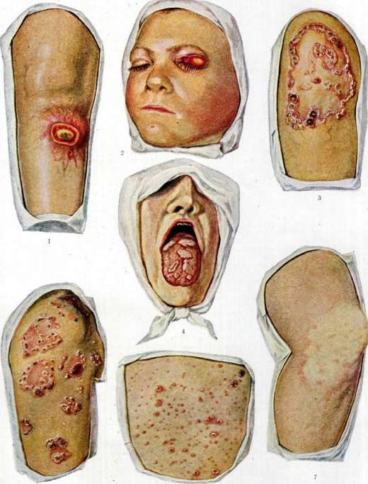
Gumma of the skin Hard chancre Fig. 3. Pustular syphilid (pustular). Fig. 4. Syphilitic papule
Pustular and papular secondary syphilis (so-called secondary eruptive syphilis). 7. Papular (tertiary) rash, the size of a lentil, and sometimes larger. Usually the size of individual elements is uniform in each case, which is why one distinguishes between micropapular, micropapular, micropustular syphilid or macropapular, macropapular, macropustular. The surface of syphilids is smooth until secondary changes appear in the form of scales, crusts, etc. The localization of rash elements is characteristic of fresh forms of S: lateral surfaces of the trunk and flexor surfaces of the limbs with frequent extension to the palms and soles with symmetrical arrangement of the rash; the face is affected less frequently; in recurrent forms, especially in late recurrences, the rash is more sparse and its localization becomes atypical (sometimes on the extensor surfaces of the limbs), and symmetry is often lost. The presence of various rash elements in each case (polymorphism of the rash) is considered characteristic of syphilis, for example, along with spotted rash, papular rash (syphilis cutanea maculo-papulosa), or papular and pustular rash (syph. cutanea papulo-pustulosa), or all three types of secondary rash (syph. cutanea maculo-papulo-pustulosa). In addition to true polymorphism, one should note the so-called false polymorphism, when with the presence of uniform syphilitic efflorescences, secondary changes (crusts, scales, etc.) are noted on some of them. The course of syphilids varies depending on various conditions. Sometimes the rash elements disappear spontaneously: roseola - without a trace, papules - leaving behind pigment spots, in shape and size fully corresponding to the former papules. In other cases, the rash elements persist, sometimes increasing in size and taking hypertrophic forms or giving vegetations. Under specific treatment, they quickly undergo regression, and often at the very beginning of treatment, especially after the use of salvarsan, a reaction is noted from the side of the rash elements in the sense of their more distinct, sharp protrusion; they become brighter, roseola sometimes takes on an urticarial character, new rash elements that were previously unnoticeable appear. This reaction (Jarisch-Herxheimer reaction) usually occurs within the first day from the start of treatment and lasts for several hours or about a day, and then disappears, and the rash elements subsequently quickly begin to undergo regression. The spotted, or erythematous, syphilid is characterized by the appearance of a spotted rash or syphilitic roseola (syphilis cutanea maculosa, s. roseola syphilitica) [see separate table (pp. 527-528), figure 1]. These spots appear weakly expressed, reddish in color with indistinct borders, irregularly round in shape, in some cases only a few millimeters in diameter (small-spotted roseola), in others of larger size (large-spotted roseola). The former is usually found in fresh secondary S, the latter in recurrences. On mucous membranes, the spotted rash is clearly noticeable in the form of sharply defined red spots. Varieties of syphilitic roseola: roseola elevata - spots are slightly elevated above the level of the skin like wheals (often roseola becomes such during the Jarisch-Herxheimer reaction); roseola annularis - spots of ring-shaped outlines (usually in recurrences); roseola configurata - spots in the form of an unclosed ring or oval, forming various figures, touching their ends with neighboring spots; roseola granulata - a roseola spot on the surface of which an abundance of the smallest nodules is noted in the area of skin follicles. The diagnosis of spotted S. is based on the morphol. features of syphilitic roseola, typical localization, absence of subjective symptoms, and presence of other signs of S. (remains of primary sclerosis, scleradenitis, etc.). Differential diagnosis with acute infectious diseases (measles, rubella, typhus, etc.) is easily carried out on the basis of the nature of the rash elements and data on the general state of health. Toxic or drug rashes are easily distinguished by their morphol. features, preferred localization on the extensor surfaces of the limbs, general phenomena, anamnesis data, and absence of indications of S. Urticaria (urticaria) differs from wheal-like roseola by the variability and inconstancy of the rash, disordered localization, presence of burning, itching, and dermographism. Blue spots (maculae coeruleae) from flea bites are easily distinguished from roseolas by their color, they do not disappear from pressure, the cause is easily found, i.e., fleas. Pityriasis rosea Gibert (pityriasis rosea) although in its localization it can fully correspond to syphilitic roseola, it is easily distinguished from it by the variety of size of the rash elements, bay-like outlines of the spots, presence of a central scale and mother plaque. Marbling of the skin (cutis marmorata) only by its bluish tint can resemble syphilitic roseola, but it gives a picture of a peculiar looped network, which is not even present in annular roseola. Papular syphilid (syphilis cutanea papulosa) is most often found in recurrences of secondary S. and is characterized by the appearance of infiltrated roundish nodules of reddish-brown color [see separate table (pp. 551-552), fig. 6]. Over time, scaling (syph. cutanea papulo-squamosa) may appear on the surface of the nodules, sometimes crusts (syph. cutanea papulo-crustosa) or pustules (syph. cutanea papulo-pustulosa) form, and other changes (see below) are also noted depending mainly on localization. The main varieties of papular S. are small- and large-papular syphilids. Small-papular syphilid (syphilis cutanea papulosa miliaris, s. lichen syphiliticus) is characterized by the appearance of nodules the size of a poppy or millet seed, sometimes somewhat larger, usually grouped in different parts of the body surface, mainly on the trunk. The nodules are quite dense in consistency, located in the area of hair follicles; on top, scales or crusts or small pustules are noted. This syphilid is more often found as a recurrence and usually in persons with scrofuloderma or tbc, resembling scrofulous lichen. Large-papular, or lenticular, syphilid (syphilis cutanea papulosa lenticularis) is characterized by the appearance of infiltrated nodules of brownish-red color, the size from a hemp seed to a lentil seed or larger, in abundant quantity or scattered single elements, sometimes in combination with roseola in fresh secondary or early recurrent S. In late recurrences, this syphilid gives a grouped arrangement of papules in the form of rings, arcs, etc. In the further course, scales, crusts may appear on the nodules or pustules may form. Varieties of papular syphilid depend on various causes and in particular on localization: syph. cutanea papulosa nummularis - large papules the size of a 10-20 kopeck coin; syph. cutanea psoriasiformis - abundantly scaling papular elements of large size, resembling psoriatic foci; syph. cutanea papulosa corymbiformis - grouped arrangement of papules, with a large papule in the center of the group and numerous small ones around it like a halo (cockade syphilid, or Bombensyphilid); papulae orbiculares - in persons with seborrhea sometimes in the form of pinkish circles with a slightly elevated periphery and a sunken center with radial folds, sometimes in the form of larger rings covered with greasy scales; sometimes in the form of elongated nodules (e.g., in the folds of the wings of the nose), also covered with greasy scales; sometimes in the form of flat nodules of a whitish, pearly tint - papulae nitidae; syph. cutanea verrucosa - papules with papillary growths on the surface, covered with abundant horny deposits; syph. cutanea framboeiformis - vegetating papules with a red, shiny moist surface, resembling raspberries; psoriasis palmaris et plantaris - papular syphilid of the palms and soles, scaling on the surface; sometimes in the central parts the scales fall off, exposing the red surface of the skin, and in the peripheral parts the scales remain in the form of a rim or cuff, overhanging the exposed part; clavi syphilitici - nodular elements on the palms and soles, covered with tightly fitting horny masses; keratodermia syphilitica - keratinization on extensive infiltrates with scalloped outlines at the periphery (grouped arrangement of papules, merged with neighboring ones), also located on the palms and soles. Palmar and plantar syphilids usually appear in recurrent S, sometimes being the only but very characteristic sign of syphilis.
On the hairy parts of the skin, the following forms of papular syphilid are distinguished: impetigo capitis specifica - papules covered with crusts; syph. papillomatosa capillitii - vegetating papules. On 'moist areas of the skin', subject to the irritating influence of sweat, urine, etc., papules take a weeping character or an erosive appearance due to maceration and shedding of the horny layer of the epidermis. Often they grow, taking on a hypertrophic character - papulae hypertrophicae, s. condylomata lata. Such papules are usually located in the area of the anus and genitals, less frequently on other places of contacting skin surfaces [see separate table (pp. 527-528), fig. 4]. On mucous membranes, flat papules with a whitish surface (plaques muqueuses), erosive and hypertrophic papules are encountered; sometimes the surface of the papules breaks down and they are found covered with a dirty-gray coating (diphtheroid papules) [see separate table (pp. 551-552), fig. 4]. The diagnosis of papular syphilid is based on clinical data of both the rash elements themselves and the accompanying signs of S. (polyadenitis, etc.). In difficult cases, bacterioscopic examinations for the pale spirochete of the discharge from weeping papules or the expressed juice from the papule after scraping off the horny layer of the epidermis are resorted to. Serological examination of the blood is also performed. The differential diagnosis of micropapular syphilid and scrofulous lichen (lichen scrofulosorum) is carried out on the basis of the distinctive features of the latter: yellowish-red color, slight infiltration of the nodules, location in groups of round outlines, localization usually on the trunk and extremely rarely on the extremities, uniformity of individual elements and absence of pseudopolymorphism. The course of scrofulous lichen, with its simultaneous appearance of groups of nodules, lack of tendency to form pustular elements, and simultaneous regression without leaving pigment spots, also serves as an essential sign for differential diagnosis. Scrofulous lichen usually appears in childhood, while in adults it occurs as an exception. The macropapular syphilid must be differentiated from flat red lichen (lichen ruber planus), which resembles papular syphilid in its localization and the pigment spots it leaves behind, but differs from it in yellowish- or bluish-red color, shiny surface, polygonal outlines of the nodules with depression of the center, grouping, etc. When localized on the genitals, red lichen often gives ring-shaped formations simulating recurrent papular syphilid, but differing from it in the absence of scalloped outlines, pearly color, and the presence of polygonal nodules arranged in a ring. It is somewhat more difficult to distinguish hypertrophic red lichen (e.g., in the femoral-genital folds) from hypertrophic papules, but even in such cases, the bluish-red color with opalescent papillae on the surface and lack of tendency to weeping speak for red lichen. Psoriasis vulgaris differs from scaling syphilitic papules in the absence of a brownish tint and noticeable infiltration, as well as the presence of characteristic symptoms of psoriasis and the abundance of silvery-white small scales, easily removable by scraping with the phenomena of 'stearin spot' and 'pointed capillary bleeding'. Parapsoriasis (parapsoriasis en gouttes) is very similar to papular syphilid, but the absence of other signs of S. and the presence of characteristic scales in the form of 'wafers' (especially distinct when scraped with a nail), as well as the appearance of petechiae in the surrounding skin give grounds for the diagnosis of parapsoriasis. Pigment urticaria (urticaria pigmentosa) is not accompanied by infiltration, rubbing with the finger gives a characteristic wheal-like protrusion of the skin in the area of the pigment spot. Papulonecrotic tuberculid (folliculitis, acnitis) with its chronic course, presence of superficial necrosis and characteristic scars is easily distinguished from papular syphilid. Weeping papules from erosions of other origins differ by the presence of infiltrate, regional adenitis and examination of the discharge for the pale spirochete. Hypertrophic papules from pointed condylomas (condylomata acuminata) are easily distinguished by their broad base, relatively smooth surface, color, round or scalloped outlines of the base. Vegetating pemphigus (pemphigus vegetans) develops in the place of a former blister, the remains of which are often visible in the form of a peripheral cuff; loose vegetations and the presence of initial blisters in the absence of signs of S. serve as distinguishing features of vegetating pemphigus. Bromoderma (bromoderma tuberosum) and iododerma (iododerma tuberosum) with their nodular-tumor-like character, soft consistency, papillary proliferations, rapid onset with growth at the periphery and tendency to merge with neighboring formations, as well as diverse localization are easily distinguished from broad condylomas. Papillary carcinoma (carcinoma papillaris) with its slow course, singularity of the lesion, massive appearance and occurrence in elderly people is easily distinguished from broad condylomas. In the differential diagnosis of mucous membrane papules, their clinical features must be taken into account. Erosions of bullous lichen, due to their origin from a group of closely located blisters, usually have scalloped outlines, they are painful, and at the periphery there are remains of blister covers. Erosions after other bullous lesions (erythema exud. multiforme, pemphigus) are diagnosed by the remains of blister covers and bullous elements on other parts of the mucous membranes or skin. Aphthae and aphthous stomatitis (aphthae, stomatitis aphthosa) differ from syphilitic papules in their morphological features (aphthae have the appearance of superficial ulcers with sharply defined edges, yellowish bottom and bright inflammatory halo around, painful). Accidental erosions and ulcers (burns, trauma) have irregular outlines, are painful, and at them the producing moment is noted (caries tooth, pertussis - with ulcers of the frenulum of the tongue in children, etc.). Flat red lichen, usually located on the buccal mucosa, dorsum of the tongue, etc., differs from syphilitic papules in its shagreen-like surface, ring-shaped or figurative outlines and whitish, shiny color. Leukoplakia (leukoplakia) differs in localization (on the buccal mucosa along the line of tooth closure, on the lateral surfaces of the tongue in the anterior two-thirds, its dorsum, on the lip mucosa, absent on the soft palate, arches and uvula), diverse form with distinct winding borders, milky-white color, slow development and chronic course. Geographic tongue (lingua geographica) is easily diagnosed due to ring-shaped or figurative outlines with constant variability of configuration and chronic course. Other diseases of the mucous membranes differ from syphilitic papules in clinical phenomena, course, and in difficult cases, bacterioscopic examination of the discharge or histological examination of biopsied tissue pieces. Pustular syphilis (syphilis cutanea pustulosa) usually is an expression of a more severe course of S. It usually arises by pustulation, due to suppuration in the initially appearing papular rash. Depending on the size of the pustular elements, small-pustular and large-pustular syphilis are distinguished. Small-pustular, or acneiform syphilis (syphilis cutanea acneiformis, s. acne syphilitica) is characterized by the appearance of small pustules the size of a pinhead on a dense infiltrated base, having the skin follicle area as the starting point. Initially, a nodule (papule) appears here, which in 2-3 days turns into a pustule in its upper part, which rather quickly dries into a crust. Sometimes the phenomena develop acutely with an increase in t°, giving abundant widespread eruption (acne syphilitica conferta), located preferably in those places that are favorite for vulgar acneiform rash (face, chest, scapular area), but often also involving other places as well as extremities. Large-pustular syphilis (syphilis cutanea pustulosa varioliformis, s. variola syphilitica) is characterized by the appearance of large pustules resembling smallpox pustules, sitting on an infiltrated base. Sometimes an abundant rash appears with an increase in t° (up to 40°). Subsequently, new and new pustular elements appear, while the old ones dry into crusts, and then heal, leaving a pigment spot in their place. Impetiginous syphilid (impetigo syphilitica), like pustular syphilis, generally has a papule as its starting point, which breaks down with the formation of a pustule that dries into a crust [see separate table (pp. 551-552), fig. 5].
Often occurs in fresh secondary S. on the hairy part of the head (impetigo capitis specifica).-- Syphilitic ecthyma (ecthyma syphiliticum) is essentially a pustular element of larger size than pustular elements, dried into a crust. Under the crust, an ulcerative surface is exposed, healing with scar formation.- Syphilitic rupia (rupia syphilitica) is also a syphilitic pustule, gradually growing at the periphery and in depth, while the forming crust gradually increases and takes on a layered character, resembling a column of coins gradually increasing in size downward (rupia-coin). Under the crust is a fairly deep ulcer, leaving a scar upon healing. The diagnosis of syphilitic pustule is based on the morphological features of the elements and accompanying signs of S. Differential diagnosis of small-pustular syphilis and vulgar acneiform rash is easy. Vulgar acne have a comedo as their starting point, upon suppuration of which fairly significant inflammatory redness appears; they appear in certain places, usually at a young age, and have a chronic course. Drug acne (from iodide or bromide preparations) and professional acne (from lubricating oils, etc.) also have a sebaceous plug as their starting point. Anamnesis and absence of indications of S. provide grounds for the diagnosis of non-syphilitic acne. Differential diagnosis of large-pustular syphilis and smallpox is carried out on the basis of the characteristic features of each disease. Moreover, in syphilis there is false polymorphism, whereas in smallpox all elements are in the same stage of development. The localization of the rash elements in general also has great diagnostic significance (in smallpox, pustules prefer the extensor surfaces of the limbs, including the back of the hands and feet, as well as the face, which is not characteristic of syphilitic pustules). Chickenpox is easily distinguished from syphilis by its clinical signs and course. Vulgar pustular rash (pyodermia) with acute inflammatory phenomena, diversity in size of individual elements and course, as well as random or focal localization, is also easily distinguished from syphilitic pustule. Differential diagnosis of impetiginous syphilis from vulgar impetiginous rash (impetigo vulgaris) is carried out on the basis of the absence of island-inflammatory phenomena (characteristic of the latter), uniform size of individual elements and the presence of a dense brown infiltrate, characteristic of syphilitic elements. Contagious pustular rash (impetigo contagiosa) begins with the formation of a vesicle, which quickly dries into a crust, has a tendency to peripheral growth and random appearance of new elements, often merging with each other, which distinguishes it from impetiginous syphilis. Impetiginous eczema (eczema impetiginosum) with its diffuse inflammatory process with sharp reactive phenomena is easily distinguished from impetiginous syphilis. Differential diagnosis of syphilitic ecthyma and vulgar ecthyma (ecthyma vulgare) is carried out on the basis of the inflammatory phenomena characteristic of the latter and the absence of a brown infiltrate at the base. Differential diagnosis of syphilitic rupia and an atypical form of psoriasis (psoriasis rupioides) is easy, since in the latter there is not a true crust, but the so-called crusta lamellosa. Under the crust is not an ulcerative, but a surface characteristic of psoriasis. Late secondary S., i.e., late recurrent manifestations of it, is characterized by a tendency to group arrangement of rash elements in the form of rings, arcs, and such groups are often located asymmetrically and not in typical places for fresh S. Late roseola often has a ring-shaped form [see separate table (pp. 551-552), fig. 7], papular rash sometimes has the character of an abortive, weakly expressed rash, and sometimes papules have a character resembling tuberculous syphilid of the tertiary period of S. (so-called tertiary papulae-papulae tertiariae). Late pustular rash occurs less frequently and has no special features. The tertiary period of syphilis is characterized by the appearance of nodular lesions of the skin and deeper tissues and organs (subcutaneous tissue, periosteum, bones, internal organs and central nervous system). From the side of the skin, two main forms are distinguished: 1) superficial skin gummas or so-called tuberculous syphilid (syphilis cutanea tuberculosa, s* tuberosa) and 2) deep gummas (gumma syphilit.), arising in the subcutaneous tissue or in deeper tissues. Tuberculous syphilid represents dense, sharply limited infiltrates the size of a hemp seed to a lentil grain, located in the thickness of the skin. The color of the nodes is red-brown. These nodes are located singly or in groups. In their course, they often break down in the central parts, and then resolve, leaving some depression of the skin (subcutaneous scarring) and brown pigmentation, or the broken-down node opens outward, forming ulcers (syphilis cutanea tubero-ulcerosa). Usually tuberculous elements are located in groups, and previously appeared tubercles may undergo reverse development, and new tubercular elements arise at their periphery and the process takes on a serpiginous character (syphilis cutanea tubero-serpiginosa). Sometimes such serpiginous spread is also noted in breaking down nodes with ulcer formation (syphilis cutanea tubero-ulcerosa serpiginosa), and sometimes broken-down nodes give an ulcer that heals partially and at the same time grows in the opposite direction, giving a kidney-shaped ulcer form, which can also take on a serpiginous character (syphilis cutanea ulcero-serpiginosa). In some cases, tuberculous elements give rise to vegetations on their surface (syphilis cutanea tuberculosa vegetans). Sometimes the tubercles are of small size (dwarf) with sufficient density, and sometimes they are soft, resembling true lupus tubercles (syphilis cutanea tuberculosa lupoides). On the mucous membranes, tuberculous syphilid in general can give pictures of the same character as on the skin. Diagnosis of tuberculous and tuberculo-ulcerative syphilis in typical forms is easy (presence of characteristic elements, located in groups with festooned borders or serpiginous spread; brownish pigmentation, dense infiltrate, etc.) [see separate table (pp. 551-552), fig. 3]. Differential diagnosis with vulgar lupus (especially the lupoid form of tuberculous syphilis) is based on the features of clinical signs, more chronic course and tendency for vulgar lupus to appear in childhood. The character of lupus scars (delicate scars penetrated by telangiectasias and often the presence of new lupoid nodules on them) also sharply differs from the scars of tuberculo-ulcerative syphilis [see separate table (pp. 527-528), fig. 5]. The localization also has essential significance (e.g., in the area of the nose, lupus begins with the skin coverings, while tuberculous syphilid-more often from the side of the mucous membrane). Tuberculoid leprosy (lepra tuberosa) is easily distinguished from tuberculous syphilis by the presence of soft tubercles of normal skin color or a yellowish tint, extremely slowly developing without tendency to ulcerative breakdown and with characteristic localization. Chancre (ulcus rodens) differs from ulcerative-serpiginous syphilis by the singularity of the lesion, easily bleeding bottom, peripheral roll of the new growth, slow course and tendency to appear in certain places in elderly persons. Papulonecrotic tuberculid (folliclis, acnitis) is easily distinguished from tuberculous syphilis by the absence of group arrangement and serpiginous spread, the presence of necrosis on the surface and characteristic scars. Gummatous syphilid and gummatous ulcer. Syphilitic gumma of the skin appears as a deep node, somewhat flattened spherical in shape, the size of a pea to a walnut, with a tendency to break down in the central part, and then of the entire node, with outward opening through one perforation, gradually increasing in size and turning into a crater-like ulcer leading into the cavity of the broken-down node [see separate table (pp. 551-552), fig. 1]. The ulcer further increases in size, sometimes far beyond the limits of the original node, sometimes, healing with a scar in one place, it spreads serpiginously in the opposite direction. Gummatous nodes are usually single, and their starting point is the subcutaneous tissue, periosteum, lymph glands, etc. The breakdown of the node can lead to the destruction of the periosteum with the formation of a sequestrum of the underlying bone. Gummatous lesions are often located in the area of the nose (see), soft palate (see Pharynx).
The diagnosis of gummatous syphilids and other nodular skin lesions is based on the clinical data of these diseases.--The differential diagnosis of gummatous and scrofulous nodules and ulcers (scrofuloderma) is based on the fact that the scrofulous ulcer breaks down in different places and opens to the outside through multiple perforating holes, the softened nodule does not have a dense infiltrate around it; scars of healed scrofulous ulcers include remnants of undestroyed skin in the form of papillae, bridges or beams, which is the result of multiple perforating holes that did not completely destroy the skin in the area of the softened nodule. The slow course and young age of the patients are also distinctive features of scrofulous lesions. Varicose ulcers of the leg (ulcus cruris chronicum), cancerous ulcers (carcinoma cutis), ulcers of blastomycosis (blastomycosis), sporotrichosis (sporotrichosis), actinomycosis (actinomycosis), leishmaniasis (leishmaniasis), Bazin's erythema (erythema induratum Bazin), etc., differ from gummatous ulcers in clinical signs and course; in difficult cases, diagnosis is decided by bacteriological, serological and histological examinations of biopsied tissue. It is also necessary to make a diagnosis in relation to gummatous nodules and inflammatory processes of the skin (e.g. rhinoscleroma) and neoplasms (lipomas, atheromas, fibromas, sarcomas, etc.). Syphilitic lesions of the nails are observed as lesions in the area of the nail folds (see Onychia).--Syphilitic lesions of the hair see Alopecia.-Changes in skin pigmentation in S. are observed either as hyperpigmentation in places of former syphilitic manifestations, or around scars after syphilitic ulcers, or in the form of the so-called syphilitic micoderm (see).-On S. of individual organs see the corresponding Organs.
M. Demyanovich. Laboratory diagnosis of S.- see Wassermann reaction, Precipitation and Luetin reaction. VIII. Visceral syphilis. Visceral syphilis combines lesions of internal organs from syphilitic infection, in contrast to lesions of S. of the skin and mucous membranes, on the one hand, and of the nervous system (neurolues)-on the other. Such a division of a single process-syphilitic infection of the body-into three departments naturally does not correspond to modern concepts of infection as a disease of the entire organism and entirely reflects both the historical stages in the development of the doctrine of S, and the localistic thinking of Morgagni and Virchow (Morgagni, Virchow), which excluded diseases common to the entire organism. 1. History and evolution of the doctrine of visceral S. In the 15th and 16th centuries, when S. was raging epidemically in Europe, it was described only as a disease of the skin and bones, and Paracelsus in 1536 was perhaps the first to mention syphilitic lesions of the liver and syphilitic jaundice. Lieutaud (1770), studying path.-anat. material, was the first to substantiate morphol. lesions of internal organs from S, which is why in the 17th and 18th centuries physicians admitted the possibility of visceral syphilitic diseases. Subsequently, these concepts were for a long time destroyed by the doctrine of Hunter, who taught that S. affects only the skin, mucous membranes and bones and does not touch the internal organs. The authority of Hunter was so great that it took almost a whole century until Ricord, and then Fournier, having accumulated a large number of path.-anat. and clin. facts, showed the artificiality of Hunter's doctrine and refuted it. However, even after this, starting from the end of the 19th century, first a doctrine was created about syphilitic lesions of the nervous system T while the doctrine of visceral syphilis was limited only to the accumulation of morphol. and clin. observations and received its full justification only after the discovery of Schaudinn's spirochete made it possible to experimentally study the evolution of syphilitic infection in the animal organism and to prove the specificity of the process in individual organs in humans. And yet to this day no coherent doctrine of visceral S. and its evolution has been created, starting from the moment of infection, as has been done for the skin and mucous membranes, on the one hand, and for the nervous system-on the other. This primarily explains why, when speaking of visceral S., one still has in mind only the gross morphol. changes of internal organs of the late, tertiary period, where sometimes decades separate the disease of one or another organ from the moment of general infection. These late syphilitic lesions of internal organs-gummatous and sclerogummatous-and often already sclerotic consequences as a result of the reverse development of the process that destroyed part of the organ, are most often the subject of study by pathologists and clinicians. We still know relatively little about the early and reversible processes of syphilitic infection, and the functional disorders of organs in the early stages of S. have been very little studied. Therefore, we know much more about the statics of visceral S., 588 syphilis than about its evolution and dynamics from the point of view of the modern doctrine of functional pathology, which establishes the clinic of diseases of internal organs and systems in the closest connection with the endocrine, autonomic and central nervous systems. 2. Pathology of visceral S. The clinic and path. anatomy of syphilitic lesions of individual organs are described in the corresponding articles, and here we will dwell only on the path. and clin. data concerning visceral S. KH. PC of general infection of the body. From the gate of infection, the pathogen very early penetrates into the general bloodstream through lymphatic pathways; spirochetemia immediately leads to the generalization of S, and the appearance of 'minimal early diseases' (Golay) is possible. Even at this stage, the syphilitic virus can become fixed in one or another organ and cause corresponding reactive phenomena in it. As is known', in congenital S. in the fetus, the skin and mucous membranes are affected to the same extent, on the one hand, and internal organs-on the other, with individual internal organs, for example the liver, being predominantly affected. When infected with S. after extrauterine life, the organism, although entirely exposed to the infection, but visible clin. lesions are localized mainly on the skin and mucous membranes. This, of course, does not mean that the internal organs remain spared by the infection. We have a number of path.-anat., experimental and clin. data, indicating that from the very beginning syphilitic infection affects all systems and organs and localizes in them. Not to mention that in the earliest stages of S. such general reactions of the organism as feverish rises in temperature, changes in the blood picture, chemistry of gastric secretion, etc., have been noted, there are quite definite observations about early rashes on the mucous opQHxB, in the liver, about early albuminurias as an expression of nephropathies, etc. However, it is not these early forms of infection that are typical for visceral S. The clin. concept of visceral S. includes mainly path. processes developing as a result of late multiplication of the spirochete in one or another internal organ when the early localizations of infection in the skin and mucous membranes have long since disappeared or even been forgotten. Visceral S. is still considered today as diseases of the tertiary period or metasyphilitic processes. Under this name one must understand the sum of reactive processes from the mesenchyme, leading to sclerotic phenomena with the destruction of the specific parenchyma of the affected organ. Thus, S. of internal organs manifests itself: 1) in early specific diseases of the primary and secondary stages of infection, 2) in late specific gummatous, sclerogummatous lesions and 3) in non-specific connective tissue lesions of the cirrhosis type. It is not without interest that the condition of internal organs can in turn have a certain influence on the manifestation and course of syphilitic infection; thus, in thyroidectomized animals Pearce and Allen (Pearce, Allen) saw a significantly more severe course of experimental S. than in controls; similar observations have been made in relation to the extirpation of the thyroid gland. It is very probable that the different results of treatment of S. in humans are connected with this influence of the endocrine apparatus on the course of infection. It is also not without interest that the course of syphilitic infection is influenced by other diseases of internal organs. Thus, tuberculosis worsens the course of S. and significantly complicates its treatment, on the other hand, acute infectious diseases, such as influenza, scarlet fever, have a favorable effect on the course of syphilitic infection, with rashes disappearing and the Wassermann reaction may temporarily become negative. Clin. observations on the treatment of some forms of syphilitic infection with malaria and relapsing fever speak of a certain antagonism between S. and acute infections. As for the localization of the process in visceral S., we have data only for the late, tertiary forms of syphilitic infection. There are only fragmentary clin. observations on the early stages of visceral S., while the path.-anat. picture has been described only rarely and as a great rarity. In the study of 414 cases of visceral S., Schrumpf found lesions of the cardiovascular system and especially of the aorta in 56.76%, lesions of the nervous system in 23.43%, liver-8.45%, lungs-3.14%, arthritis-0.72%, kidneys-0.48% (without definite localization-2.9%). In 280 patients with visceral S., Luria saw diseases of the circulatory organs in 38.3%, nervous system-19.7%, liver-18.9%; without definite clin. localization-11.1%, arthritis-2.6%, kidney diseases-3.1%, gastrointest. tract-2.6%. However relative these data may be, all authors unanimously note that in the late stages visceral S. is most frequently localized in the cardiovascular system, mainly in the aorta and in the liver; gummatous lesions also occur in all other organs, but much less frequently.-Some interest is presented by a comparison of the frequency of lesions of internal organs from S. with the results of experiment. The influence of syphilitic infection on them is not exhausted, as we have seen, by specific lesions of internal organs; it is also necessary to keep in mind the non-specific influence of the virus and its toxins on tissues, i.e., not purely syphilitic, but syphilogenic processes of chronic infection of internal organs with various morphol. their changes.-The significance of S. in the etiology of diseases of internal organs, according to Wittgenstein and Brodnitz (Wittgenstein, Brodnitz), is exceptionally great; thus, in 90,553 patients of the Charité outpatient clinic in Berlin, they found syphilitic infection as the cause of internal diseases in 7.54%.
Leredde, investigating the cause of mortality in the population of Paris, found that out of 45,814 deaths in 3,414 cases, i.e., in 7%, S. was the cause of death due to lesions of internal organs, and in 3/4 of cases angina pectoris was noted, in half of the cases vascular diseases, in 1/3 of cases organic diseases of the heart, in 1/6 of cases death resulted from chronic nephritis and cirrhosis of the liver. Bayet gives for Brussels 11% mortality from visceral S. According to data from the Gotha Life Insurance Society based on material from 44 years, mortality among syphilitics was 68% higher than among non-syphilitics; according to data from American insurance societies, mortality among insured patients with S. was at least 50% higher than among the non-infected. These facts are quite sufficient to illustrate the enormous role of visceral S. as a social disease. 3. The clinical picture of visceral S. is extremely varied and diverse as a result of a whole series of very different pathological processes caused by the pale spirochete in different stages of the disease. Already the specific manifestations of S., starting from allergic processes in the early stages of the disease to syphilides and gummatous infiltration in later stages, reactions on the part of the mesenchyme—all these closely interwoven pathological processes cannot give a uniform clinical picture. To this must be added the typical for S. pluriviscerality, i.e., the multiplicity of localization simultaneously in a number of organs and systems, including the endocrine glands, in the autonomic and central nervous system. All this taken together creates conditions for the most varied and at times quite bizarre combinations of organic and functional disorders, which in turn are inextricably linked and very closely interwoven. This is what imprints a stamp of uniqueness on the clinical picture of visceral S. and does not make it possible to draw a general clinical picture for all these diseases. Descriptions of individual diseases of organs as a result of syphilitic infection are given in the corresponding chapters; they are developed mainly with regard to the late, tertiary, or metasyphilitic forms; the clinical picture of early lesions of internal organs disappears from the view of the therapist precisely because observations are conducted by a syphilidologist specialist, mainly engaged in the cutaneous localization of the infection. These early forms of syphilitic infection of internal organs are especially difficult to recognize, since it is almost exclusively a matter of functional disorders, of changes in the organ's behavior. Thus, on the part of the cardiovascular system, sensations of pressure in the heart area, palpitations, precordial anxiety are observed, and objectively—tachycardia or, conversely, bradycardia and disorders of heart rhythm. These phenomena some are inclined to consider not so much as a result of anatomical changes, but as functional ones as a result of toxemia (Citron); however, it is hardly possible in the secondary period to completely deny the possibility of the appearance of organic lesions of the heart—myocarditis, or more correctly pericarditis (Pletnev). As for the vessels, if one takes into account the vasotropism of the syphilitic virus, one can sometimes observe in the early stages of infection endoarteritis and especially endophlebitis, and syphilitic phlebitis occurs more often in women than in men. It is precisely these early lesions of the vascular system that explain the early appearance in syphilitics of deep circulatory disorders in the heart—angina pectoris, myocardial degeneration—or in the central nervous system—thrombi and hemorrhages in the brain. On the part of the respiratory apparatus, besides early exanthemas in the larynx, specific bronchitis and pleurisy with a positive Wassermann reaction in the punctate appear; radiologically, lesions of lung tissue in the form of small foci of consolidation have been established already in the secondary period (Groedel). As for the digestive tract, there are data on gastric processes in the early stages of infection, expressed besides dyspeptic phenomena in a decrease in the secretory activity of the stomach up to achylia. It is very interesting that these inflammatory and functional lesions of the stomach in the early stages are reversible under the influence of specific therapy (Gausman, Luria). Will found in the intestine in the secondary period roseolas and papules, Hayem and Tissier—ulcerative processes; that is why it is not surprising that in these early stages of infection one can expect the appearance of diarrhea of both functional (allergic) and organic origin (enterocolitis). Of particular interest are the lesions of S. of the liver in the early periods of infection and first of all the early syphilitic jaundice (icterus syphiliticus praecox). This form of jaundice can appear already during the primary affection; however, it is more often encountered with the first exanthemas. Of great interest are the early syphilitic diseases of the kidneys (see Kidneys, syphilis of the kidneys). With such a disease totius substantiae as S., the diseases of the endocrine apparatus are of exceptional interest, however, data on their behavior in the early stages of infection are still very insufficient. That these glands, as having an especially abundant network of vessels, should be affected during spirochetemia, can hardly be doubted; thus, there are indications of early strumites, as well as phenomena of irritation and swelling of the thyroid gland in the initial stages. Data on early lesions of S. of other glands are still very scarce. From this brief enumeration of lesions of internal organs in the early stages of syphilitic infection, it is quite obvious how incorrect it is to consider visceral S. a disease only of the late, tertiary stage of S, and the task of the therapist is precisely in the first weeks and months of the infection to look for those often subtle functional disorders of the body that are the result of shallow, and therefore reversible, syphilitic and syphilogenic lesions of internal organs. Organizationally this requires the closest coordination of the work of the venereologist and therapist, starting from the moment of the patient's infection with S. In view of the fact that the lesions of internal organs of the tertiary period of S. and their metasyphilitic diseases are described in detail in the corresponding chapters, we will dwell here only on two syndromes of visceral S, representing its reflection as malum totius substantiae—on changes in the blood and on syphilitic fever. Changes in the blood in S. have been known for a long time, and in the early stages of infection, old physicians described under the name of 'syphilitic chlorosis' a specific anemia. In the primary stage, a decrease in hemoglobin without oligocytopenia is described, in the secondary—phenomena of typical secondary anemia with oligochromia and oligocytopenia, disappearing when S. passes into the latent form. Specific, especially mercurial treatment improves the blood picture. On the part of the white blood, a slight leukocytosis is noted, with the number of neutrophils decreased, mononuclears increased; in the late stages of S, as well as in other chronic infections, a relative mononucleosis is observed. Great controversy is aroused by the question of the relationship between S. and hyperchromic anemias of the type anaemia perniciosa, and the opinions of authors on the role of S. in the etiopathogenesis of pernicious anemia differ greatly. While some authors (Fr. Miiller, Grawitz, Schlesinger and others) believe that the toxic effect of the syphilitic virus on the bone marrow causes the picture of progressive pernicious anemia, others (Ehrlich, Lazarus, Pappenheim) deny the genetic connection of pernicious anemia with S. Although individual observations speak in favor of the fact that blood transfusion followed by anti-syphilitic treatment gives exceptionally brilliant results in almost hopeless cases of anaemia perniciosa (Schlesinger), this question still remains open, and all authors unanimously speak against vigorous specific treatment for anaemia perniciosa and require great caution in dosing, especially of salvarsan. Among the general phenomena caused by syphilitic infection, fever deserves attention; according to clinical manifestations, the following forms are distinguished: 1) Fever accompanying syphilitic exanthemas in the secondary and tertiary periods of infection; it is especially pronounced in severe cases of S. and more often occurs with pustular efflorescences of the skin. 2) Fever appearing in gummatous lesions of individual organs. This includes first of all S. of the liver, very often accompanied by very persistent feverish movements t° (according to Talkvist in 50% of all cases); the fever here is periodic, sometimes intermittent, giving grounds to speak of malaria, typhus and septic processes, more rarely it has a remittent character. S. of the spleen, accompanied by fever, has more than once been confused with malaria, Malta fever and kala-azar; S. of the lungs very often occurs with fever and has therefore long been considered tbc; syphilitic lesions of the peritoneum with fever have been described, also mistaken for tbc of the peritoneum.
Finally, late syphilitic joint lesions (arthrolues tardiva) often proceed with constant, remittent, and even hectic fever, lasting for weeks and months, giving grounds for erroneously assuming true rheumatism or sepsis. 3) Fever resulting from therapeutic intervention that provokes a Jarisch-Herxheimer reaction; it sometimes occurs after the first injections of mercury, salvarsan, bismuth after 3-10 hours and lasts 2, 3, or even 4 days; reaching 39-40°. 4) Finally, the last and most interesting form is essential syphilitic fever; numerous cases of prolonged feverish course of the disease lasting for months have been described, which led to various diagnoses and were cured by specific therapy. These cases represent great theoretical interest and are explained by pathological processes in the nervous and endocrine systems that are not subject to localization. At the same time, they always present very great diagnostic difficulties. 4. Recognition of visceral S. in the early stages of infection presents great difficulties, since functional disorders of the organs in most cases do not differ from pathological conditions of other etiology, and only in individual cases during endoscopy (bronchi, rectum, and sigmoid colon) is it possible to see typical syphilitic eruptions. Recognition of late forms of visceral S. presents significantly fewer difficulties, especially when it is localized in organs most frequently affected by the sclerogummous process, such as the cardiovascular system or liver. However, even in these cases, differential diagnosis with malignant neoplasms is not always possible even during laparotomy, when the surgeon can both see and feel the pathological process, or in the lungs, when it can be easily fixed on an X-ray. In addition to local symptoms that provide points of reference for differential diagnosis (see Stomach, Lungs, Liver and other organs), the clinical picture of visceral S. nevertheless presents a number of features, knowledge of which significantly facilitates the recognition of syphilitic lesions of internal organs. Visceral S. proceeds atypically, often not fitting into the usual forms of internal organ diseases for physicians. This gave rise to numerous and very successful clinical aphorisms, both old ones: 'indubio suspice luem' (in doubtful cases suspect S.), and later ones: 'la verolle vieille a la mine honnete' (Ricord) (syphilis, growing old, takes on a decent appearance), or-as Zigmund said-'die Syphilis ist die Affe unter den Krankheiten' (syphilis is the monkey among diseases). All these empirically developed clinical rules fully correspond to the clinical picture and course of visceral S. and find their explanation in the combination of very various pathological-histological processes in the organs: gummous lesions in various stages-formation, softening, scarring of the gumma,-death of the parenchymal tissue of the organ and sclerotic processes of the mesenchyme, lesion of the organ's vascular system-endarteritis, endophlebitis,-specific and non-specific changes in nervous tissue, etc. On the other hand, this atypicality of the clinical picture of visceral S. is explained by the typical plurivisceral nature of the disease, when a number of organs and systems are affected simultaneously precisely because S. is a disease not of one organ, but a malum totius substantiae. Thus, syphilitic diseases of the aorta in no less than 1/3 of cases are combined with neurosyphilis and in 1/2 of cases with tabes; according to Pletnev, 40% of S. of the cardiovascular system are combined with lesions of S. of other organs; Luria in 48 cases of S. of the heart and vessels saw 26 lesions of the nervous system, 21 lesions of the liver, 3 lesions of the endocrine glands; in a number of diseases of the stomach of syphilitic origin, diseases of the heart, liver, and central nervous system were simultaneously noted. Of course, in addition to multiple lesions of internal organs, the matter may also concern traces of lesions of the skin and bones of a tertiary nature-periostitis, scars, etc. But, as is known, in recent decades S., especially in cities, has lost its dermotropic character and therefore combinations of visceral and skin-bone S. are encountered less and less frequently, which may be connected with the immunobiological properties of the skin in terms of ezophylaxis (Hoffmann). All the more important is the study of small but essential symptoms resulting from previous lesions of other systems. Here primarily belong isolated symptoms from the eyes- anisocoria, anomalies of the pupillary reflex to light with preservation of their reaction during convergence (Argyll-Robertson symptom) and fading of the iris (Darkshevich). In a number of diseases of internal organs, these isolated symptoms from the eyes without other visible lesions of the central nervous system were observed in various forms of visceral S. and were a symptom that led to the correct recognition of the nature of the disease (Luria); this symptom is stable, does not disappear after specific treatment and is the result of either a latently proceeding basal meningitis in the secondary stage of infection (Forster) or a specific lesion of the autonomic system (Laignel-Lavastine). These pupillary symptoms, on the other hand, are valuable confirmation for the diagnosis of S. of internal organs. The presence of a positive RW, of course, indicates syphilitic infection, but does not yet decide the syphilitic nature of this visceral disease, all the more so that so-called hybrid forms, i.e., banal diseases in syphilitics-cancer, stomach ulcer, nephropathies, tbc of the lungs, etc., are not infrequently encountered. On the other hand, a negative RW does not yet give grounds to exclude visceral S., especially if the matter concerns the reaction only in the blood and not in the cerebrospinal fluid; thus, Stokes-Brown found in visceral S. a positive RW in the liquor in 59%, whereas in the blood it was obtained only in 44% of all cases; on the other hand, cases of negative RW with the presence of Treponema pallidum in the tissues have been described. The Wassermann reaction in pleural, ascitic fluid and effusions in the joints, unfortunately rarely practiced, also has great importance for diagnosis. Thus, the Wassermann reaction for the diagnosis of visceral S. has only relative importance, all the more so that in a number of cases it appears only during or even after specific treatment. The anamnesis has an even more conditional value for the diagnosis of visceral syphilitic diseases. If the patient had S., this only makes the physician think of the possibility of visceral syphilitic processes; but if there is no S. in the anamnesis, then in 57.4% of women the matter may concern syphilis ignore (Marschalko, Fournier). The doctrine of 'silent' infection and the multiplied observations of cases of unquestionable S. without definite localization of the primary infect give sufficient theoretical justification for this fact. The old method of recognizing visceral S. based on the results of trial specific treatment is permissible, but with essential reservations of careful study of the patient during treatment; brilliant results of treatment often confirm the diagnosis of visceral S., and the appearance of a previously absent positive RW makes the assumption already an unquestionable clinical fact. Thus, the atypicality of the clinical picture, the plurivisceral nature of the diseases, the presence of isolated symptoms from the eyes, the Wassermann reaction in the blood, in the cerebrospinal fluid, in effusions, the study of the anamnesis, and sometimes the results of specific treatment-these are the points of reference for the diagnosis of visceral S. The prognosis in visceral S. depends, of course, primarily on the early recognition of the true nature of the disease, its syphilitic origin; in the late stages of syphilitic infection, it is especially important to apply treatment before the fibroplastic irreversible process is completed. S. of the heart and large vessels gives an especially unfavorable prognosis and often an unexpected fatal outcome. 5. The treatment of visceral S. is in most cases a grateful task and is facilitated by the fact that the localization of the syphilitic process plays a subordinate role here, because antisyphilitic agents do not act directly on the local process but by mobilizing the immunobiological adaptations of the organism. However, in the treatment of visceral syphilis, even less than in the treatment of its skin manifestations, a standard, identical for all cases system of treatment is permissible. For example, in syphilitic diseases of the kidneys, caution is required in prescribing mercury-indifferently in the form of injections, rubs or taking mercury preparations internally; the same applies to bismuth preparations and salvarsan, and therefore treatment must begin with the prescription of iodine preparations internally or in the form of injections in relatively small doses, and only after the disappearance of edemas should one cautiously proceed to treatment with neosalvarsan.
In syphilis of the liver, preparations of iodine in large doses are well tolerated, mercury in small and medium doses also gives good results, as do preparations of bismuth (bijochinol); however, it is better to avoid preparations of salvarsan, as it often causes a severe Jarisch-Herxheimer reaction here. Syphilis of the stomach and intestines responds well to any antisyphilitic treatment. Incidentally, preparations of iodine, which are poorly tolerated by patients with common stomach diseases, are well tolerated by them in stomach diseases of syphilitic origin, which can sometimes be used for diagnostic purposes. When treating with salvarsan, great caution is required due to the possibility of profuse gastric bleeding. Therapy for cardiovascular syphilitic diseases requires special caution; there are many known cases of severe complications and even sudden death when large doses of salvarsan are administered immediately. This caution in treating visceral syphilis with specific agents is dictated by the fact that the physician cannot in each individual case assess the magnitude of the Herxheimer reaction and therefore the volume and results of the loss of function of the patient's organ. According to experience, in addition to modern methods of treating syphilis, visceral diseases often respond well to old methods of therapy, especially to the rubbing in of sulfur-mercury ointment in combination with large doses of iodine internally. Very good results have also been described when prescribing the old Zitmanovskiy decoction and sarsaparilla decoction. There are indications of a favorable effect on the course of visceral syphilis from malaria vaccinations, however, anginal phenomena with fatal outcomes have already been noted, e.g., in the treatment of syphilitic mzaortitis with these vaccinations (Jagic, Spengler). In addition to specific therapy for visceral syphilis, balneological treatment is indicated, mainly resorts with sulfur springs (Pyatigorsk, Sergiyevskie mineral waters, Matesta, Sernovodsk), however, taking into account all anatomical and functional disorders and with strict individualization of each case. Specific and resort treatment for visceral syphilis, of course, must be combined with dietetic, physiotherapeutic, and medicinal therapy as indicated in each individual case.
r. Luria. IX. Treatment of acquired syphilis. In the treatment of S., so-called specific means are mainly used, which include arsenic, mercury, bismuth, and iodine preparations. Experimental data and clinical observations at the present time increasingly strengthen the view that specific means are not directly spirocheticidal. Acting on the cells and tissues of the organism, they produce changes in their biochemical state, stimulating immunobiological reactions to the introduced infection. For this reason, the condition of individual organs and systems during the course and treatment of S. has colossal significance, and in cases of violation of their functional activity of a non-specific nature, it is necessary simultaneously with specific treatment to conduct non-specific treatment for the purpose of restoring the impaired function. Although in everyday practice we do see patients in whom, despite inadequate treatment or its complete absence, S. does not manifest itself for decades, still the majority of them either serologically or from the side of the liquor are found to be diseased, women give birth to diseased with S. children, and mainly from this contingent of diseased are compiled aortics, tabetics, progressive paralytics, and other diseased with late manifestation of visceral S. and S. of the central nervous system. Thus, immunobiological processes alone are not sufficient to fight against syphilitic infection, and only intensive and systematic treatment with specific means can give complete cure of S. and is the best prevention of visceral S. and of the nervous system. The discovery of the pale spirochete, the serological reaction of the blood, and the introduction into therapy of syphilis of salvarsan, especially in combination with mercury and bismuth, made it possible with early and intensive treatment to abort S., which is why modern therapy of S. is based on the principle of possibly early, intensive, and systematic treatment, conducted without violation of the general condition of the organism and of the functions of its individual organs. I It is unnecessary to cite all existing variants of treatment of S., developed by individual representatives of syphilidology on the basis of personal observations. The difference consists mainly in the application of single and total doses of the used medications in various combinations of them and various intervals between injections and individual courses, and in the total duration of treatment. It is necessary to note only the main provisions of the methodology of treatment and application of specific means in different periods of S. They consist of the following: 1) Specific treatment must be begun as early as possible, in the primary seronegative period, immediately after establishing the bacterioscopic diagnosis. The earlier treatment is begun, the greater the chances for complete sterilization of the organism. 2) The less time has passed since the infection, the more intensive should be the treatment in regard to the application of single and total doses per course and the shorter should be the intervals between courses. The longer the duration of infection, the less intensive should be the specific treatment in regard to an individual course, but in general more prolonged. 3) Total doses of specific means in the first courses of treatment of fresh S. should be brought to the limits of the organism's endurance, but not to the detriment of its general condition. For an adult person, the average therapeutic dose of neosalvarsan is 0.1 pro die, mercury--0.01 Hg met., bismuth--0.02 Bi met. The course dose of neosalvarsan on average is 5.0-6.0, mercury--0.3-0.5-1.0 Hg met., bismuth--1.0-1.5 Bi met. In the tertiary period of S. these doses may be reduced. 4) In the primary seronegative period of S. treatment can begin directly with the infusion of neosalvarsan, in the subsequent periods to avoid sometimes severe phenomena of the Herxheimer reaction--with injections of bismuth or mercury preparations, and in the tertiary--with the use of iodine preparations either in pure form or together with mercury or bismuth preparations. 5) Since in each specific means there may be hidden refractoriness of the syphilitic virus, the most powerful treatment is alternating mixed salvarsan-bismuth and salvarsan-mercury courses. Even in those cases where salvarsan proves ineffective, success can be obtained from mercury and bismuth and in the tertiary period--from iodine. For the purpose of longer therapeutic action in combined salvarsan-mercury treatment, it is better to use insoluble preparations of mercury. In one mercury treatment of fresh S. with contagious manifestations, it is better to begin treatment with injections of soluble preparations or even intravenous infusions in view of their more rapid action with this method of administration. After the elimination of contagious manifestations of S., it is advisable to switch to insoluble preparations acting more durably. Soluble preparations as more easily tolerated and more uniformly and rapidly eliminated than suspensions are more applicable to weak organisms or in violation of the function of visceral organs. In view of the therapeutic significance of local application of specific means in the secondary period of S., one or two courses of treatment are advisable to be conducted with friction of mercury ointments, with specific lesion of the gastro-intestinal tract--by taking mercury preparations internally. 6) Intervals between courses, if there are no special contraindications for this, should not exceed 4-5 weeks in the treatment of fresh S. and 2-3 months--of tertiary S. Systematic courses with small intervals most ensure the absence of clinical recurrences, stable negative serological reactions, and normal liquor. 7) Iodine therapy is usually prescribed from the second year of infection, and in old age at its very beginning. In the tertiary period of S. it should be the most intensive and prolonged. Mixed treatment with iodine with salvarsan, bismuth or mercury exerts a stronger therapeutic action than each means separately, but also one iodine treatment in the tertiary period of S., applied after the completion of salvarsan-bismuth-mercury treatment, has great preventive significance. 8) Treatment should strive to achieve the stable disappearance of clinical symptoms, stable negative serological reactions, and improvement of the liquor, which is why it must be conducted under the control of serological reactions of the blood and examination of the liquor. After the disappearance of clinical and serological phenomena of S., treatment should be continued in the form of so-called consolidating courses in the greater number, the later it was begun. 9) If after systematic intensive treatment in the amount of not less than 5-6 courses serological reactions remain positive, salvarsan, bismuth, and mercury treatment should be discontinued, leaving only iodine treatment in the amount of 2-3 courses a year. It is very desirable in such cases to add physio-, balneo-, aerotherapy. 10) In pathological liquor in cases similar to the previous ones, it is advisable to prescribe 1 or 2 salvarsan-bismuth or mercury courses a year and in the intervals between them iodine treatment. Such cases serve as an indication for the application, where the condition of the organism permits, of malaria therapy together with salvarsan-bismuth (on the treatment with malaria--see Progressive paralysis). 11) Upon the appearance of clinical symptoms of S. in the cases indicated in paragraphs 9 and 10, systematic mixed treatment in the amount of not less than three courses is conducted, according to paragraphs 5 and 6. 12) The application of the above provisions must be strictly coordinated with the individual endurance of the organism in relation to the used medications and their doses, with contraindications to their prescription, age, general condition, and functional activity of individual organs. Patients before prescription of treatment must be thoroughly examined and treatment must be individualized in relation to each patient. In all cases of relative contraindications to the use of specific means, they are prescribed in reduced doses both single and total, the intervals free from treatment are increased, and non-specific therapy is applied, strengthening the general condition of the organism and correcting the impaired function of this or that organ. 13) Overfatigue, prolonged nervous tension, sexual excesses, alcoholic beverages, narcotics adversely affect the course of S., lowering the natural immunity of the organism and causing intolerance to specific preparations, especially to salvarsan. Moderate physical labor, gymnastics, light sports as improving metabolism are very desirable. 14) At the conclusion of treatment the patient must remain under clinical, serological, and from the side of the liquor control and the longer, the later treatment was begun. Of the methods of treatment of S. used at the present time, the following can be indicated: ^Application of only one of the specific means, mainly salvarsan and its derivatives, mainly in abortive treatment of S., in the amount of one or several courses. Against pure salvarsan treatment can be put first the rapid elimination of salvarsan from the organism and consequently the short duration of its therapeutic action and secondly the possibility of relative difficultly perceptible refractoriness to it of spirochetes.
As a result of this, with only Salvarsan treatment alone, clinical, serological, and cerebrospinal fluid recurrences are observed significantly more frequently than after combined treatment. Treating fresh S. with mercury alone must be considered insufficient due to its weak therapeutic effect at present. Only a few authors consider it possible to use only bismuth. 2) Combined treatment with Salvarsan, bismuth, mercury, and iodine preparations in various combinations depending on the period of S, the general condition of the body, and its individual organs. From this group, the 'chronically intermittent,' or 'preventive' method of treatment, developed by Fournier and Neisser in the middle of the 19th century, is more widespread. This method compensates for the insufficient treatment of the once popular 'symptomatic method,' enhancing it with repeated courses during the latent stage of the disease. At the same time, due to the intervals between courses, it does not disrupt the general condition of the body, as was the case with the 'continuous,' or 'tonic' method. The principles of this method are still recognized by most syphilologists. Some authors, following this method, conduct combined courses, alternating single doses of Salvarsan, bismuth, or mercury, while others alternate courses, each conducted with one of these preparations. In both cases, breaks are made between courses to avoid an excessively large burden on the body from the administered preparations and consequently a decrease in its resistance to the virus. 3) The permanent method of treatment (Almkvist, MHNap). It involves the use of Salvarsan, bismuth, and mercury in the form of separate courses, following one after another without interruption and constituting separate cycles. Such cycles are repeated without interruption depending on the period of S. for several months (from 31/2 to 14 according to Almkvist). The method of continuous application of specific agents is based on not allowing spirochetes to become activated, which, in the opinion of the authors who use it, occurs during the treatment breaks in Fournier-Neisser's chronic intermittent method. 4) The method of treatment known as 'shock' therapy, consisting of Salvarsan and bismuth (Scholtz, Spiethoff) or mercury (Stern, Silberstein, Pollitzer) series. In this method, Salvarsan preparations are used daily for several consecutive days in increased doses compared to the usual. These series are repeated several times with different rest intervals depending on the course of S. and its periods. The method is based, in the opinion of the authors who use it, on the fact that the body reacts more strongly to a stronger impact of specific agents, contributing to a faster and stronger therapeutic effect. Due to the high intensity of this method, it is applicable only to strong, young patients with normal visceral organ functions and mainly in abortive treatment. 5) The method of treatment consisting of the simultaneous combined application of preparations like Neosalvarsan and soluble mercury in one syringe (Linser's method), is based on the assumption that mercury preparations activate the therapeutic effect of Salvarsan. Preventive treatment of syphilis, i.e., the application of treatment in cases where infection is suspected but has not yet manifested either clinically or serologically, became possible only thanks to Salvarsan. The principle of preventive treatment is the same as in abortive treatment: early therapeutic intervention that most ensures the sterilization of the body. The main motivation to begin treatment before clinical manifestations of the disease appear is the significant percentage of failures of abortive treatment, which according to some authors reaches (15%), even when started during the seronegative chancre stage. The following cases of probable infection should be considered suitable for preventive treatment: 1) after intercourse with syphilitic individuals with contagious manifestations on the genitals. Preventive treatment is not indicated only in cases of positive serological reactions without manifestations of S. on the skin or mucous membranes of the partner. 2) After a healthy woman breastfeeds a child with active manifestations of S. on the face or in the oral cavity, or a child breastfeeding from a syphilitic woman with contagious manifestations on the mammary glands. 3) For public health prevention, in cases of close non-sexual contact with individuals with contagious manifestations of S. when there is reason to suspect the possibility of transmission of infection. Preventive treatment should not be given to persons frequently exposed to the possibility of infection with S. Clinical experience shows that the earlier preventive treatment is started, the greater the chances of success. Simon recognizes preventive treatment as effective if it is started within 3 days after probable infection, Meshchersky-up to 6 days, Nikolsky and Kozhevnikov-up to 7 days, Nicolau considers 8-9 days after infection as the most favorable period, Brin and Legrain-8-10 days, Gallais extends it to 14 days. Reports of failures of preventive treatment are still rare, but the later it is started and the weaker it is conducted, the more frequent the failures. It is more correct to limit the time for cases suitable for preventive treatment to 6-7 days from the suspected infection. After this period, patients should be left untreated but under careful clinical and, due to the possibility of 1. ignorata (especially in women), serological observation for 3-4 months. For preventive treatment, the most potent antisphilitic agents should be used-arsenical preparations mainly in the form of injections of Neoarsenol 3.0-6.0 (in larger quantities, the more time has passed since the start of infection). In cases where injections cannot be given, intramuscular or subcutaneous injections of Myosalvarsan or Sulfarsenol or preparations like Osarsol in an amount of 30.0-40.0 for a full course can be used. Most authors limit preventive treatment to one Salvarsan course. Some recommend performing several pure Salvarsan courses (G. I. Meshchersky) or a combined bismuth-Salvarsan course (A. P. Iordan, Gougerot, etc.). Abortive treatment. All attempts to abort syphilis through local measures-excision of the chancre (Tarnovsky, Pospelov, Fournier, etc.) and even regional glands, the use of mercury preparations in the form of injections into the chancre tissue or between the chancre and the groin (Hunter) or arsenical ones (Hallopeau)-are based mainly on the mistaken belief that during the primary manifestation the virus remains localized only in it and the surrounding tissues and the nearest glands, and have been completely unsuccessful or have given a very small percentage of successes, which would more correctly be attributed to the latent state of infection. Numerous attempts made before the appearance of Salvarsan to abort S. with general mercury treatment, and later with bismuth, also remained fruitless. With the discovery of Salvarsan, a new era began in the abortive treatment of S. The hope for abortion was so great that attempts to treat with this drug took on the character of 'therapia magna sterilisans.' From the very beginning of Salvarsan's appearance, it was considered possible to abort S. not only in the seronegative stage of primary manifestation but also in the seropositive stage and in cases of fresh S. with secondary manifestations. However, further observations, which counted a large number of recurrences in the last two stages of S, forced to narrow the scope of cases suitable for abortive treatment to the seronegative primary manifestation. According to material from an international questionnaire of the State Venereal Institute on abortive treatment of S., processed by V. I. Feldman, the best moment for applying abortive treatment by most authors is considered to be the primary seronegative chancre with the duration of infection from 4 to 5 weeks and no more than 5-7 days of the chancre's existence, provided that the seronegative reaction persists during the first weeks of treatment, especially after the first injections of Salvarsan. Since regional glands are the most refractory to specific agents, it is quite understandable that the condition for abortive treatment-the absence of their enlargement-has very significant importance. Most observed failures of abortive treatment occur in cases with a pronounced enlargement of the glands. The general condition of the body, its ability to react to the introduced infection, is also of great importance. The greatest success can be expected in the age group of 20-30 years (Iordan). At present, most authors consider it necessary to conduct combined, alternating Salvarsan-bismuth or Salvarsan-mercury treatment with several (3-4) intensive courses by Fournier-Neisser's intermittent method. But besides this method, other methods can also be successfully applied, among which one can mention the permanent method of Almkvist-Scholtz and the method of maximum early treatment of Hoffmann (see below). In view of the fact that even after several courses of combined treatment, virulent spirochetes may remain at the site of the primary lesion, it is advisable, where possible according to the topography of the chancre, to excise it or at least apply thorough local treatment.
Abortive treatment should be accompanied by serological studies before, during, and after its completion, for at least 3 years, and in case of even a temporary positive seroreaction, it should be considered unsuccessful. At the end of the 2nd year of the disease, it is necessary to examine the cerebrospinal fluid. Treatment schemes. Although treatment should be carried out individually for each patient according to his general condition and the condition of individual organs, taking into account the environment and living conditions of the patient, but due to the large disagreement in the treatment methods among various authors, it is advisable in practical terms to develop schemes applicable to most patients whose health condition does not deviate from the norm. Brychev adheres to the following treatment regimen for S. in its various periods, conducted by him in the clinic of the State Venereal Institute: S. I seronegative. With the duration of infection not more than 4-5 weeks, chancre-10 days, absence or weakly expressed regional glands, abortive treatment is carried out according to the chronic intermittent method in 3 courses. 1st course - combined treatment with neosalvarsan and bismuth. Treatment begins with the administration of an initial dose of neosalvarsan 0.3; on the 4th day - 0.45; then every sixth day 0.6 until a total dose of 6.0 for men. In women, the initial dose is 0.15; the highest does not exceed 0.45 and the total is 5.0. Injections of bismuth suspensions (biochinol, >77 bismoverol, etc.) are given in the intervals between neosalvarsan infusions; in strong people with healthy visceral organs, they can be given simultaneously. The total dose per course should be brought to 1.0-1.5 Bi met.. Single doses and intervals between injections - see Bismuth. After 0.5-0.6 Bi met., it is recommended to prevent kidney irritation to take a break in injections for 7-10 days, continuing the neosalvarsan infusion. At the end of the course, a treatment break is made for 3-4 weeks. The 2nd course consists of neosalvarsan infusions in the same doses as in the previous course, and mercury injections. For mercury injections, it is better to use insoluble preparations - a 10% suspension of mercury salicylate, 1.5-2.0 of the salt (0.8-1.0 Hydr. met.) per course, divided into 15-20 or more injections at a rate of 0.02 of the salt per day, or a 10% suspension of calomel 0.5 - 0.9 of the salt (0.42 - 0.76 mercury), divided into 10-15 injections at a rate of 0.02-0.015 of the salt per day. If patients do not tolerate insoluble preparations, soluble salts with a higher percentage content can be used: 40-45 injections of a one-percent solution of Hydr. sublimati no 1 cm3 daily or 20-25 injections of a 2% solution of the same salt at 1 cm3 every other day (0.29--0.37 Hydr. met. per course), 40-45 injections of a 1% solution of Hydr. oxycyanati daily at 1 cm3 or 20 injections of a 2% solution of the same salt at 1 cm3 every other day (0.34 Hydr. met.), 40 injections of 2% Hydr. bijodati at 1 cm3 daily or every other day (0.34 Hydr. met. per course). Rubbing with sulfur ointment during abortive treatment may only be allowed in extreme cases, provided it is done by experienced, specially trained medical personnel. When using soluble mercury salts or rubbing with sulfur ointment, it is better to do 1-2 more courses than with injections of insoluble salts. It is advisable to adhere to the alternating scheme of bismuth in the 1st course and mercury in the 2nd, but with poor tolerance of one of these drugs, both courses can be conducted with one of them. 3rd course after 1-1.5 months after the completion of the 2nd course. Combined treatment with neosalvarsan and bismuth or mercury is carried out in the same doses as in previous courses. Due to the possibility of serological reactions appearing during the treatment course, it is advisable to examine the blood serum after the 1st and 2nd infusions, in the middle and at the end of the course. In cases of absence of positive seroreactions, absence of increase in regional glands and hardening at the site of the chancre, treatment after the 3rd course ends and the patient is subsequently subjected to clinical and serological observation for at least 3 years. At the end of the second year of the disease, it is necessary to examine the cerebrospinal fluid, which it is desirable to repeat in the third or fourth year of the disease. If before the start of treatment the duration of infection in patients with primary affection exceeded 4-5 weeks, chancre-10 days, and in cases of extensive and sharply expressed sclerosis or the presence of enlarged regional glands remaining after treatment, 1 or 2 more courses of the same intensity are added with intervals between them of 2-3 months. In the 1 MMI in the clinic of Prof. G. I. Meshchersky, the following treatment scheme is carried out: Days Preparations Dose Days Preparations Dose 9 12 14 16 19 21 Neo..... Biochinol. . Neo..... Biochinol. . Neo ..... Biochinol. . » . . Neo ..... Biochinol. . » . . Neo ..... Biochinol. . 0.3 1 cm3 0.45 1.5 cm3 0.45 2 cm3 0.60 2.5 cm3 2.5 cm3 0.60 2.5 cm3 2.5 cm3 0.60 2.5 cm3 27 28 30 34 35 37 40 41 43 47 48 50 Biochinol Biochinol » Neo . . . 1.5 cm3 0.60 2.5 cm3 1.5 cm3 0.60 2.5 cm3 2.5 cm3 0.60 2.5 cm3 2.5 cm3 0.60 3 cm3 Biochinol Neo . . . Biochinol » Neo . . . Biochinol Total 9 infusions of neo in a dose of about 5 g and 15 injections of biochinol in a total dose of about 35 cm3.-Abortive treatment of S. according to the Almqvist method see below. The abortive method of treating S. according to Scholtz's method consists of the following: on the 1st day, an infusion of Alt-Salvarsan is given - 0.2-0.3, on the 2nd day - 0.3, on the 3rd day - 0.2-0.3. On the 5th day, injections of Bismogenol or Spirobismol are given for 2 days in a row at double the usual dose; the injections are repeated after 8 and 10 days another 2 times in the same doses (Bismogenol at 1.5-2 cm3 and Spirobismol at 2-2.5 cm3). Then the salvarsan series is resumed in the same doses, after which 2 or 3 bismuth shocks are repeated with 14-day intervals and finally another salvarsan course. If positive Wassermann reactions appear, another series of bismuth and a series of salvarsan are added. With his method, Scholtz had 100% cures in private practice and 85% in the clinic. Spiet-hoff gives an infusion of Neosalvarsan in a dose of 0.45 daily for 3 days in a row and combines it with bismuth. Polizer uses 0.9 for 3 days in a row, after which follows a mercury treatment course - 40 daily injections of a 1% solution of neutral mercury salicylate at 1 cm3. Werther (J. Werther) gives an infusion of 0.45 three times within 24 hours. Every 8 days, the 2nd and 3rd similar series are repeated. Only when a positive RW appears, he uses bismuth between salvarsan series. In the seronegative stage of primary S., Hoffman conducts one combined salvarsan-bismuth treatment course, which consists of 10-12 infusions of neo or sodium salvarsan in single doses of 0.6-0.75 for men and 0.45-0.6 for women and 12 injections of bismuth (0.8 mesurol or 1.0-1.25 bismogenol), and both the neo infusions and injections occur simultaneously on the same day about 2 times a week. In the seropositive period of primary affection, after 5 weeks, a second such course is made. In secondary syphilis - after 2-3 months, a third course. In case of serological or clinical relapse, malaria therapy is prescribed with a new cycle of maximum courses. A somewhat modified method of Scholtz regarding the technique of using bismuth (Biochinol) was tested in the syphilidological department of the State Venereal Institute. Good tolerance by patients, more rapid disappearance of spirochetes and resorption of infiltrates of primary lesions and regional glands were noted compared to conventional use. The treatment scheme is given on p. 579. Thus, for the entire course, 3.15-3.6 neosalvarsan and 45 cm3 Biochinol are used. In case of a positive seroreaction, another series of neosalvarsan and a series of biochinol are added. On average, within half a year, patients treated by this method remained without clinical and serological relapses. Days Preparations Dose Days Preparations Dose Neosalvarsan 0.3 Biochinol 5 cm3 » 0.45 » 5 cm3 » 0.45 (0.3) 5 » 5 cm3 Biochinol 10%-Hg suspension Neosalvarsan 0.3 » 5 cm3 » 0.45 » 5 cm3 » 0.45(0.3) Neosalvarsan 0.3 Biochinol 5 cm3 » 0.45 » 5 cm3 ; » 0.45 (0.3) h » 5 cm3 To convert positive serological reactions to negative and to heal the pathological liquor, a larger number of treatment courses is usually required than in S. II recens. In relapses on the skin, it is desirable to conduct 1 or 2 courses of treatment with friction of mercury sulfur ointment. In cases of persistent positive serological reactions, it is recommended to use injections of calomel suspension. In general, the provisions set forth for S. II recens are followed. Iodine treatment after salvarsan-bismuth-mercury is better carried out for 3-5 years.
If positive serological reactions or pathological cerebrospinal fluid do not respond to vigorous specific treatment, it should be discontinued and general strengthening resort treatment should be administered or physiotherapy applied, after which renewed specific treatment may have greater success. In recent years, especially in such cases, treatment with malaria inoculation in combination with salvarsan-bismuth preparations is recommended. The clinical, serological, and cerebrospinal fluid control is the same as in S. II recens. S. II seronegativa. If treatment was clearly insufficient in terms of the doses used, the length of treatment, and its lack of systematization, it should be supplemented with two, three, or more systematic courses of mixed treatment depending on previous treatment and subsequent treatment with iodine. S. III manifest, S. III latens seropositive, or with pathological cerebrospinal fluid. The rule of beginning treatment with preparations that do not cause a sharp Jarisch-Herxheimer reaction must be followed especially strictly. In cases of specific lesions of visceral organs or the central nervous system, it is better to begin treatment with iodine and soluble mercury preparations and to conduct treatment under constant control of urine, general condition, and weight of the patient. If treatment is well tolerated, one can proceed to suspensions of bismuth, mercury, and neosalvarsan. There should not be too large intervals between individual courses. The conversion of positive serological reactions to negative and the normalization of cerebrospinal fluid are achieved more quickly and more frequently with more condensed courses. Bichev recommends that for the first three courses, intervals should not be more than 1½-2 months, during which iodine treatment is prescribed. In subsequent treatment, intervals can be increased regardless of the condition of serological reactions and cerebrospinal fluid. After the disappearance of clinical manifestations of S., positive serological reactions, and normalization of cerebrospinal fluid, it is necessary to perform at least three or four more neosalvarsan-bismuth-mercury courses. In the best case, treatment should consist of at least 6 systematic mixed treatment courses with mercury, bismuth, and neosalvarsan in various combinations and doses, depending on tolerance of the preparations, general condition of the body, and clinical manifestations of the disease. If during this treatment the blood serological reactions remain positive, then with healthy cerebrospinal fluid and absence of clinical manifestations of S., one can subsequently be limited to intermittent iodine treatment, the use of physiotherapy and resort treatment. In cases where cerebrospinal fluid remains positive, Bichev recommends for the prevention of clinical manifestations of S. Basic principles of treatment of S. in various periods. S. I seropositive (this category also includes cases in which a positive seroreaction appeared only during treatment). The specific preparations, doses, and methods of their application are the same as in the treatment of S. I seronegative. Treatment must be continued until negative serological reactions are obtained. In addition, 3-4巩固ing courses must be conducted. Serological reactions should be performed after each course, if possible in the middle of it, and before the next course, and subsequently after treatment, at least 3 times a year for 1-2 years, preferably after reactivation according to Milyan-Gennerich. Control of cerebrospinal fluid is performed after treatment and after 3-4 years. S. II recens. To avoid the Jarisch-Herxheimer reaction, treatment should begin with the administration of several (2-3) injections of bismuth or mercury preparations. Subsequently, combined treatment of the same intensity as in S. I seropositive. After achieving negative serological reactions, another 4-5 courses are given, of which 1 or 2 it is desirable to conduct with friction of mercury sulfur ointment (40 frictions of 3.0-5.0 each), if only it can be hoped that they will be done with sufficient intensity. In cases of pronounced prodromal phenomena, general malaise, headache, joint pains, fever in the first course, iodine treatment is added. In general, it is better to do not less than 6 courses, since for most patients to convert RW to negative requires not less than 2 courses. In the intervals between the 4th and 5th and 6th courses, iodine treatment is prescribed for 1½-2 months, which must also be conducted after the main treatment with salvarsan, bismuth, and mercury for 2-3 years, 2 times a year. Serological control is performed as in S. I seropositive, but for a somewhat longer period, approximately for 5 years after treatment. Examination of cerebrospinal fluid is performed after salvarsan-bismuth-mercury treatment and is repeated in the fourth year of the disease and in the years of most frequent development of aortitis, tabes, and progressive paralysis, in the 8th-12th year. S. II recidivamanifesta, S. III latens seropositive or with pathological cerebrospinal fluid. Treatment is the same as in S. II recens. Iodine treatment begins either simultaneously with the first neosalvarsan-bismuth or mercury course or after it and is prescribed in each interval. For the central nervous system, in addition to iodine treatment, the use of bismuth and mercury in medium doses in the amount of one or two courses per year. In view of observed cases of death directly related to malaria in the treatment of its inoculation in late stages of S. in elderly patients with marantic heart and signs of its insufficiency, as well as in diseases of the liver and kidneys, one must be very cautious. Treatment with malaria should be limited for now to special indications: for pathological cerebrospinal fluid that persistently does not respond to specific and non-specific therapy, for progressive paralysis, and for the initial stage of tabes dorsalis. S. III latens seronegativa. Taking into account that with negative serological reactions, even after several decades, severe manifestations of S. of visceral organs and the nervous system may occur, patients in whom treatment was clearly insufficient should undergo specific treatment. It is necessary for them to undergo 3-4 mixed salvarsan-mercury-bismuth courses with small intervals, iodine treatment between these courses and after them, on average for 5-6 years. In cases where it is impossible to conduct treatment with injections of neosalvarsan, it can be replaced by osarsol, a preparation manufactured in the USSR by VOHIM-FARM for internal use, analogous to the French stovarsol and German sporocid. In view of the fact that osarsol, according to its therapeutic effect, is close to neosalvarsan, it is used in the same doses and according to the same scheme. Every five days a break of 3-5 days is made until a total dose of 30.0-40.0 is reached. Each dose of osarsol is taken with a small amount of water. For side effects and contraindications to the use of osarsol, see Salvarsan. Among other methods of treatment, the permanent method developed by Almkvist deserves attention. The principle of this method, in contrast to the chronically intermittent one, is that treatment is conducted continuously in separate courses with various antisyphilitic preparations. Treatment begins with neosalvarsan in the amount of 10 infusions over 1½ months, starting with a dose of 0.3 or 0.45 and reaching 0.9 (according to the latest instruction of the People's Commissariat of Health of the USSR on the use of neosalvarsan in the USSR, the initial dose should not exceed 0.15 and the maximum 0.6 for men and 0.45 for women). Immediately following this, a bismuth or mercury course of one month's duration is conducted in usual dosages. For the mercury course, Almkvist prefers the suspension of Hydr. salicylici. Sometimes bismuth and mercury courses follow one immediately after the other without intervals. Then follows again salvarsan-bismuth and mercury courses. Such alternating courses continue for various periods depending on the duration of infection and the condition of serological reactions. At the end of such treatment, Almkvist sometimes prescribes a course of injections of Ol. cinerei, with the aim of achieving a longer therapeutic effect. In conclusion, the Scheme of permanent treatment of S. of various durations according to Almkvist. Neosalvarsan 10 infusions 1½ months. Bismuth 10 injections 1 month. Salicyl. mercury 10 injections 1 month. Abortive treatment 3½ months Neosalvarsan 10 infusions 1½ months. Bismuth 10 injections 1 month. Salicyl. mercury 10 injections 1 month. Neosalvarsan 10 infusions 1½ months. Bismuth 10 injections 1 month. Salicyl. mercury 10 injections 1 month. Short permanent treatment 7 months. Neosalvarsan 10 infusions 1½ months. Bismuth 10 injections 1 month. Salicyl. mercury 10 injections 1 month. Neosalvarsan 10 infusions 1½ months. Bismuth 10 injections 1 month. Salicyl. mercury 10 injections 1 month. Neosalvarsan 10 infusions 1½ months. Bismuth 10 injections 1 month. Salicyl. mercury 10 injections 1 month. Permanent treatment, medium duration 10½ months. Neosalvarsan 10 infusions 1½ months. Bismuth 10 injections 1 month. Salicyl. mercury 10 injections 1 month. Salicyl. mercury 10 injections 1 month. Neosalvarsan 10 infusions 1½ months. Bismuth 10 injections 1 month. Salicyl. mercury 10 injections 1 month. Neosalvarsan 10 infusions 1½ months. Bismuth 10 injections 1 month. Salicyl. mercury 10 injections 1 month. Neosalvarsan 10 infusions 1½ months. Bismuth 10 injections 1 month. Salicyl. mercury 10 injections 1 month. Neosalvarsan 10 infusions 1½ months. Bismuth 10 injections 1 month.
Long-term continuous treatment for 14 months has a weaker effect than neoarsenol, and in fresh forms of syphilis, when the most intensive treatment is required, it should only replace neoarsenol injections in extreme cases. The use of osarsol is most indicated in the late periods of S., especially in the tertiary period, in so-called consolidation courses, and in the treatment of children. A stronger therapeutic effect is achieved when osarsol is used together with bismuth, mercury, and iodine preparations. For adults, osarsol is prescribed on the first day 0.25, on the second day 0.25-2 times, on the third day 0.25-3 times a day on an empty stomach and 2-3 hours after meals, then 1.0 per day: in the morning on an empty stomach 0.5 and in the evening after meals 0.5. Treatment with iodine preparations, lasting two months, begins with no less than 3.0 pro die of potassium iodide. Observations of this treatment method revealed a somewhat larger number of side effects with its prolonged use compared to chronic intermittent treatment, and the impossibility of conducting continuous treatment due to living conditions, especially in the summer months. Treatment of S. according to the Linzer method. This method, proposed by the author in 1919, consists of the simultaneous combined intravenous use of soluble mercury preparations and neoarsphenamine. The main idea was that the addition of mercury salts to arsenical preparations enhances the effect of the latter. Subsequently, this was confirmed by experiments by Colle on animals. Taking into account that soluble mercury preparations, when administered intravenously, act faster and more intensely than with subcutaneous injections (Avvakov and Paskhalov, P. A. Pavlov), in the Linzer method we have the most active effect of both mercury and arsenical preparations. Clinical observations of the effect of this method in the very early periods of S. (primary and fresh secondary) are very favorable. With it, a more rapid disappearance of spirochetes from the contagious manifestations of syphilis is observed than with the use of pure neoarsphenamine in the same doses, and a more intense effect on the Wassermann reaction. Its negative side is the insufficient duration of action, causing more frequent clinical and serological relapses than with ordinary combined treatment, and a somewhat larger number of side effects from the kidneys, stomach (nausea and vomiting), and skin (dermatitis). It can only be recommended in cases where ordinary combined treatment cannot be carried out, for preventive treatment, or in the very early stages of primary seronegative chancre. Of the arsenical preparations for this method, at present, preparations of the neoarsenol type are mainly used; previously, Alt-Salvarsan, silverneoarsolan (Brychev) were used. Of the mercury preparations, those that do not coagulate protein and contain a high percentage of Hg should be preferred. Sublimate and mercuric iodide most often cause pain along the vein tract - phlebitis. The solution of mercuric chloride and sodium iodide [Hydrarg. bichlorati corrosivi 0.3, Natri jodati 1.4, Aq. dest. 30.0 (Somesin)] proposed by Scily and Haller for intravenous injection in one syringe together with a 10% solution of neoarsenobenzol also often causes thickening of the venous walls (Brychev, Grigoriev) and has no advantages over a mixture of pure mercury solutions and NS according to observations at the State Venereal Institute. According to Brychev's observations, mercuric oxycyanide and especially Novoasurol, Embarin (Herbeck) and Cyarsal (Nagel, Oelze) are well tolerated. On average, 8 to 12 mixed infusions of neoarsphenamine and mercury solutions are given per course at the usual intervals for neoarsphenamine. Neoarsphenamine preparations are used in the usual doses from 0.15 to 0.3-0.6. The single dose of a 1% solution of mercuric oxycyanide and mercuric iodide is from 0.5 to 3.0 cm3, of a 1% solution of sublimate - from 0.5 to 2.0 cm3, of soluezin - from 0.5 to 2.0 cm3, of novoazurole - from 0.5 to 2.0 cm3, of embarin - 1 cm3, of diarsal - from 0.5 to 2 cm3. To prepare the mixture, first the arsenical preparation is dissolved in 5.0-8.0 cm3 of sterilized water, then the solution of mercury preparations in the indicated doses is added to it, and the mixture is shaken. When mixed, a greenish-yellow or olive-green solution is formed, from which when using sublimate, mercuric oxycyanide and mercuric iodide, small flakes begin to precipitate quite quickly, and therefore the infusion must be administered immediately after preparing the mixture. In the intervals between infusions of the neoarsphenamine mixture, some authors intravenously administer the same mercury preparation that was used for the mixture in the usual doses for each preparation. Such courses are repeated at the usual intervals of chronic intermittent treatment or can be alternated with injections of insoluble mercury preparations, friction with sulfur ointment, or bismuth suspensions. It must be borne in mind that the schemes given should be individually modified according to the general condition of the body, its individual organs, the course of the disease, and the tolerance of individual preparations and their doses. Treatment of malignant syphilis. Before the introduction of salvarsan and bismuth into the therapy of S., in the malignant course of S., combined treatment with iodine preparations in ordinary doses and injections of inorganic arsenic preparations was used with great success, and after the general condition was restored, mercury treatment should be followed (A. I. Pospelov). The manifestations of malignant S. pass best under the influence of salvarsan preparations used in ordinary doses. The first injection in severe exhaustion should preferably be started with a dose of 0.15. According to the observations of some authors (N. Miller, Brychev), bismuth suspensions can be equated to salvarsan preparations in their effect on the manifestations of malignant S. In some cases, bismuth preparations gave good results when salvarsan had no effect whatsoever on the manifestations of malignant syphilis. In severe cachexia, it is best to use the method of continuous administration of specific agents to avoid a heavy load on the liver and kidneys: first a salvarsan course of treatment is given, followed immediately by a bismuth course. In cases of failure from specific agents, treatment with decoctions should be prescribed, of which the Citmanovsky decoction is more common, after which treatment with specific agents should be resumed. After the disappearance of the manifestations of malignant S., ordinary treatment according to the chronic intermittent method should be followed. In cases where patients cannot frequently visit medical points, the method of simultaneous administration of neoarsenol and a 20% suspension of biochinol, tested in the clinic of the Regional State Venereal Institute, can be recommended. Treatment by this method can be carried out in adults with undisturbed functions of visceral organs. With this method, patients need to visit a medical point only 9 times during the entire course of treatment. Treatment of S. is carried out according to the following schemes: Scheme 1. Primary seronegative period. 1st day biochinol 1.5 cm3 + neoarsenol 0.3 4th day biochinol 1.5 cm3 + neoarsenol 0.3 7th day biochinol 1.5 cm3 + neoarsenol 0.3 12th day biochinol 1.5 cm3 + neoarsenol 0.3 18th day biochinol 1.5 cm3 + neoarsenol 0.3 24th day biochinol 1.5 cm3 + neoarsenol 0.3 30th day biochinol 1.5 cm3 + neoarsenol 0.3 36th day biochinol 1.5 cm3 + neoarsenol 0.3 42nd day biochinol 1.5 cm3 + neoarsenol 0.6 The total dose for the course is 25.5 cm3 of 20% suspension of biochinol and 4.95 neoarsenol. Scheme 2. Primary seropositive and secondary period of S. 1st day biochinol 1.5 cm3 + neoarsenol 0.3 3rd day biochinol 1.5 cm3 + neoarsenol 0.3 7th day biochinol 1.5 cm3 + neoarsenol 0.45 12th day biochinol 1.5 cm3 + neoarsenol 0.6 18th day biochinol 1.5 cm3 + neoarsenol 0.6 24th day biochinol 1.5 cm3 + neoarsenol 0.6 30th day biochinol 1.5 cm3 + neoarsenol 0.6 36th day biochinol 1.5 cm3 + neoarsenol 0.6 42nd day biochinol 1.5 cm3 + neoarsenol 0.6 Total for the course 25.5 cm3 of 20% suspension of biochinol and 4.95 neoarsenol. Tertiary period of S. 1st day biochinol 1.5 cm3 3rd day biochinol 1.5 cm3 6th day biochinol 1.5 cm3 + neoarsenol 0.15-0.3 16th day biochinol 1.5 cm3 4th day biochinol 1.5 cm3 4th day biochinol 1.5 cm3 4th day biochinol 1.5 cm3 4th day biochinol 1.5 cm3
0.45-0.6 Total for the course 25.5 cm3 of a 20% suspension of biochinol and 2.25-3.15 novarsenol. Instead of biochinol, a 10% suspension of mercuric salicylate can be used in the amount of 1-1.5 cm3 for 6 days. When using the latter drug, it is necessary to more carefully monitor the condition of the oral cavity and to prevent gingivitis and stomatitis, prescribe the application of astringents to the gums and rinsing with disinfectants at the beginning of treatment. To this treatment, treatment with iodine preparations according to the scheme of chronic intermittent treatment can be added. Local treatment of manifestations of S. on the skin and mucous membranes. Although the visible manifestations of S. on the skin and mucous membranes disappear very quickly under the influence of general treatment with the introduction of salvarsan into therapy, nevertheless, for the purpose of possibly reducing the duration of contagious forms of S. and eliminating the source of infection, it is quite advisable to use local means that contribute to the rapid death of spirochetes and the resorption of specific infiltrates. Local treatment is especially required for manifestations of S. that persistently resist specific agents, and cases where it is contraindicated to use full therapeutic doses of them in non-specific diseases of the kidneys, liver, heart, etc. Local treatment of hard chancre a. In view of the presence of virulent spirochetes in the places of former chancres even after vigorous mixed treatment, local treatment of primary manifestations must be mandatory. In abortive treatment, when the chancre does not exceed ten days' duration, if localization permits, it is better to perform its excision, which should be done beyond the palpable induration and as deeply as possible. Otherwise, a specific infiltrate forms again in an even larger size due to the enhanced activity of spirochetes caused by traumatic irritation, remaining in large quantities in the lumen and walls of blood and lymphatic vessels beyond the excision. After removing the sutures, during the 1st course, for complete resorption of the infiltrate, local hot baths can be recommended followed by the application of a mercury plaster 1 or 2 times a day. If excision is not possible, the chancre is destroyed by cauterization with hot air, according to Hollander, or by galvanocautery. But in most cases, especially when the time for abortive treatment has been missed, they are limited to the local application of disinfectants. It must be a rule not to use disinfectants until a bacterioscopic diagnosis is established in cases that are not quite clear clinically, because later, in the absence of clinical and serological data, it is impossible to decide on the existence of infection in this subject. Before bacterioscopic examination of the ulcer, compresses from physiol. solution of NaCl should be applied. Locally on chancres, mercury preparations in the form of ointments or powders are better to apply. Carbolic-precipitate ointment according to the prescription of Prof. A. I. Pospelov can be recommended: Rp. Hydrarg pp. albi-0.3, Phenoli puri-0.2-0.5, Ung. ceti- 30.0. M.D.S. Ointment. Of powders, calomel is usually used in a mixture with talc, xeroform, dermatol, Magnes. carbon, etc. In gangrenous and phagedenic chancres, iodoform is added: Rp. Calomelanos 2.0- 3.0, Talci pulv., Magnes carbon aa 15.0. M.D.S. Powder. Rp. Jodoformii 10.0, Calomel, vap. par. 1.0. M.D.S. Powder. With localization of the chancre in the urethra, after each urination, a wick of cotton wool, coated with the precipitate ointment according to the above recipe, or a suppository with calomel is inserted into the orific. urethrae. Rp. Calomel. 0.1, Butyr. cacao q; s. ut. i. bacil. urethralis. M.D.S. 1 suppository 2-3 times a day. In cases of severe decay or sharply expressed induration, to prevent stricture formation after healing of the chancre, the use of bougies is necessary. For chancres in the oral cavity, frequent rinsing with disinfectants is recommended, preferably with solutions of sublimate 1 : 5,000-- 10,000. Rp. Hydrarg. sublimati corrosivi 0.5, Aq. menthae piperitae ad 100.0. M. D. S. A teaspoonful to a glass of water. The chancre erosion is smeared with 5-10% solutions of Arg. nitrici daily or every other day. For mixed chancre, iodoform is best used. Rp. Jodoformii, Pulv. Naph-thalini subtil, aa 0.3, Hydrarg. pp. 0.3, aa Lanolini, Vaselini aa 15.0. M.D.S. Ointment. Rp. Iodoformii, Pulv. Naphthalmi subtil, aa 10.0, Calomel, vap. par.-2.0. M.D.S. Powder. For faster resorption of the induration remaining after ulcer healing, it is recommended to apply warm local baths 2--3 times a day for 20-30 minutes followed by the application of a mercury plaster. In the presence of inflammatory indurative phimosis, washings of the preputial sac with a weak solution of sublimate (1.0 : 5,000.0) 2-3 times a day or other disinfectants are prescribed, followed by the application of precipitate ointment, and in case of suspicion of mixed chancre-ointment with the addition of iodoform. Only in those cases when the induration develops so strongly that it interferes with urination, it is necessary to perform a median incision of the prepuce along its dorsal side. For gangrenous and phagedenic chancres, accompanied by severe general phenomena and high temperature, bed rest and immediate intravenous administration of salvarsan preparations are necessary, regardless of the temperature. Locally-frequent application of warm baths and disinfectants, heating compresses, preferably from x/4% Sol. Argenti nitrici or sublimate (1.0 : 5,000.0), ointments and powders with iodoform. For chancres in the vagina or on the cervix, douching with a solution of sublimate 1:1,000-2,000 is prescribed 1 or 2 times a day, followed by the insertion of a calomel ball or one of the powders or ointments mentioned above. Rp. Calomelanos-0.2, But. cacao^-3.0.M. f. glo-bulus vaginalis D. tal. dos. N... D. S. 1 ball 1-2 times a day. Local treatment of secondary manifestations of S. To accelerate the resorption of individual papules on the skin, which is necessary in nummular papules, in papules on the palms and soles covered with a thick horny layer (psoriasis palm, et plant., papulae corneae), warm local sublimate baths are used or gray or precipitate mercury ointment is rubbed in. Erosive, ulcerative and vegetating papules on the genital organs, around the anus, in the axillary fossae, under the breasts in women or other places are dusted with calomel according to one of the formulas for treating chancre or the precipitate ointment is applied 1-2 times a day after preliminary washing or a local bath with a weak solution of potassium permanganate or other disinfectant. In very developed vegetations and organized papules, it is necessary to resort to smearing with a 10-20% solution of lunar caustic every 2-3 days or with a stick of lunar caustic. Papules on the oral mucosa are smeared with a 5-10% solution of lunar caustic or chromic acid 1 time a day or with a 1% aqueous-glycerin solution of sublimate (Pospelov) or a solution of salvarsan 1:100-300. Patients should observe oral hygiene, clean their teeth, rinse their mouth frequently with disinfectants, remove carious teeth, abstain from smoking, alcoholic beverages, spicy dishes, etc., which not only promotes the rapid absorption of specific infiltrate but also serves as a preventive measure against the formation of gingivitis and stomatitis during treatment with mercury and bismuth. For paronychia syph., which is usually accompanied by severe pain, especially in inflammatory forms and in suppuration, local warm baths are used followed by the application of a mercury plaster in dry forms, precipitate ointment or a heating compress from a solution of sublimate 1 :5,000- in erosive or ulcerative forms; Local treatment of tertiary manifestations on the skin and mucous membranes. Local and general warm baths well promote the resorption of tubercles and gummas; the latter are especially indicated in the extensive spread of tubercular S. on the trunk. On non-ulcerated tubercles and gummas, mercury plaster, local heat in the form of a hot water bottle are applied; on ulcerative syphilides, carbolic-precipitate, iodoform and other disinfectant ointments are applied. In general, it is necessary to carefully care for the cleanliness of the ulcer, which promotes faster growth of granulations and scarring. For this purpose, washing of ulcers with solutions of potassium permanganate, hydrogen peroxide, smearing with T-ra Jodi is used. For painful ulcers, carbolic oil (10-15%) recommended by A. I. Pospelov is prescribed with great success. Most often, local treatment has to be applied to ulcerated gummas surrounded by scar tissue over a large area, when even mixed salvarsan-mercury and bismuth treatment remains completely powerless. In such cases, prolonged local hot baths with disinfectants, smearing the edges of the ulcer with a stick of lunar caustic 1 or 2 times a week followed by the application of a heating compress from weak solutions of disinfectants (e.g. 5% solution of boric acid) and a hot water bottle are prescribed.
If the healing process is nevertheless slow, physical methods of treatment can be used, such as X-rays, mountain sun, and diathermy. When all the mentioned methods are ineffective, it becomes necessary to resort to scraping the ulcer and cauterizing it with a thermocautery. For ulcerative gummas on mucous membranes, it is best to apply daily lubrication of the ulcers with Lugol's solution and frequent rinsing with disinfectant solutions or lubrication with 5-10% solutions of silver nitrate or chromic acid.
A. Bichev.
B. Personal Prevention. Personal prevention of S. can be carried out by using protective means before or after sexual intercourse; the first category includes the condom (see), whose preventive value in relation to the danger of infection with S. is small, as it leaves the pubic area and thighs unprotected. However, the relative but significantly greater importance belongs to the means used after sexual intercourse. These include: washing of the genitals, thighs, and pubic area with solutions of potassium permanganate or sublimate (1:1,000); washing with sublimate should be done with double caution. After washing, the following ointments should be rubbed in: 33% calomel ointment (according to Metchnikoff and Rou: Calomel, v. h. p., Lanolini, Vaselini fl. aa 10.0); sublimate ointment (according to Neisser-Ziebert: Hydrarg. bichlor. or Hydrarg. oxy-cyan. 0.06, Lanolini pur. 15.0); 30% quinine ointment (according to Shereshewsky); calomel ointment with the addition of carbolic acid (according to Walker: 33% Ung. calomel., Ac. carbolic. 1%, Camphorae 1%). Königsberg (at the State Venereal Institute in Moscow) proposed a drug synthesized by him, oxiviridol. In connection with the introduction into the therapy of S. of drugs of the orvarsol type (see Salvarsan), some authors have recommended their use for prevention; thus, Jadasson recommends prescribing 0.25 of 3-4 tablets per day for 3 days, then repeating this amount after a 3-day break; Pincus - 4 tablets every other day for 4 days. The prescription of orvarsol for the prevention of S. requires medical supervision due to the possibility of side effects. In cases where the danger of infection with S. is aggravated by the presence of infection in one of the partners, it is recommended to carry out a preventive course of injections of neo-salvarsan in a total amount of up to 3.0-4.0 of the drug; Jordan recommends in such cases to carry out a mixed bismuth-salvarsan course conducted according to the general schemes for the treatment of S. (see above).- Medical supervision in the personal prevention of S., which is of great importance both in terms of the correct choice of method and means, and most importantly, in their timely application, is carried out in a number of cities of the USSR at so-called personal prevention points, where all preventive measures are carried out by medical personnel. The danger of simultaneous infection with gonorrhea is prevented by, in addition to the methods mentioned above, irrigations of the anterior urethra with warm solutions of potassium permanganate (1:3,000-4,000) followed by injections of 5-10% solutions of protargol-glycerin or albargin. Personal prevention of S. is most commonly used among men; in women, due to the complexity of the anatomical structure of the genital organs, the use of means for personal prevention is difficult.
S. Gadyperin. XI. Congenital syphilis. Congenital S. (syphilis hereditaire, lues congenita) became known at the end of the 15th century, when S. endemically spread throughout Europe. Scientists and physicians of that time noted the fact that children of parents with S. are born sick, and that the clinical signs of the disease in newborns, especially rashes, showed some similarity to the skin phenomena observed in S. in adults. Thus, Torella in 1498, Vella in 1508, and Cataneus in 1516 first pointed out that infection of the child occurs through the mother during childbirth or after childbirth through infected milk, and sometimes through a syphilitic breast gland. In 1529, Paracelsus expressed the opinion that S. in the child appears in the mother's womb and that infection of the fetus occurs from the father through infected semen, and sometimes the child can be born healthy. In 1533, we find indications in Ferrier that S. can be transmitted by the germinal path (from father or mother), as well as in utero (through the placenta). A little later, Rosenstein notes that parents with S. can give birth to children who appear healthy, who after some time, and sometimes after several years, show manifestations of congenital S. An interesting thought for his time was expressed by Raulen (in 1768), when he pointed out that the only preventive measure against hereditary diseases that cause miscarriages or the birth of defective children is the treatment of parents before marriage. Thus, in the 18th century, the study of the question of transmission of congenital S. was on the right path. Subsequently, however, the work of Hunter in England introduced erroneous concepts into the doctrine of congenital S. Hunter claimed that S. in newborns is possible only if the mother had a hard chancre on the genital organs during pregnancy or childbirth. A number of physicians of that time joined Hunter's opinion. The second prominent scientist, Ricord, also considered the secondary period non-contagious, however he recognized the existence of congenital S., an opinion shared by many authors despite the denial of this theory by authoritative scientists of that time. Some authors recognized the transmission of S. only from the father's side, others attributed exclusive importance to the mother in the transmission of this infection to offspring, and still others believed that the disease could be transmitted by both father and mother. The prominent German scientist in the field of congenital S., Cassovic, first recognized exclusively the germinal method of transmission, mainly from the father's side, while Fournier and his school, along with infection through the placenta, considered paternal transmission possible. In 1903, the famous work of Matzenauer appeared, who expressed the view that the only possible way of transmission of congenital S. is placental transmission by a sick mother. This opinion is now recognized by almost all authors, although such scientists as Finger, Höxzingen, Almqvist defend both the placental and germinal theories, indicating the possibility of the direct participation of the father in the transmission of infection to the child, while the mother may remain healthy. Thus, views regarding the transmission of S. to offspring have undergone constant changes over the last centuries (see Kola's law). Transmission of S. through the placenta to the child can occur in 2 ways. First, the spirochete can be introduced into the child's body in the form of an embolus through the umbilical vein. This method is the most frequent and is confirmed by patho-anatomical data, as spirochetes were often found in the lumen of umbilical vessels. In the second method (indicated by Rietschel as early as 1912), penetration of spirochetes occurs due to their ability to move into the lymphatic spaces of the umbilical vessels. On S. of the placenta - see Placenta, patho-changes in some diseases of mother and fetus [see also separate table (art. o91-92), fig. 3 and 6]. Graefenberg found the spirochete less frequently in the placenta (in 40%) than in the organs of the same fetus (in 100%); this indicates that the fetus is a better medium for the multiplication of spirochetes than the placenta. The research of Manouelian, indicating the poverty of the placenta in syphilitic virus, states the intense phagocytosis of pale treponemes in the placenta, and spirochetes can be found in the form of rings, grains, and disintegrating clumps. It is possible that destruction of spirochetes occurs in the placenta. Histopathological changes in the umbilical cord are characterized by lymphocytic infiltration of the vessel walls, mainly of the muscular layer (Thomsen). As for the spirochetes, they were found in the umbilical cord by many authors in 100%, while the presence of them in the egg membranes is noted extremely rarely (Graefenberg, Moip). According to some authors, the egg membranes give inflammatory phenomena with infiltration by large embryonic cells in the chorion and amnion. These lesions can lead to an increase or decrease in amniotic fluid. The law established by Cassovic in 1878, that the fresher the S. in the mother, the more often she gives birth to sick children and the more severe the manifestations of S. in them, remains in force now, although deviations from it are observed. Most often, S. is transmitted in the first 3 years after infection of the parents, with the maximum of the disease falling on the first year of the disease, on the secondary period of S. of the mother. At this time, the largest number of abortions and stillbirths is observed. After 3 years, the transmission of infection becomes less regular, but does not disappear completely (see Fetus, diseases of the fetus). Such circumstances are explained by treatment that is not systematic and not sufficient enough: after treatment, especially during pregnancy, it is possible to give birth to a healthy child, the next pregnancy - without specific treatment - may end in the birth of a sick child. Intensive treatment, conducted before and after pregnancy, usually gives birth to healthy children. It should be noted that there is not always a parallelism between the freshness of the mother's S. and the nature of the manifestations of S. in the child: mothers with manifestations of S. of both secondary and tertiary periods can give birth to apparently healthy children, while mothers without visible manifestations of S., but with positive RW, often give birth to children with severe symptoms of the disease. The course of S. in women who give birth to sick children is often peculiar: the disease in some women proceeds without the usual symptoms of acquired S. (hard chancre, syphilides of the skin, mucous membranes, bones, etc.), and is expressed only in prolonged RW, gradually increasing cachexia, miscarriages, premature births, stillbirths, birth of syphilitics. The peculiar course of S. in women is explained either by the method of introduction of the syphilitic virus (Rietschel) or by the nature of the tissue where the spirochete enters (Neisser). If the syphilitic poison enters the skin, the usual course of the disease is observed with the formation of a hard chancre, but if the spirochete enters the uterus lined with ciliated epithelium, then infection gives a milder course of the process. It is quite possible that for the multiplication of spirochetes, the mucous membrane of the uterus is not a favorable soil, that the well-developed network of lymph glands of the pelvis captures the spirochetes and thus the body is put in better conditions and keeps the infection in a latent stage. The question of S. of the second generation, about the possibility of transmission of infection by congenital syphilitics to offspring, is now resolved thanks to the works of Tarnovsky, Barthelemy, Finger, Jullien and Fournier. Almost all authors believe that one can speak of S. of the second generation under the following conditions: 1) that one of the parents has congenital S., 2) that the other parent has acquired S. excluded, 3) that the born child has unmistakable signs of congenital S. established soon after birth. Particularly interesting in this direction is the work of Fournier, published in 1905. The author observed 116 families of congenital syphilitics, where lues acquisita can be excluded, and on the basis of his material he believes that S. of the second generation gives greater mortality (2'5 of all offspring), gives manifestations of dystrophy, as in the first generation, in a small percentage of cases gives manifestations of florid S., and finally the number of healthy children in S. of the second generation is small - 8.4%. Thus, syphilis of both the first and second generations, according to Fournier, is of two kinds: S. with manifestations of dystrophy and S. with obvious signs; Millian adds a third form - syphilis muette (dumb S., or latent). Early congenital syphilis. Congenital S. gives lesions of a number of organs and systems, causing in a significant percentage its non-viability (see Fetus, diseases of the fetus). In those children who are born alive, clinical symptoms are comparatively rarely present at birth. In most cases, the child is born without signs of the disease, and only at the end of the first month or at the beginning of the second do symptoms appear indicating the presence of S. Most often, the appearance of symptoms refers to the first 2 months of life.
Finkel'shtein, distributing his material, notes that in 56 children, symptoms appeared in the first month 29 times and in the second month 27 times. According to data from the GNIO MM NKZ, the appearance of signs of the disease in the first month was observed in 32%, in the second - in 36%, in the third - in 16%, in the fourth - in 10%, later - in 6%, and in 3 cases, the appearance of symptoms was noted by the 8th and 9th months, and in 2 cases by the 3rd and 4th years. Skin disease in children with congenital S. occurs relatively frequently and takes the most diverse forms. Particularly characteristic and frequent rash in congenital S. is the diffuse syphilide, described in particular detail by Hochsinger. Most often, this process, according to Kassowitz and Hochsinger, is observed on the soles and palms (in 88.4%), less frequently on the face and hairy part of the head (in 47.2%), and only in 16% on the buttocks, posterior surface of the thighs and shins [see separate table (art. 407-408), fig. 4 and 5]. Among children admitted with skin manifestations to the syphilitic clinic of GNIO MM, this rash was observed in 60% in one or more of the above-mentioned locations. Diffuse syphilide usually occurs in the first half of the year, with the maximum falling on the first 3 months of life, especially the 6th and 8th weeks. The appearance of this syphilide is preceded by diffuse erythema or the appearance of individual spots that tend to merge. Particularly typical is the diffuse syphilide on the face, lips, and chin. The lips are thickened, covered with bleeding cracks that leave deep scars after healing, especially at the corners of the mouth. These scars remain for life and have great diagnostic value. On the chin, maceration and formation of crusts are often observed. The forehead and cheeks are less frequently affected, and the skin in these areas has a brownish-yellow tint. At the border of the forehead and the hairy part of the head, the skin thickening takes the form of a seborrheic disease. The involvement of the soles and palms by the diffuse process is observed more frequently than the involvement of the face and occurs at an earlier period. Usually, when there is skin involvement on the face, infiltrative processes are also noted on the soles and palms, and both the soles and palms give a uniform erythema and complete thickening. In later stages, the soles become shiny, varnished, especially on the heels. Usually, the process on the soles and palms ends with peeling. Diffuse syphilide on the buttocks, on the posterior surface of the thighs and shins is more extensive, and the skin of the scrotum and labia majora is often affected. On the affected areas, desquamation of the skin and a tendency to ulcers are observed, which merge and form bleeding areas devoid of epithelial covering. These areas often have scalloped edges.
The second typical rash for congenital S. is syphilitic pemphigus, which is observed less frequently than other rashes and may be present at birth or appears in the first days after birth. This rash is localized on the soles, palms, often extending to the shins, forearms, trunk, and face. Syphilitic pemphigus consists of small blisters - from a pea to a cherry. The contents of the blisters, initially transparent and serous, quickly become cloudy and purulent, and sometimes take on a hemorrhagic character. These blisters are surrounded by an inflammatory border, have a tendency to merge, and when they burst, form erosive surfaces. At mm &ЙГ&3 щрх J*JbY« D".1:^ *!_. V £>1 - ^^*tJ5VT!ii ,r' P .£-- 5i'-i>:^fe5!5?£ Ж^^^З* 2Ж-«Ш ; ^*b^rr> 5^\
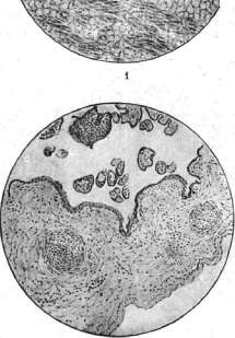
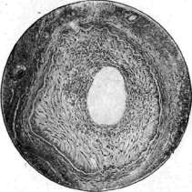
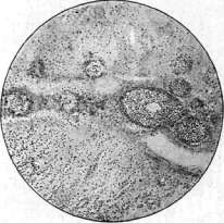
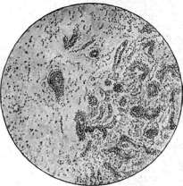
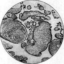
Fig. 1. Fatty infiltration of the myocardium. Figure 2. Endarteritis in syphilis. Figure 3. Obliterating
syphilitic endarteritis of the placenta. Figure 4. Gummatous encephalo-endo- and periarteritis. Fig. 5. Gummatous arteritis, arteritis of the brain. Fig. 6. Villous 'abscesses' of the chorionic villi. During healing, abundant lamellar scaling is usually noted. This rash contains a large number of spirochetes and is observed simultaneously with lesions of internal organs, especially the liver and spleen. Most children with pemphygus syphiliticus are born prematurely and die (without treatment) in the first weeks of life. In addition to the skin lesions described above, papular rash is often encountered in congenital S., usually 4-6 and 8 weeks after birth, more rarely after 3 months of life. This rash is pink in color, most often localized on the face, limbs, and buttocks, also frequently on the body, in the form of small flat nodules rising above the surface of the skin, moderately indurated and characteristically shiny. They tend to scale, which begins in the center, leaving a clean pink rim at the edges; on the face the rash often turns into pustular, while on the buttocks and in skin folds it takes a weeping character, which gives it a resemblance to intertrigo, and sometimes only the presence of other specific changes determines the true nature of the condition. Papular rash leaves behind pigmentation in the form of brownish spots, which later disappear. A characteristic feature of congenital S. is the various stages of development of papular rash in the same child and its polymorphic character. - Spotted rash is encountered in lues congenita in early childhood quite rarely, it appears on the body, face, and limbs. Its color is pink, often with a yellowish tint. Sometimes the rash develops in the first days after birth. Syphilides of the skin are often polymorphic. Skin changes are not always present in children with congenital S. Müller determines the frequency of skin lesions at 84%, Werner at 65%, Still at 69%. According to data from the GNIOMM NKZ, the percentage of children with skin manifestations reaches 68. - Changes in the nails in children with congenital S. occur (according to Müller) in 25% and consist, on the one hand, of disease of the nail itself (tubular nails, nail brittleness, transverse and longitudinal striation, division of the nail by the cuticle into 2 parts), and on the other hand, of lesions of the nail folds and nail bed, leading to the formation of cracks and ulcers - syphilitic paronychia, which can be observed on several fingers simultaneously. - Changes in the hair cover in congenital S. consist of bald patches in areas or general thinning of hair. In addition, in infants as a result of diffuse syphilid on the face, absence of eyebrows and improper positioning of eyelashes are noted. - Recurrent phenomena on the skin are usually observed in poorly treated children and have a large papular and vesicular character. The appearance of rashes in children with congenital S. is often accompanied by an increase in temperature and worsening of the general condition. Spirochetes are found in skin changes frequently (according to V. R. Sklyar in 95%). Disease of the mucous membranes. Of the mucous membranes in congenital S., the nasal mucosa is most frequently affected. Syphilitic rhinitis is one of the early and frequent signs of the disease; it is most often detected in the first weeks of life and often already at birth. According to observations of the GNIOMM NKZ, syphilitic rhinitis occurs in 70% of congenital S., most often developing in the first 4 weeks of life. As autopsies of stillborn syphilitics show, rhinitis begins to develop in the child still in the mother's womb. Syphilitic rhinitis is a chronic disease characterized by inflammation of the nasal mucosa with simultaneous involvement of the nasal turbinates. Its basis is infiltration of the mucosa, leading to narrowing of the nasal passage, and as a result the characteristic snuffling during breathing, which is often audible from the next room. Mucopurulent or serous discharge leads to the formation of crusts blocking the nasal passage, which prevents the child from breastfeeding. Syphilitic rhinitis is characterized by slow progression and is resistant to treatment; it rarely disappears after one course of treatment, more often only after 2-3 courses. In untreated or poorly treated cases, the process spreads to the cartilaginous and bony parts of the nose, their partial destruction, which often gives the nose a characteristic saddle-shaped form. Deformation of the nose can take more severe forms, especially in the presence of perforation of the nasal septum and loss of the entire bony-cartilaginous skeleton. In these cases, atrophic catarrh of the nose is usually observed, often accompanied by a strong odor. Particularly characteristic of congenital S. in infancy are the small size of the nose, depression and widening of the bridge of the nose, and the transition of the bony part into the cartilaginous part in the form of a step. Sometimes syphilitic rhinitis is the first and only sign of congenital S. and is therefore decisive in making the diagnosis. Spirochetes on the nasal mucosa (Sklyar) were found mainly in cases of simultaneous skin involvement by a specific process. In addition to the nasal mucosa in children with congenital S., the laryngeal mucosa also becomes diseased, which manifests as a hoarse voice, aphonia, and in rarer cases, laryngeal stenosis. Syphilitic stenoses in early childhood children can simulate diphtheritic stenosis; the duration of the course speaks for the syphilitic nature of the condition. These stenoses, according to the material of the GNIOMM NKZ, were noted in 1% of cases. - Lesions of the oral and pharyngeal mucosa in congenital S. are encountered relatively rarely; they are observed in the form of erosions and superficial ulcers. These ulcers are occasionally noted on the hard palate and in untreated cases the process from the mucosa spreads to the bone, causing destructive processes ending in perforation of the hard palate. - Disease of the anal mucosa in early childhood children is observed in the form of condylomas and is usually encountered in poorly treated cases. Fissures around the anus most often accompany diffuse process lesions of the buttocks. Disease of internal organs, which is one of the early signs of congenital S., is characterized mainly by a process that leads to diffuse infiltration and proliferation of cellular elements, which is clearly evident when studying the changes in visceral organs of syphilitic fetuses, newborns, and infants with lues congenita. Acquired S., as well as late congenital, on the contrary, tend to give focal formations. In addition to diffuse changes in the organs of syphilitic children, the presence of gummas is noted, which occur more often simultaneously with diffuse infiltration, more rarely - independently. Gummas have been described (Kraus, Aschoff, Ribbert, Tikhomirov and others) in the liver, kidneys, lungs, and spleen. Lesion of the liver in children with congenital S. occurs relatively early and can often be detected already at birth. Spirochetes enter the child's body mainly through the umbilical vein, which explains the frequent disease of the liver. Among the clinical symptoms, enlargement of the liver should be placed in the first place, followed by its density. Hozzinger in 148 children with congenital S. in the first year of life was able to clinically confirm enlargement of the liver 46 times, i.e. in 31%. According to data from the GNIOMM, enlargement of the liver was noted in 80%, and in children under 4 months of age this sign was relatively rarely absent. However, the liver was not always sharply indurated - in 30% an enlarged but not dense liver could be noted, and after a course of treatment its dimensions returned to normal. The surface of the liver is usually smooth. Liver disease, often accompanied by other specific symptoms on the skin and in the skeletal system, does not present diagnostic difficulties. In the absence of skin manifestations, specific rhinitis, as well as enlargement of the spleen and changes in the blood, are almost always present. A dense liver, being one of the signs of visceral S., is resistant to treatment. Along with enlargement of the liver, Zhukovsky described cases of atrophic cirrhosis. These are usually severely ill children, not viable. The spleen in lues congenita is affected as often as the liver, and as Marfan indicates, its enlargement is constant and one of the early symptoms of congenital S. According to Finkelstein it is enlarged in 77%, but it is not always possible to palpate it immediately. Usually enlargement of the spleen is accompanied by induration of it, its lower edge is sharp, 2-3 cm extends from under the costal arch; in more severe cases of visceral S. the spleen reaches the navel and can be palpated in the pelvis. These cases are almost always accompanied by enlargement of the liver and severe changes in the blood. Sometimes during palpation of the lower edge of the spleen, irregularities and roughness can be determined, indicating the presence of inflammatory processes in the capsule of the spleen; Parro and Finkelstein pointed to these phenomena in their time. The histopathological process in the spleen, according to Prof. Nikolaev, has an interstitial character or is reduced to the formation of miliary gummas. Around the follicles and vessel walls, the development of connective tissue is observed, which contains a large number of cellular elements.
Lung disease in infants with congenital syphilis is of little importance to the clinician, as most children with this condition die in utero or shortly after birth. They rarely live to be 3 months old. Children with syphilitic pneumonia appear to be severely ill. The clinical symptoms of the disease—shortness of breath and cyanosis—do not correspond to the findings of percussion and auscultation. Here, as a rule, there is neither dullness nor altered breathing. There are no phenomena indicating inflammation of the bronchi. The observed microscopic proliferation of connective tissue occurs in small areas around the alveoli, leading to their narrowing and reducing air content, without giving any changes either on the side of the lung sound or on the side of respiratory noises. Miller's works, based on material from the Moscow Foundling Home for the period from 1880 to 1890, indicate the rarity of lung damage by a specific process in infants. Out of 14,411 cases of pneumonia in infancy with a fatal outcome, verified at autopsy, only 0.7% showed syphilitic changes in lung tissue. Kidney damage in children with congenital syphilis occurs, according to data from the GNIOMM NKZ, in 30%. Often in children with specific rashes, symptoms of nephrosis-nephritis are noted, which, under the influence of treatment, disappear along with the disappearing rash. Protein and formed elements are usually found in the urine, edema is often observed, and in severe cases, uremia. A characteristic of specifically affected kidneys is a high percentage of protein in the urine, reaching 12-14%, in the absence of formed elements. Usually, under the influence of treatment with salvarsan or mercury preparations, these phenomena quickly resolve, unless the kidney disease is one of the symptoms of general visceral syphilis, which significantly worsens the prognosis. It should also be noted that on the basis of congenital syphilis, underdevelopment of kidney parenchyma is observed. Histopathologically, in kidney damage, interstitial processes and rarely gummas are noted (see Kidneys).-Disease of the cardiovascular apparatus is noted in early childhood in children with lues congenita, and (as Hutinel points out) the basis of heart damage lies: 1) pathological processes in the heart muscle, both interstitial and parenchymatous, 2) changes in the endocardium, leading to valve damage, and 3) disease of the pericardial sac. Vascular damage, which plays a major role in the late stages of syphilis, is found in infants more often than was previously thought. Autopsies and histopathological studies made on material from GNIOMM (Tikhomirov) show vascular damage in 94%, with changes in small arteries in 58%, expressed in obliterative proliferation of the endothelium of the intima. In addition, in 70%, inflammatory-infiltrative changes were found in the walls of veins, especially in the liver and adrenal glands. Disease of the pancreas on the basis of congenital syphilis gives enlargement of the organ, which is based on the proliferation of connective tissue rich in cells in the lobules themselves, around them, and around the ducts. The thyroid gland gives the same picture. Of other endocrine glands, the adrenal glands are most often affected; according to Levaditi, spirochetes are very often found here. In children with congenital syphilis, shortly after birth (in 20%, according to GNIOMM data), testicular disease is noted. This phenomenon has been described by both German (Genoch, Hochsinger) and French (Marfan and Vallery-Radot) authors. Both Hochsinger and Marfan point to the frequent connection between hydrocele of the testicle and congenital syphilis, even in cases where there is no disease of the testicular parenchyma. The testicle is usually enlarged and its density is striking. Anatomically, the process amounts to round-cell infiltration, proliferation of perivascular tissue, which initially leads to enlargement, and subsequently to shrinkage of the organ.-Enlargement and density of peripheral lymph glands are noted in children with lues congenita relatively often, in about 70% (GNIOMM data). However, literary data on this issue are quite contradictory. While syphilidologists believe that polyadenitis, which is a characteristic symptom in adults, can often be absent in children with congenital syphilis, pediatricians, such as Genoch and Doyen, note the relative frequency of this sign in children with lues congenita. The elbow glands are enlarged, according to Hochsinger's data, in 90%, according to GNIOMM data, in 50%. Damage to the bone system - see Bone, clinical diseases of bone, Osteochondritis, Periostitis. The central nervous system suffers in infants with congenital syphilis, as Hochsinger indicates, significantly more often than is generally thought. Almost always, children with fresh manifestations of syphilis give symptoms of irritation from the meninges. The rigidity of the neck and rapidly passing convulsions often observed in this period disappear under the influence of treatment along with the skin manifestations. On the other hand, cases are not uncommon where in newborns and infants, in the absence of clinical symptoms from the nervous system, severe inflammatory changes with softening as in the meninges and in the brain tissue were found at autopsy, and in some cases spirochetes were found. One of the early symptoms of irritation of the nervous system is the crying of the child, both day and night, which responds well to specific therapy. Relatively early, sometimes in the first weeks of life, convulsions are noted—rapidly passing, sometimes resembling petit mal. The often occurring dilation of veins at this time indicates an increase in intracranial pressure. Spinal puncture gives fluid under high pressure. These convulsions in early childhood are harbingers of more severe damage to the nervous system in older children. The cardinal symptom in lesions of the central nervous system in infants on the basis of congenital syphilis is hydrocephalus or dropsy of the brain, which can occur acutely and chronically, be congenital or develop some time after birth (see Hydrocephalus). Hydrocephalus is often accompanied by other specific lesions, among which rhinitis and a sunken nose are particularly often present. Among other diseases of the central nervous system, syphilitic meningitis can be noted, developing, like tuberculous, on the basis of the brain. The diagnosis is determined by examination of the spinal fluid. In addition to meningitis, meningo-encephalitis are also observed, giving hemiparesis and hemiplegia, which do not always yield to specific therapy. Of the sense organs in infants, the eye and fundus oculi are most often affected. Relatively early, often already at birth, the retina and choroid, as well as the optic nerve, are changed. These lesions were first described in detail by Hirschberg in 1895. Syphilitic chorioretinitis is characterized by the presence of small yellow spots on the fundus with pigment foci in the form of salt and pepper or 'snuff'. These changes are localized in the area of the equator and at the periphery of the fundus and are especially typical for congenital syphilis of early childhood. Vision is not affected. Disease of the optic nerve gives blurred discs and unclear outlines, and these phenomena can be observed simultaneously with disease of the retina. Dangerous in terms of loss of vision is atrophy of the optic nerve, which can be observed even in children under 3 months of life. However, it is more common after a year and in older children. Inflammation of the iris is less commonly observed in infants. In addition, in early childhood, parenchymatous keratitis is noted, albeit rarely (according to TNIOMM NKZ data, in 1%) (see Keratitis). The pathomorphological picture of the blood in children with congenital syphilis relatively often gives phenomena of secondary anemia, the severity of which is closely related to the damage of visceral organs and the bone system (see Anemia, anemias in children). Physicochemical properties of the blood also give deviations from the norm. Blood enzymes catalase and protease (Walter) give low figures, especially in children with visceral syphilis. The erythrocyte sedimentation reaction in children with congenital syphilis almost as a rule gives acceleration (Weiss), which is directly dependent on the severity of the process, the sharpness of the manifestations, and the variety of affected organs. The Wassermann reaction in children with congenital syphilis gives a positive result in a higher percentage of cases shortly after birth. Cases with skin manifestations almost always proceed with a positive RW. Diseases of the bone system sometimes give a negative RW in the presence of a specific process (in 6%) (see Wassermann reaction). Late congenital syphilis. Late congenital syphilis (syphilis hereditaria tarda, lues congenita tarda) was particularly studied and described by A. Fournier. Many authors, including Zappert, consider late congenital syphilis as a relapse of early syphilis, the manifestations of which were overlooked. Others, such as A. and E. Fournier, believe that congenital infection can remain in a hidden clinical state for a long time and manifest, albeit rarely, not only in adulthood but also in old age.
According to existing observations, the earliest age at which manifestations of late congenital syphilis have been noted is considered to be the 4th year of life, according to data from the GNIOMM, 21/2 years. However, late congenital S. is most commonly encountered between the ages of 8 and 14 years, although there are many observations where the first signs of fresh late S. appeared in older children. Patho-anatomically, late congenital S. is characterized by chronic inflammatory, gummatous, and degenerative lesions of organs and systems with changes in blood vessels. These lesions resemble the changes observed in the tertiary period of acquired S. and are sometimes difficult to distinguish from them. Late congenital S., like early congenital S., can affect all organs and systems of the child's body, and the manifestations can be divided into 2 groups. The first group includes phenomena that arose in early childhood, the so-called stigmata, which serve as an important aid in recognizing late congenital S. (e.g., scars at the corners of the mouth, deformation of the nose). The second group includes signs that appeared at a later age. The skin is affected specifically in late congenital S. less frequently than in infancy, and the changes are more persistent and more severe. Characteristic of this period are: 1) gummas, which more often occur on the extremities, generally having a benign course and sometimes giving a great resemblance to scrofuloderma; the diagnosis is then decided by RW or the beneficial effect of specific treatment; 2) tubercular syphilides of the ulcerative type, tending to serpiginization, leaving characteristic scars with scalloped edges, white in the center and pigmented at the periphery, not uniformly deep everywhere. The nasal mucosa is not often affected in the form of atrophic catarrh and sometimes is accompanied by perforation of the nasal septum with deformation of the nose, more pronounced than in early childhood. Gummatous processes with disintegration are found on the soft palate, on the tonsils and around them, and often cause perforation of the soft palate as well as the hard palate. Gummatous infiltrates with a tendency to disintegration are also observed on the tongue. The lymph glands in late congenital S. are often changed, especially in the cervical and submandibular areas; inguinal and axillary lymphadenitis are observed less frequently. These lesions often simulate tuberculous lymphadenitis. As Meshchersky points out, specific lymphadenitis can occur without skin lesions, giving slowly developing tumors of the cellular tissue; they can resemble scrofuloderma with subsequent softening, formation of fistulas, periadenitis (sometimes this form represents a hybrid syphilitic-tuberculous lesion of the glands); they can be accompanied by extensive ulcers and represent gummatous glands, softened and suppurating. This form often occurs with splenomegaly and anemia. In late congenital S., bones (osteomyelitis and osteoperiostitis) are particularly often affected. In addition, osteosclerosis is noted on the long tubular bones. The tibia is affected more often than others, which takes a saber-like shape, pathognomonic for late congenital S. The same shape is observed on the bones of the forearm. Gummatous processes with the formation of fistulas are also noted on the bones of the skull. As in early congenital S., multiple bone lesions are encountered here, and simultaneously with the phenomena of neoplasms (periostitis and osteosclerosis), phenomena of destruction (gummas with the formation of sequestra) are also noted on X-rays. Some authors have described syphilitic Pott's disease, simulating tuberculosis, as well as Paget's disease, in connection with late congenital S. (see Deforming osteitis). Of the bone dystrophies, dystrophies of the skull should be noted: the Olympian forehead, a skull with prominent frontal and parietal tubercles, and a pear-shaped skull. In connection with late congenital S., the congenital absence of the xiphoid process of the sternum has also been described (Landa, Institute named after Glavche). Joints. A characteristic form of late congenital S. is the painless accumulation of fluid in both knee joints. The joints swell slowly, and movements are not impaired. The diagnosis is made on the basis of RW and other specific lesions. In addition, massive swellings of the joints, especially the knees, resembling tumor albus and accompanied by bone thickening, are also observed; on X-rays, phenomena of osteosclerosis and osteoporosis are sometimes noted. S. is indicated by slight pain and minimal impairment of joint function. Of the joint dystrophies, congenital dislocation of the hip has been described (Meshchersky). Distrophies of the teeth are particularly frequent and varied in late congenital S., especially characteristic are Hutchinson's teeth (see Hutchinson's triad). Of other dental dystrophies, atrophy of permanent molars should be noted, as well as cup-shaped atrophy of the first large molar. In addition, teeth in late congenital S. change their shape, have an atypical appearance, differ in small size, and sometimes in excessive size; the absence of one or both upper incisors, as well as anomalies in the arrangement of teeth, has also been described. The eye is affected more often than any other organ in late congenital S., and parenchymatous keratitis takes first place, which is pathognomonic for this disease and is included by Hutchinson in his triad (see Hutchinson's triad). Of other eye diseases based on congenital S., iritis, iridocyclitis, choroidoretinitis, atrophy of the optic nerve in connection with S. of the central nervous system, strabismus, congenital nystagmus, and dacryocystitis (Oditsov), arising on the basis of specific bone lesions, can be noted. The labyrinth is affected in late congenital S. less frequently than the eye, but disease of the labyrinth and the auditory nerve is as typical as parenchymatous keratitis (see Hutchinson's triad). Hutchinson's triad is observed comparatively rarely. Most often, as Meshchersky points out, deafness is absent from the triad. Deafness gives a poor prognosis, as it hardly responds to specific therapy. Late syphilitic lesions of the larynx are not uncommon and give rise to small tumors, especially on the epiglottis; sometimes perichondrites with destruction of individual parts of the larynx and edema of the vocal cords are noted. Clinically, hoarseness and difficult breathing are observed. Disease of the lungs is rare and difficult to diagnose. Pulmonary and pleural fibrosis based on S. in children has been described by Hutinel. Cardiac diseases in children with late congenital S. have been described in the form of valve insufficiency, myocarditis, and endocarditis. There are also indications that congenital narrowing of the aorta, stenosis valvulae mitralis, and stenosis a. pulmonalis are characteristic of late congenital S. Lesions of the liver and spleen are observed in late congenital S. as a syndrome associated with Banti's disease (see Banti disease). Specific cirrhosis of the liver also occurs, and in severe cases, bumpy swellings are observed, indicating the presence of limited hepatitis and gumma. Disease of the kidneys in late congenital S. is rare and gives no characteristic signs. Hutinel points to the connection that exists between aplasia of the kidneys and late congenital S. Sometimes in such children, orthostatic albuminuria is noted. The paroxysmal hemoglobinuria observed in this disease responds well to specific therapy (Meshchersky, Rar). The blood gives phenomena of secondary anemia in late congenital S. Polyglobulia, which improves under the influence of salvarsan treatment, has also been described (Shulman) as a symptom of late congenital S. Disease of the testicle and ovaries is rare. In some connection with the disease of the sex glands in late congenital S., infantilism, more often in girls, which is characterized by delayed menstruation and slow sexual development, is observed. Late congenital S. comparatively often gives disease of the nervous system, which sometimes remains the only sign of the disease and is characterized by the persistence of symptoms and the duration of the course. As harbingers of severe brain diseases, Fournier points to headaches, as well as general convulsions observed at the beginning of the disease. Jacksonian epilepsy, characterized by convulsive seizures of one limb, less often of half the face, indicates local cortical disease (Fischl, Erlenmeyer), based on which is a gummatous process or limited meningitis. In children with late congenital S., mental retardation and mild forms of dementia are also noted. According to data from E. Müller, among children with congenita tarda, 1/3 have normal mentality, 1/3 have weakened mentality, and 1/3 show a significant decrease in intelligence. Disease of the brain and spinal cord in late congenital S. gives phenomena of hemiplegia (Marie), as well as often spastic paralysis or Little's disease (Babonneix, Tenissier) (see Children's paralyses). Tabes dorsalis in children with late congenital S. gives mainly symptoms from the eyes, atrophy of the optic nerve, Argyll-Robertson symptom with mild disorders from the motor and sensory spheres.
Romberg's symptom is usually absent in children. Urinary incontinence is often noted. Progressive paralysis in children can be observed as early as five years of age (Zappert) and is characterized by dementia (disorders of memory, speech, delusional ideas), symptoms from the eyes (immobility of the pupil, atrophy of the optic nerve), later paralysis and seizures, which are often the cause of death. Diseases of the endocrine glands in children, as a number of authors indicate, in some cases also have late congenital syphilis as their basis. However, it should not be forgotten that a number of dystrophies affecting individual organs and systems, noted in connection with congenital syphilis, may occur: 1) on the basis of other chronic infections, such as tuberculosis, alcoholism, and lead poisoning, 2) they can be observed in families where there are no indications of syphilis and the above-mentioned infections. However, according to statistics, the largest percentage of dystrophies occurs in children with congenital syphilis. Fournier divides dystrophies into 6 categories: dystrophies involving: 1) the skull, 2) teeth, 3) eyes, 4) the nervous system, 5) endocrine glands, and 6) rachitic-type dystrophies. These dystrophies must be taken into account along with other phenomena. Diagnosis of congenital syphilis. Congenital syphilis does not always give rise to obvious specific changes in organs and systems, and therefore its recognition can present great difficulties even for an experienced physician. Meanwhile, early diagnosis is of great importance not only for the child but also for the mother, who often does not suspect the existence of this disease in herself. Obvious symptoms of early congenital syphilis include rashes, specific rhinitis, a large dense liver and spleen (Parrot's disease). Auxiliary data for diagnosis include a suspicious obstetric history (late miscarriages, premature births, stillbirths), data from the birth period (large placenta, hydramnios), as well as signs concerning the child itself: a) lack of appetite, b) slow increase in weight or its stoppage during breastfeeding and in the absence of disorders from the gastrointestinal tract, c) presence of cubital glands, d) presence of a dense enlarged testicle, sometimes simultaneously with hydrocele of the testicle, unilateral, long-lasting, e) a number of phenomena from the nervous system, mentioned above. The combination of several of the listed signs allows for diagnosis and to begin treatment. For the diagnosis of late congenital syphilis, among other signs, the stigmas remaining after congenital syphilis in early childhood are also of great importance: 1) scars at the corners of the mouth, 2) deformation of the nose, 3) eye phenomena (parenchymatous keratitis), 4) saber-shaped shin, 5) dystrophies of the teeth and skull, 6) Hutchinson's triad, 7) serpiginous and deep skin ulcers, etc. In the diagnosis of congenital syphilis, the following are also of great importance: 1) laboratory examinations - RW, blood sedimentation reactions, morphological blood picture (anemia) and its physicochemical properties, as well as examination of cerebrospinal fluid; 2) examination of the bone system by X-rays (see above); 3) examination of the eyes and fundus of the eye. The prognosis of congenital syphilis, both early and late, depends on a number of conditions. The following significantly worsen the prognosis: 1) recent syphilis in parents, 2) their poor treatment before conception of the child and insufficient treatment of the mother during pregnancy, 3) early appearance of signs of syphilis in the child, as well as their intensity (especially visceral and nervous syphilis). Breastfeeding and hygienic living conditions significantly improve the prognosis. Early and vigorous treatment of the syphilitic child, both with and without manifestations, is of great importance for the prognosis. Prevention. The decrease in congenital syphilis in the USSR in recent years is closely related to the decrease in syphilis among adults, however, the percentage reduction in contagious forms of congenital syphilis, according to Volpian's data (Moscow), lags significantly behind that of adult syphilis. This is explained by the circumstance that poorly treated latent syphilitics often give birth to sick children with obvious signs of syphilis. Thus, prevention of congenital syphilis should be reduced to: 1. Vigorous systematic treatment of future parents before conception of the child. Women with syphilis should be treated even in the absence of clinical symptoms. 2. Early detection of syphilis in pregnant women and their early and vigorous treatment (with salvarsan + Bi preparations) (2-3 courses), which guarantees the birth of a healthy child. Work on detecting syphilis in pregnant women is carried out in women's consultation clinics. 3. Early and vigorous treatment of children not only with manifestations but also without visible signs, born from poorly treated syphilitics, especially if the mother did not receive courses during pregnancy. Only early and vigorous treatment (with salvarsan + Bi + Hg preparations) protects children from manifestations of late congenital syphilis, protects from infection and the collective (nurseries, children's homes) where the child of syphilitic parents goes. 4. Finally, it is necessary to strictly monitor the implementation of the decree on compulsory treatment. Treatment. In the treatment of both infants and older children, neosalvarsan (neoarsenol), bichinol, mercury, and iodine are used, with the best results being obtained from combined courses. Each course consists of 10-12 infusions of neosalvarsan and 12-15-20 injections of bichinol or mercury. Neosalvarsan is administered to infants at a rate of 0.01-0.03 per 1 kg of the child's weight, with the dosage gradually increasing. For children over 3 years of age, infusions begin with 0.1 and are increased to 0.3; after 12 years, a single dose can be increased to 0.4 if neosalvarsan is well tolerated. Infusions are made into the veins of the head, cubital, and jugular veins. This method of administration is the best for children due to its painlessness, but it can sometimes be difficult in mass treatment. Then one has to resort, especially in infants, to intramuscular and suprafascial administration of neosalvarsan. The intervals between infusions are 4 days. Bichinol is administered into the buttock muscles at a rate of 0.01, mercury at 0.001 per 1 kg of the child's weight. For children over 3 years of age, injections of bichinol are given at 1.0, 15-20 times. During treatment, it is necessary to strictly monitor the urine, examining it no less than once every ten days, since bismuth and mercury preparations often give complications from the kidneys, more often in the form of albuminuria. Bismuth, like mercury, is administered to children every other day. Mercury is administered intramuscularly in the form of an injection of a 1% solution of sublimate or 1% Hydrarg. cyanati, since oral administration and rubbing are methods of low intensity. Complications observed under the influence of treatment are reduced to the appearance of protein in the urine, a slight increase in temperature, and disorders of the gastrointestinal tract. All these complications are usually eliminated within a few days and little interfere with treatment. Contraindications in children for infusions of neosalvarsan are acute infectious diseases, frequent stools with admixtures of blood and mucus; in kidney disease, it is recommended to use mercury preparations or small doses of neosalvarsan. The above-mentioned combined courses (10-12 neosalvarsan + 12-15-20 Bi or Hg), a child with congenital syphilis receives from 4 to 6 depending on the intensity of the manifestations and the course of the process. In the intervals between courses, children over 3 years of age may receive iodine preparations. Neosalvarsan can sometimes be replaced by osarsol, giving it in combination with bichinol or mercury. Under the influence of the above-mentioned treatment, skin manifestations disappear relatively quickly; where there are no changes in internal organs, the general condition quickly improves, weight increases, the color of the skin becomes less pale; hoarseness, loss of voice and stenosis phenomena also decrease after 2-3 infusions of neosalvarsan. Syphilitic rhinitis is long and stubbornly resistant to treatment, but here too at the end of the course the discharge decreases, and snoring breathing disappears, sometimes only after 2 courses of treatment. As slowly as rhinitis, the size of the liver and spleen decrease in visceral syphilis, and such children develop poorly and their weight increases only at the end of the first course or after its completion. Lesions of the bone system, severe gummatous processes, erosion of joint ends respond well to treatment. Diseases of the nervous system also show improvement by the end of the first course, provided there are no deep destructions in brain tissue. The blood composition and its physical properties, which give severe changes in congenital syphilis, improve under the influence of treatment in most cases.
m. Raii.
Related articles
Cite this page
“Syphilis.” Soviet Medical Encyclopedia. English translation of Bolshaya Meditsinskaya Entsiklopediya, 1st ed. (Moscow, 1928–1936), ed. N. A. Semashko. https://sovietmedicalencyclopedia.pages.dev/article/syphilis/