Syphacia Obvelata
Historical document, translated for reference. It reflects medical knowledge of the 1920s–30s and is not medical advice.
Summary
Syphacia obvelata is a nematode parasite commonly found in rodents, with a single reported human case from the Philippines. The article describes its morphology, biology, and discusses the controversy regarding its potential parasitism in humans.
Encyclopedia article (1928–1936)
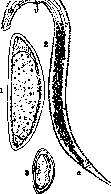
SYPHACIA OBVELATA (Rud, 1802). A nematode of the subfamily Syphaciinae (order Oxyurata, family Oxyuridae), a frequent parasite of rodents. Cosmopolitan. In humans, it was discovered once in the intestines of a child from the Philippine Islands (2 females). The biology is similar to Enterobius vermicularis of humans (see Pinworms). Skryabin and Schultz (1931) express doubt about the parasitism of S. o. in humans, allowing for the possibility that the material delivered to Dr. Riley was contaminated with mouse feces. Description of the species: male 1.3 mm in length. The esophagus, including the bulb, is 4/7 the length of the body. The excretory opening is 0.09 mm posterior to the bulb. The length of the tail end is 0.13 mm; there are three medioventral ridges; the spicule is 0.035 mm in length. Female 3.5-6 mm in length with a maximum width of 0.275-0.340 mm. The elongated tail constitutes 1/6-1/7 of the body length. Two lateral wings extend in a narrow strip along almost the entire length of the body. The vulva is located in the anterior part of the body, usually on a cuticular elevation. Eggs 0.115-1.140 mm in length, 0.035 mm in width, flattened on one side and arranged in pairs, touching with their flat bases; in this case, the ends of both eggs do not coincide. Localization: small and large intestines, more often the cecum. Despite its wide distribution among rodents, S. o. in humans has never been recorded in the USSR.
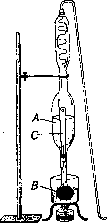
SYPHILIMETRY (la syphilimetrie, a name given by Professor Chantemesse), in the narrow sense means serological measurement of the degree of syphilitic infection, but in essence it represents a fully developed theory and practice of syphilidology, created and led by the French syphilidologist and serologist Arthur Vernes (A. Vernes). The starting point of Vernes' entire concept was the serological study of syphilis. As early as 1913, Vernes came to the following conclusions: 1) in the humoral environment of the body, one must seek the slightest reflections of active syphilis; 2) the RW (Wassermann reaction) is technically complex and often gives errors; 3) the correct approach to the patient requires not only detecting a positive or negative reaction, but above all, the dynamics of the course of syphilis; 4) to determine the activity of syphilis and the effect of treatment, it is not so much an isolated positive and negative reaction result that matters, but the possibility, when many sequentially performed reactions on the same patient, to be able to compare them, in other words, to be able to trace their dynamics, evolution; 5) this requirement can be implemented only by the colorimetric method of evaluating reaction results, proposed by Vernes, and by the graphical curves obtained through the application of this method; 6) the seroreaction curve (then the Wassermann reaction) should become a factor guiding the treatment of the patient; 7) 'over this or that medicine, over the present technical difficulties, there is therefore a guiding idea, on which, at least for the present, the future of syphilis treatment depends' (Vernes). These principles of Vernes' initial work served as the basis for his subsequent research, crowned with a series of most valuable discoveries in the field of serodiagnosis, as well as in the field of generally diagnostic and therapeutic methodology of syphilis. Vernes' work lead the author to the conclusion that 'all serological reactions of syphilis are merely a question of precipitate formation' and that 'certain physical conditions reveal special changes in the serum of a syphilitic patient, which without possible error distinguish the serum of a syphilitic patient from normal and which can be traced and measured during the course of syphilitic infection'. The essence of the matter is that syphilis enhances the ability of normal serum, when mixed with colloidal suspensions, to form flakes, to flocculate. The question therefore concerns the degree of enhancement of a certain physical property of normal serum caused by syphilitic disease. The RW also has as its mechanism the phenomenon of flocculation, which is in no way dependent on the erroneous Ehrlichian theory - the so-called fixation of alexins or deviation of complement. Vernes' experiments led him to the conviction that in the RW, the 'antigen' is nothing other than an empirically chosen suspension of a colloidal substance, devoid of any 'specificity' and with a far from regulated, not clarified physical state, which is the cause of many erroneous reaction results. Vernes explains the errors of RW results as being due to nothing other than biological factors in determining its results, the unnecessary inclusion in the reaction system of substances that do not possess the constancy of properties necessary for the reaction, such as: sera and red blood cells of animals (guinea pig, sheep, etc.), and thus representing a constant source of uncertainty in the hemolytic evaluation of the reaction. Discarding the biological elements of determining the reaction, Vernes transfers the operation from 'the area of biology to the area of physics,' ensuring the possibility of creating uniform reaction conditions. Thus, as we noted above, Vernes takes as the basis for differentiating normal serum from syphilitic serum the difference in their stability with respect to the same factor - reagent (colloidal suspension). Vernes' method consists precisely in finding the conditions for determining that zone in which syphilitic serum gives hyperflocculation, while normal serum does not. To achieve this goal, the following points become of great importance: a) the correct choice of the so-called antigen; b) the possibility of preparing from it a colloidal suspension of constant stability; c) the development of precise methods for maintaining uniform reaction technique; d) the creation of conditions by refining the technique to possibly protect the reaction from the individual qualities of the operator - the 'authoritative serologist,' who proclaims the position that 'every serum appears to the serologist as a patient does to the clinician'. Vernes uses for his reaction as antigen an extract of horse heart muscle, the so-called perethynol (a term derived from the first syllables of the names of the ingredients used to prepare this reagent: per(chlorure) ethy(lene) + (alcoh)ol = perethynol). Technique for preparing perethynol. Fresh horse hearts are thoroughly cleaned of tendons and fat and thoroughly minced in a meat grinder, placed in 95% alcohol, stirred and kept in it for 1 hour; then the muscle pulp is slightly squeezed through gauze and again placed for 10 min. in 95% alcohol (ethyl). After this, it is thoroughly squeezed and dried on glass plates at 37° until completely dry and ground in a coffee mill into powder. 200 g of this powder are taken, and, mixing with 300 g of quartz sand thoroughly washed sequentially in water and 95% alcohol, are poured into the thimble of a Soxhlet apparatus (Fig. 1); into the flask of the apparatus, 750 g of pure tetrachloroacetylene-C2H2Cl4 (tetrachloroethane, or ethylene perchloride) is poured. The apparatus is hermetically ground and connected to a vacuum pump, manometer, and a pressure-regulating flask. The flask of the Soxhlet apparatus is immersed in a water bath, in which the temperature is maintained at 63-68°. Then the air is pumped out of the entire system by the pump until the C2H2Cl4 begins to boil, the vapors of which rise through tube C, and, cooling, drip onto the mixture of muscle with sand A and then flow back into the flask. Such extraction continues for 12 hours straight. After the extraction is complete, the powder is dried at 37° to dryness and again subjected to extraction with absolute alcohol, also under reduced pressure and at a temperature of 50-56° for 12 hours. The alcoholic extract is the antigen - perethynol. It is subjected to filtration, then standing for 20 days; then follows filtration and again standing for 20 days, then checking it against the standard and pouring into bottles. Perethynol is an alcoholic solution of 15 g of dry extract of horse heart in 1,000 g of absolute alcohol. In France, the preparation of perethynol is centralized in the laboratory of the Preventive Institute in Paris. Perethynol is also manufactured in America; in the USSR - in the Leningrad Control Serological Laboratory and in Tiflis in the biochemical laboratory of the railway. The required constancy of the physical properties of perethynol is achieved with the help of a special apparatus - a mixer (melangeur) (Fig. 2), which allows: a) maximum precision in the amount of perethynol (3 cm3); b) precise control of the flow rate of perethynol into bidistilled water (1 cm3 per minute); c) maintaining the same number of rotations of the mixer rod, intended for mixing perethynol with bidistilled water (200 rotations per minute, determined by a tachometer), etc. To obtain the suspension, 3 cm3 of perethynol and 16.5 cm3 of bidistilled water are taken, i.e., 1:5.5 or perethynol
Figure 1. Soxhlet apparatus.
Figure 2. Mixer apparatus.
__ 1 __ 3 total quantity of suspension-- 6.5 19.5' The prepared colloidal suspension is used for the reaction only within two hours after preparation. Vern reaction technique. Production of the reaction: 1) Distribution of serum into test tubes (13 x 60) with labels indicating the patients' surnames. For each reaction, six test tubes are taken, arranged in four rows of special racks, in the first two rows one each, in the others two each; the test tubes of the first row are centrifuged for 10-15 minutes, after which they are
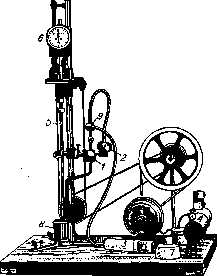
Figure 2. Mixing apparatus: 1-capillary graduated pipette for peretinol with a capacity of 3 drops; 2-flow rate regulator for peretinol; 3-syringe for drawing peretinol into the pipette; 4-rheostat; 5-glass rotating rod for mixing liquids; 6-tachometer determining the rotation speed of this rod; 7-rheostat plug; 8-cylindrical glass with a flat bottom, in which the colloidal suspension is prepared; 9-faucet regulating the flow of liquid.
carefully poured into empty test tubes of the 2nd row, avoiding mixing the precipitate with the serum. The test tubes are stoppered with ordinary stoppers that do not touch the surface of the contents. 2) The test tubes with serum are placed for inactivation in a special water bath at a temperature of 55° for 30 minutes. 3) During this time, the peretinol suspension is prepared. 4) After 10 minutes have passed since the serum was removed from the bath, 0.8 cm3* of it is distributed into four test tubes of the 3rd and 4th rows by a regulated automatic Vern rheometer-syringe (fig. 3). In each row, one test tube is for the reaction, the other for control (two rows for mutual verification of reaction results). After 30 minutes from the moment the serum was removed from the bath, 0.4 cm3 of the peretinol suspension is added to the serum tubes for reactions of both rows by the rheometer, and 0.4 cm3 of an alcoholic solution is added to the control sera of these same rows. The test tubes are gently shaken for better mixing. 5) Then these test tubes with the mixtures, stoppered with rubber stoppers, are again placed in a bath at a constant temperature of 25° for four hours. During this time, flocculation occurs. 6) After this time, the sera are removed from the bath and the results of the reaction are read visually and with the Vern, Brik, and Ivon photometer. If the reaction is positive, the reaction serum already shows significant cloudiness compared to the control visually. In cases of normal sera, the contents of both test tubes remain equally transparent. But the main thing in evaluating the reaction results lies in the research of the photo
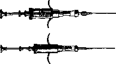
Figure 3. Vern rheometer.
meter, by striking the prism (T2), are directed into the telescope (V) in the form of two lateral semicircular bands, separated in the middle by an unilluminated band corresponding to the opaque strip (W) of the prism (C). These two beams of light rays, passing through the serum being examined (X), determine variations in the intensity of its cloudiness, giving a corresponding intensity of light display on the objective (O5). The task of the second beam of light rays, obtained simultaneously from the same light source (J), is precisely to measure the degree of the above-mentioned optical variations of the serum being examined. This beam (black line), passing by the prism P1, is deflected by another counter prism (P2) to cross specially prepared smoked glass or gelatin (N). Then by means of the prisms (C and T2), occupying the space between the two lateral light bands of the photometer (fig. 4), an apparatus that determines the optical density of the liquid being examined, depending on the degree of flocculation. 1. Internal construction of the photometer (fig. 5). An electric lamp (J) passes into the apparatus a bundle of parallel rays. Half of them, due to the pentagonal prism (P1) in their path, are deflected at a right angle and cross the frame (X), designed to receive the liquid being examined in a special cuvette. After passing through the serum, it strikes the prism (P1), is again deflected at a right angle, in order to cross two prisms (C) joined along their hypotenuse surfaces. Here the middle band of this beam of rays is blocked by an opaque strip (W). The two lateral beams of rays, after
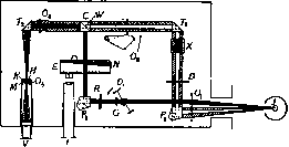
Figure 5. Photometer (internal construction).
passing through the serum, are directed into the telescope (V), reflected side by side on the red filter diaphragm at its end. At this, it must be emphasized that the optical density (logarithm of the ratio between the amount of received and transmitted light) throughout the entire wedge-shaped smoked glass (N) is precisely determined and expressed in numbers on a special scale (E) and can be read through a magnifying glass (L). By moving the aforementioned glass (N) with a micrometer screw toward either a smaller or larger diameter, complete uniformity in the intensity of coloring is achieved for both the middle [rays crossing the glass (N)] and lateral (rays passing through the serum) bands of the diaphragm visible in the telescope (V) of the photometer (fig. 6). After establishing equal coloring, the operator looks into the magnifying glass (L) of the photometer and notes the number on the scale (E), determining the degree of optical density of the glass at this diameter and thereby the optical density
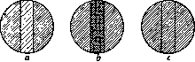
Figure 6. Light beams in the telescope of the photometer.
density of the serum. Photometric determination of the reaction results includes determining the optical density, on the one hand, of the serum with the colloidal suspension of peretinol, and on the other hand, of the serum in mixture with an alcoholic solution. Normal serum in both cases gives the same figures. With syphilitic serum, however, high figures (say '150') are obtained in the first case, and low figures (approximately '90') in the mixture with alcohol. The difference between these figures (150-90 = 60) is the so-called syphilitic index of the given serum, the most important constituent element of the theory of syphilitry. Using the photometer is extremely simple. In recent times, in addition to the widespread photometer with electric lighting for provincial laboratories, a photometer with attachments for kerosene lighting has also been constructed. 2. Photometric scale and the idea of measuring flocculation. Three methods of determining the degree of flocculation and generally the results of seroreaction were successively replaced in Vern's research: first the colorimetric, then the gravimetric, and finally the optical methods. The colorimetric method (recommended together with Jansel before 1913 and applied in many laboratories) is based on the principle that a number of substances, in particular pig serum, possesses in mixture with colloidal suspensions a dissociating, anti-flocculating ability. These substances also exhibit a hemolytic action on sheep red blood cells. At the same time, an extremely curious phenomenon was noted in these two properties of the serum—anti-flocculating and hemolytic—namely, when one of these properties (anti-flocculating) is used up, the serum loses the other (hemolytic) as well. From what has been said, it follows that when pig serum has completely used up its anti-flocculating strength, there is also a complete absence of hemolysis (syphilitic serum); in the case where the entire reserve of this strength is preserved, on the contrary, there is clear hemolysis (healthy serum). In the colorimetric method, therefore, the hemolysis of sheep red blood cells is only an indirect factor in determining flocculation. Vern introduced into practice a colorimetric scale for determining the degree of hemolysis with nine divisions (from 0 to 8) corresponding to the degrees of serum formation depending on the intensity of the hemolysis itself. Here '0' indicates syphilitic serum, and '8' indicates normal serum. The optical, or photometric, scale currently accepted consists of more than 150 divisions (from 0 to 150 and above). 'Increasing, in a certain way, like a microscope, the visibility of small objects, the photometer allows expanding the field of observation of flocculation'. Unlike the vague terminology of the Wassermann reaction and other reactions, which has no comparative measuring value and is limited mainly to the mere statement of the presence or absence of the disease, the Vern scale makes it possible to measure the degree of intensity of the flooding of the body with disease-causing agents. The extensive photometric scale makes it possible to detect the actual and inevitably existing difference in the intensity of impregnation with the infectious agent in different individuals. For example, as shown in Fig. 7, 188 RW, evaluated with the same designation (+ + + +), gave figures from '4' to '150' and more divisions on the photometric scale, which reflects the individualization of the serological picture of the patient, while also making it possible to detect fluctuations in the intensity of infection in the same patient from one examination to another. The photometer, assisting the vision and sensations of the physician, on the one hand, makes it possible to detect weak degrees of reaction, and on the other, to objectively distribute the serums according to the intensity of the flocculate formed, thereby eliminating the usual difficulties in differentiating by eye the various gradations of changes in the serum. Bern and other authors give the following interpretation to the numerical indicators of the scale, i.e., the syphilitic indices: '0'—completely normal serum; '1-2'—negligible suspicion; '3-4'—out of 100 serums, 25 cases of syphilis and 75 normal; '5-6'—approximately 50% syphilitic and 50% normal; '7; 8; 9; 10; 11'—out of 2,000 serums, 1,999 cases of syphilis and 1 normal; 'from 12 to 18'—out of 10,000, 9,999 syphilitic and 1 healthy; 'from 19 to 27'—out of 650,000 serums, only one case with such an indicator occurred in a healthy person, the other 649,999 in known syphilitics. Thus, the figures from 1 to 6 are suspicious, from 7 to 27 (with rarest exceptions)—positive. The occurrence of an indicator above '27' was never observed in a healthy person. Shaverdov's observations, covering about 10,000 cases over six years, do not contradict this interpretation of the indicators. 3. The significance of S. for the therapy and diagnosis of syphilis. 'To measure syphilitic infection,' wrote Bern, 'means to be master of syphilis'. The photometric scale, having 150 divisions, serves to realize this idea—sero-measurement of infection. At the same time, it should be noted that high and low readings of the scale by no means are synonyms of malignancy or benignity of the disease. They indicate only the intensity of saturation of the body's humoral medium with products of the life activity of the pathogenic agent. In the outcome and character of the disease, besides this factor, the localization of the process, the virulence of the spirochetes, the general condition of the patient's body and other factors play a large role, the detection of which is, of course, beyond the objective possibilities of the photometer itself. On the basis of measuring the degree of reaction and the numerical expression of results, it becomes possible, through successive, multiple examinations, to plot curves of fluctuations of the specific for syphilis flocc
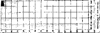
Photometric scale expressing numerically the flocculation activity of the serum and laboratory changes. Just as temperature curves, which together with clinical symptoms are one of the important diagnostic means, so do the curves of serological changes along with clinical manifestations of the disease (if such exist at all, and in syphilis the latter are very often absent) have the most important diagnostic and therapeutic value in syphilis. In this case, the flocculation curves at high and low numbers on the photometric scale have somewhat different meanings. This follows from the fact that with any method of serological research, even entirely negative single indicators, not to mention doubtful low figures (from 0 to 10 on the photometric scale), cannot in themselves serve as an absolute denial of the presence of syphilis in a given subject. From an etiological point of view, only a positive reaction result is important for us. A single high figure (above 10) on the photometric scale may be sufficient for diagnosis. In these cases, syphilitic curves turn into an aid mainly in the area of guiding the treatment of the patient. In negative and low values, they additionally acquire diagnostic value. Character of the chsyphilimetric curves. Distinctive features of curves of normal sera: a) a smooth, horizontal, straight line of flocculation; b) the low position of this line at the level of indicators from 0 to 2 or 3, in extremely rare cases and somewhat higher. Curves of syphilitic sera (Fig. 8) differ: a) increased flocculation indicators; b) a zigzag form of the flocculation line with smaller or larger amplitude of fluctuations, which is explained by the nature of the interaction between the pathogenic principle and the organism. These fluctuations do not diminish the importance of syphilitic curves and photometry. Curves of S. both in their position on the Vern scale and in the nature of their zigzags, length of resistance by other properties of the organism, relationship to treatment and give a completely individual physiognomy for each patient, just as the course of the disease is individual for each individual. It is also clear that a single, isolated figure from the curve cannot satisfy the syphilologist, just as a single figure from the patient's temperature curve does not satisfy the therapist. Based on the zigzag nature of the syphilis curve picture, the error of the statement about the sensitivity of the reaction based only on single studies without serological assessment of the dynamic picture of the disease, i.e., syphilitic curves, becomes understandable. Observation of the curves should show whether the disease is developing, whether it is falling under the influence of the therapeutic intervention applied or not, whether it disappears altogether or persists with respect to a given drug. Hence the great prognostic value of syphilitic curves. To obtain a picture of serological changes of S., it is considered absolutely necessary to clarify the following points: 1) optical density (O.D.) of blood serum; 2) optical density of cerebrospinal fluid (a special technique for the latter was developed by Vern); 3) albuminosis (normal 0.3 per 1,000) and 4) leukocytosis (normal 2 per mm3), since cases are not uncommon where with a negative serological reaction of blood serum and absence of clinical symptoms, an elevated triad of Vern is observed in the cerebrospinal fluid. Cerebrospinal fluid and blood are not always together and equally affected, being in the body in different, so to speak, independent containers. In the therapy of syphilis, along with the decisive role of the treatment rhythm, the rhythm of serological control also acquires great importance. The methodology of S., rejecting standard treatment schemes developed outside of a given patient, outside of a specific study of the course of the disease, establishes a system of treatment and its duration exclusively in the process of observation of a given individual, in the process of dynamic study of his serological changes by syphilitic curves, in other words, strictly individualizing it. This is the essence of syphilitry and its main difference from syphiligraphy. Vern's law of eight months. To clarify the question of the final recovery of the patient, Vern proposed his so-called "law of 8 months". Its essence is as follows. After a straight line characteristic of healthy serum has been established, for eight months after the last provocative injection of arsenic preparations, the patient remains under serological control. The serum is examined monthly. All 8 examinations must give "0" optical density. After 8 months, a spinal puncture should reveal a normal state of the liquor from the point of view of optical density as well as leukocytosis and albuminosis. In these cases, the given individual is considered sterile and practically recovered. S. is applied besides France in Italy, England, USA; in the USSR the Vern reaction was performed by Shaverdov and Saradzhishvili, Paliashvili and Tkeshelashvili, Shirvindt, Alanaknyan, Demidov, Hamburger.
A. Shaverdov. The significance of syphacimetry. The possibility of a systematic mathematical accounting of colloidal reactions, which are quite variable in intensity according to the variable properties of the reagents involved, is disputed by some authors. Objections to S., that is, to the possibility of measuring at any given moment the intensity of the syphilitic infection of the organism, its condition in relation to the pale spirochete, are basically reduced to the following. The intensity of the syphilitic infection in the organism depends primarily on the virulence of the spirochetes, their quantity in the organs (localization of the spirochetes), and the quantity of toxins excreted by the spirochetes; on the basis of these relationships between the organism and the microorganism, certain diagnostic, prognostic, and therapeutic data are derived. Is this possible with the current level of our knowledge about the pale spirochete, serology, and pathology of syphilis? We know of many attempts at the quantitative study of the strength of the seroreaction [Wassermann reaction, flocculation (for syphilis), Citron-Plaut's plus system, Madsen's colorimetric method, Finkelstein's sedimentimetric method, agglutinoscopic method, nephelometric method, etc.], but all of them are characterized by their authors not as a mathematical measure of the strength of the infection, but only as an approximate numerical expression of the strength of the reaction in vitro with a given serum. All so-called quantitative methods (incidentally including the quantitative Kahn reaction) give only a relative representation of the physical condition (dispersity) of the complex-syphilitic serum plus antigen; these reactions do not provide a measure for determining the basic (specific) principle of all seroreactions—the strength of the chemical affinity between the mentioned ingredients of the seroreaction; this essentially explains the absence to this day of a truly standard serodiagnostic method, to which serology has vainly striven to this day. All serological reactions for syphilis, in case of positivity, only speak of the presence 'somewhere' in the organism of the pale spirochete, in case of negativity these reactions indicate that the colloidal state of the serum is close to normal, but they do not with absolute certainty prove the absence of the pale spirochete in the organism. For the given moment there are no theoretical or clinical grounds in the entire concept of Verné that would place this method above other seroreactions in the indicated sense. We primarily proceed from analogies with other chronic infections, such as: tuberculosis, gonorrhea, in which sera undergo physicochemical shifts approaching in a certain sense those in syphilis: however, neither the so-called method of Besredka nor the very mature in practical terms Bordet-Gengou reaction claim the name 'tuberculinometry', 'gonococcometry' in the mathematical sense. The same can be said of other serological methods, such as agglutination (typhoid, paratyphoid, rhinoscleroma), which play a major role in the diagnosis of these diseases, but by no means determine mathematically the degree of the disease and therefore have no prognostic and therapeutic significance. Proceeding from general considerations about colloidal reactions, it should be pointed out that one cannot in general put an absolute sign of equality between the course of physicochemical reactions in vitro and the course of biological reactions in the infected organism; here only a distant, very cautious analogy is possible, but there can be no question of the identity of phenomena. Clinical observations of the Verné reaction deny the possibility of replacing clinical methods of S. in the matter of prognosis and grading of syphilis therapy. Thus, in the works of the 2nd Copenhagen serological conference (1928) the following data are given: in cases of anamnestically established syphilis, the Kahn reaction gave 62% positive results, the Verné reaction only 38% positive reactions; in cases of definitely normal sera, the Kahn reaction gave 0% positivity, while the Verné reaction gave 0.5% positivity; in a series of studies of so-called doubtful sera, the Kahn reaction gave 1% positivity, while the Verné reaction gave 9.5% doubtful reactions; clinical examination of these cases confirmed the correctness of the Kahn reaction results; thus, the Kahn reaction, not accounting exactly mathematically for its results, proved in practice to be more specific than S. of Verné. Finally, S. of Verné excludes complex serodiagnosis, which, in the opinion of the Copenhagen conference, is the only one that can ensure clinically acceptable results. Furthermore, it should be noted that Berné, considering negative indicators of S. (with cerebrospinal fluid and serum) as a sign of absolute recovery, i.e., absence of spirochetes in this organism, did not attempt to confirm his opinion experimentally on animals (rabbit, mouse), whereas in the literature there are facts of the opposite order: Uhlengut obtained a positive result when inoculating rabbits with serum from latent syphilitics with negative RW, and Yu. Finkelstein successfully inoculated a healthy rabbit with cerebrospinal fluid from a syphilitic rabbit that was negative in all respects (negative RW, negative Pandy reaction, negative Sachs-Georgi reaction, absence of pleocytosis, etc.). Along with a number of positive reports from American, Italian, and Soviet clinicians, there are also negative reports. Thus, Bergeron and Demanch showed that syphilis of the same localization gave different photometric indicators in the same stage of the disease; cases of syphilis of the same localization but different duration often gave the same indicators. On the other hand, sometimes more severe clinical forms were accompanied by lower photometric indicators than cases clinically milder. These data show that the photometric indicators in these cases did not express the intensity of the syphilitic infection, and therefore did not have the character of S. in the sense of Verné, and consequently could not be guiding factors for prognosis and therapy. No less critical is the attitude to S. of a number of other syphilologists, who say that the curves of syphacimetry do not run parallel with the evolution of syphilis.
Related articles
Cite this page
“Syphacia Obvelata.” Soviet Medical Encyclopedia. English translation of Bolshaya Meditsinskaya Entsiklopediya, 1st ed. (Moscow, 1928–1936), ed. N. A. Semashko. https://sovietmedicalencyclopedia.pages.dev/article/syphacia-obvelata/