Pinworms
Historical document, translated for reference. It reflects medical knowledge of the 1920s–30s and is not medical advice.
Summary
This article from the 1928–1936 Great Medical Encyclopedia describes the biological characteristics, classification, and life cycle of the pinworm (Enterobius vermicularis). It details the parasite's morphology, its localization in the human digestive tract, and the mechanisms of egg deposition.
Encyclopedia article (1928–1936)
PINWORMS, nematodes (Nematoda) from the family Oxyuridae Cobbold, 1864, are small or medium-sized parasites. There is one or two spicules; in the latter case, they are always equal. The female (Fig. 1) has two ovaries. They parasitize the digestive tract of mammals, reptiles, and amphibians. The family Oxyuridae is subdivided into 4 subfamilies: Oxyurinae Hall, 1916 (one spicule or absent, no gubernaculum), Syphaciinae Railliet, 1916 (one spicule and a gubernaculum is present), Cosmocercinae Railliet, 1916 (two spicules, gubernaculum present), Oxysomatinae Railliet, 1916 (two spicules, gubernaculum absent). The human pinworm—Enterobius vermicularis (old name, Oxyuris vermicularis)—belongs to the subfamily Oxyurinae. The helminthiasis caused by pinworms is called enterobiasis, named after the genus of the pathogen. Description of the parasite. Small nematodes, tapering at the ends. The head end is surrounded by a cuticular vesicle. The mouth is surrounded by three lips, the tips of which are studded along the edge with a denser cuticular base. The oral opening (Fig. 2) leads into the esophagus, which ends in a special expansion in the form of a muscular organ, the bulb, in which chitinous chewing plates are embedded. The male (Fig. 3) is 2 to 5 mm long. The tail end (Figs. 4 and 5) is equipped with lateral cuticular wings. Postanally, there are three pairs of genital papillae. There is one spicule, the length of which in its chitinized part is 0.090–0.093 mm. A gubernaculum may or may not be present. A preanal sucker is absent. The female is 9–12 mm long. The vulva is located at the border of the anterior and middle thirds of the body length. The tail end is awl-shaped and pointed. The eggs (Fig. 6) are elongated-oval, slightly asymmetrical; one side is flattened, the other convex. The length of the eggs is 0.050–0.060 mm and the width is 0.020–0.032 mm. A freshly laid egg contains a "tadpole-like" larva, which, a few hours after the egg has lain in the external environment, turns into a normal nematode-like larva. Enterobius vermicularis parasitizes only humans; all cases of the discovery of this parasite in other animals (dogs, cats) are based on errors. Pinworms are found all over the globe, being the most frequent parasite everywhere. According to studies conducted in the USSR, children older than 1–2 years are infected with pinworms in 25–100% of cases; the infection rate among adults is somewhat lower.
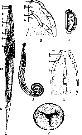
Figure 1. Mature female: 1—cephalic vesicle; 2—nerve ring; 3—esophagus; 4—bulb; 5—intestine; 6—excretory pore; 7 and 8—two ovaries; 9—vulva; 10—uterus; 11—anus. Figure 2. Head end, apical view. Figure 3. Male: 1—testis. Figures 4 and 5. Tail end of the male (ventral and lateral): 1, 2, 3, and 4—genital papillae; 5—spicule. Figure 6. Egg.
Localization of the human pinworm. Pinworms spend the greater part of their life in the lower section of the small intestine, in the cecum, the initial section of the colon, and the vermiform appendix; here the parasites grow to the sexually mature stage, after which the females, filled with eggs (in each female, according to Leuckart, there are about 12,000 eggs), descend to the anus and exit into the external environment to lay their eggs. In the human intestine, one to many thousands of specimens of pinworms can be found. The mature female actively exits through the anal opening, crawls along the perianal folds and perineum, and in women, it often crawls into the genital slit and vagina. The exit of the females occurs predominantly in the evening or at night, 1/2–3 hours after beginning to stay in bed. Upon exiting the anus, the pinworms begin to lay eggs; a factor that accelerates the process of egg-laying is drying. After 15–30 minutes, the female is usually already freed of eggs and dies, turning into a shapeless lump. Thus, egg-laying, as a rule, occurs 161
PINWORMS
Pinworms do not move in the intestinal lumen, but upon exiting the anal opening into the external environment, on the surface of the skin, in the perianal region, perineum, on underwear, etc. This explains why pinworms in most cases are not diagnosed by standard methods of helminth-coprological analysis, but require special research methods for the detection of eggs (mainly methods of anal or perianal scrapings). Skrjabin suggests performing a scraping with a diagonally cut matchstick, moistened in a 1/2% soda solution. The resulting scraping is transferred to a glass slide in a drop of soda solution or a 50% glycerin solution for microscopy. The same method is recommended for examining scrapings from the perineum, vagina, nail phalanges of the fingers, as well as the contents of the subungual spaces. The development of pinworm eggs in the external environment under suitable temperature conditions (about 35-36°) occurs very quickly: already after 6 hours a larva matures in the egg, and it becomes invasive, i.e., capable of causing infection. At low temperatures (+20° and below) the development of eggs is suspended and resumes after being transferred to conditions of higher temperature. Pinworm eggs possess several shells (four, according to Zavadovsky and Shalimov, 1928), which to a significant degree protect them from the harmful effects of physical and chemical agents; for example, pinworm eggs can retain viability even in 3-4% formalin solutions. These shells are at the same time permeable to O2, which is necessary for the eggs for their further development, i.e., for the formation of the larva. Development of pinworms in the human body. From eggs that have entered the human digestive tract, larvae hatch, which stop in the upper section of the large intestine, the vermiform appendix, and the lower section of the small intestine, and here within 2-4 weeks grow into a sexually mature individual. Fixation of pinworms to the intestinal wall occurs, as is believed, due to the suction action of the bulb, and the strength of the suction is facilitated by the tight adherence of the vesicle head to the intestinal mucosa. The exit of sexually mature females (according to Koch) occurs due to the overfilling of the uteri with eggs, which compress the bulb and thereby disrupt its suction function. Having detached from the intestinal wall, the females are then passively carried into the rectum, where they usually accumulate in large quantities before actively exiting outward. Already in the middle of the 19th century, the question was raised as to whether new generations of parasites could develop from eggs released by pinworms in the intestine without the eggs being carried into the external environment. The main motive for posing such a question (and its resolution by some authors in the affirmative) was the lack of success in therapy observed in some cases of persistent enterobiasis. Koch (1925) attempted to resolve this question experimentally, coming to the conclusion that from eggs sometimes released by the female into the intestinal lumen, larvae can hatch right there and grow into sexually mature individuals. However, Koch's conclusions cannot be accepted, as they proceed from incorrect premises: pinworm eggs cannot develop to the invasive stage without access to oxygen, as proven by Zavadovsky and Shalimov (1929), while Koch arbitrarily assumes the opposite. The experimental setup suffers from serious methodological errors. Thus, at the present time, there are no sufficient scientific grounds to admit 'intra-intestinal' development of new generations of pinworms, and the previous position remains in force that the young generation of pinworms can develop only from eggs carried into the external environment and subjected to the action of oxygen. Pinworms of domestic animals. Many species of animals have their own specific forms of pinworms. In monkeys, various species of the genus Enterobius parasitize [E. Anthropopitheci (Gedoelst, 1916) and others]. Horses have the largest of all pinworms—Oxyuris equi (Schrank, 1788); sheep and goats have Skrjabinema ovis (Skriabin, 1915); rabbits have Passalurus ambiguus (Rud., 1802); mice (wild and laboratory) are parasitized by two species of pinworms: Aspiculuris tetraptera (Nitzsch, 1821) and Syphacia obvelata (Rud., 1802). The latter species can also transfer to humans. P. E. Shultz. Clinical aspects of enterobiasis. The resistance of eggs to external influences, especially drying, explains the ease of infection in places with large gatherings of children: in orphanages, playgrounds, schools, public toilets, etc. Oleinikov found pinworm eggs on windowsills and school desks, and under the desks of infected girls he found more eggs than under those of boys. Wilhelmi and Quast, who examined the dirt under the fingernails of 1,000 schoolchildren, found pinworm eggs in 71.4% of boys and 64% of girls, and in 16% of younger children also in nasal mucus. The introduction of pinworm eggs into the human intestine is facilitated by the consumption of contaminated food products, unwashed fruits, raw vegetables, and in general, any kind of personal and public untidiness. Drinking water apparently does not play such a role, as pinworm eggs quickly die in water (Trumpp). Age, degree of immunity, state of the nervous system, and other factors play an undoubted role in the development of enterobiasis, which is especially developed in view of the lesser cleanliness at a young age. According to Rudnev, pinworms are more often encountered in asthenics (73%) and less often in digestive types (16%). Feeding on blood, pinworms not only strongly attach themselves to the intestinal mucosa but often penetrate deep into the mucous membrane, as well as into the Lieberkühn glands, causing minute tissue wounds with subsequent small hemorrhages and facilitating the inoculation of pathogenic microbes. With massive infestation, it is quite understandable that not only secondary anemia develops on this basis, but also all kinds of nervous phenomena caused by the introduction of poisonous products of the parasite itself directly into the human blood. That pinworms produce a toxin was proven by Brünings, who obtained a reaction analogous to tuberculin with an extract from pinworms, as well as by Cherkasov and Peiper, who observed eosinophilia of up to 8-16% in enterobiasis. Heubner and Meyer observed prolonged inflammation of the cornea (keratitis dendritica). P. Nonay reports a case of neuritis optica with weakened vision, which disappeared after the expulsion of the pinworms. Massive invasion of pinworms sometimes causes catarrhal inflammation of the rectal mucosa with subsequent disorders of defecation, tenesmus, proctitis, rectal prolapse, and the development of nodular varicose veins. The presence of pinworms in removed appendices, sometimes in huge quantities up to the point of blocking the lumen, is not a rare finding. Fecal stones with a pinworm as a core in the center have also been found in them. Irritation of the appendix mucosa by pinworms and their toxins can manifest in a whole series of painful symptoms and sensations known as appendicopathy. Whether pinworms can be the direct cause of the development of acute appendicitis has not yet been finally decided, although many authors speak positively in this sense. Sometimes penetrating deep into the intestinal wall, pinworms can become encapsulated there through calcification. Skrjabin speaks of tumor-like growths in connection with the penetration of ascarids; a case has been described of finding on a cadaver in the wall of the rectum such tumors covered with mucosa, the size of up to a walnut, overgrown with connective tissue, inside which was a mass of pinworm eggs. Nathan found in the spleen of a person who died from liver cirrhosis a dense connective tissue focus the size of a cherry with a female pinworm enclosed in it, undoubtedly carried there by a hematogenous route. The most characteristic symptom of enterobiasis is the itching or burning sensation around the anus caused by pinworms and their toxins, which induces a reflex need to scratch. On this basis, all kinds of skin lesions can develop (erythema, secondary eczema, abscesses, etc.), sometimes spreading far beyond the perineum. The intensity of the itching depends to a significant degree on the number of crawling-out females; there can sometimes be a colossal number of them. Pod'yapolskaya confirmed on a cadaver in the intestine of one child 2,751 specimens of pinworms. Pinworms sometimes penetrate in boys into the prepuce sac, and in girls not only into the vagina but also higher, through the Fallopian tubes into the Douglas pouch, where they have been found encapsulated on the peritoneum. The possibility of them carrying pathogenic microbes there is not excluded. Strong irritation of the urogenital sphere by them leads to the early awakening of the sexual instinct, to masturbation, dysuria, leukorrhea, balanitis, prostatorrhea, etc., with all the consequences flowing from this. With prolonged enterobiasis, neuropathic children become even more irritable and nervous, lose their appetite, lose weight, and lag in growth; they may even develop epileptiform seizures. The described pathogenesis of enterobiasis, this typical disease of everyday life, is possible only with massive and prolonged infestation. With a small number of pinworms in the intestine, usually, no painful symptoms are noted.
Prophylaxis, based mainly on a correct understanding of the life cycle of pinworms, boils down primarily to measures of personal and public hygiene, namely: it is recommended to wash hands thoroughly before eating, not to bite nails or clean subungual dirt with teeth, to sleep at night in tightly sewn pants, to wash the perineum daily and necessarily use toilet paper; to change underwear and bed linen frequently; not to consume contaminated and insufficiently disinfected food; to observe mutual cleanliness in the family; to keep children's rooms, toys, public premises for children, as well as toilets, tidy, and most importantly - broad sanitary education. The fight against pinworms, in view of the constant possibility of reinvasion, is a rather difficult task, but not unsuccessful, and here the strict implementation of all those preventive measures that hinder and prevent pinworms from re-entering the human body stands in the foreground. The principle of successful dehelminthization boils down mainly to the correct combination of medicinal treatment with enemas and laxatives. Enemas are prescribed mainly for itching, before bedtime, to prevent the crawling out of females and the scattering of eggs by them. For the same purpose, smearing the anus with gray mercury ointment mixed with Vaseline or the introduction of suppositories with the same ointment is prescribed. Frequent enemas are harmful, especially enemas with mercuric chloride or carbolic acid. Many prescribe enemas of soapy water, garlic, a 0.5-1% solution of lysol, or 0.5-1% vinegar. Any medication treatment must be preceded by a laxative, which is necessarily repeated after the completed administration of the drug. Among the numerous remedies against pinworms, the combination of naphthalene with santonin (3-4 powders over 2-3 days) enjoys a well-deserved reputation. Bismuth carbonate and aluminum salts (Gelonida alum. subacetici) are also recommended, as well as thymol, which is little suited for children due to its burning taste. Trump suggests kamala, Heubner suggests fern extract. When prescribing the latter against tapeworms in children, Scholler repeatedly observed the simultaneous discharge of a large number of both mature and immature pinworms. Consuming large amounts of onions, garlic, and raw carrots contributes to the expulsion of pinworms. The newest remedies include: a preparation from garlic - Allisatin-Sandoz and a 2% dialysate from carrots - Daucarysatum-Burger. Washing the intestine using a duodenal tube with a solution of Aluminii subacetici is hardly applicable in children. For successful treatment, it is necessary to identify all worm carriers in the family and achieve their simultaneous dehelminthization; otherwise, each untreated person will remain a constant source for new infections of the other members of the family. G. Sholpo.
Related articles
Mentioned in
Cite this page
“Pinworms.” Soviet Medical Encyclopedia. English translation of Bolshaya Meditsinskaya Entsiklopediya, 1st ed. (Moscow, 1928–1936), ed. N. A. Semashko. https://sovietmedicalencyclopedia.pages.dev/article/pinworms/