Ancylostoma
Historical document, translated for reference. It reflects medical knowledge of the 1920s–30s and is not medical advice.
Summary
This 1930s encyclopedia article describes Ancylostoma duodenale, a parasitic roundworm affecting humans, detailing its morphology, developmental cycle, modes of infection, and the disease it causes, ancylostomiasis.
Encyclopedia article (1928–1936)
ANKYLOSTOMA, Ankylostoma duodenale, a nematode (roundworm) from the family Ankylostomidae Looss. Males are 7 to 11.2 mm long, females from 8 to 18 mm. The buccal capsule (see figure 2), curved dorsally, is armed with 3 pairs of teeth, hooked inward. The male has a special copulatory bursa at the posterior end and 2 spicules 2 mm long (see figure 1). The vagina (vulva) in the female is located at the border of the middle and posterior third of the body. Eggs are oval, with a thin, transparent shell, with 2, 4, or 8 cleavage spheres (see figure 3), 0.056 to 0.063 mm long and 0.036 to 0.040 mm wide. The habitat of sexually mature individuals is the small intestines of man. Development cycle: eggs laid by females in the human intestine (one female can excrete more than 1,000 eggs per day) are carried out into the external environment with feces, where, under suitable temperature conditions (25-30°), humidity, and with access to oxygen, a larva is formed in the egg; the latter, upon emerging from the egg, representing a "rhabditiform form" (see figure 4), characterized by 2 dilations of the esophagus, lives in the upper layers of the soil for some time; after undergoing two molts in about five days, the larva turns into a filariform larva (with a simple esophagus) and becomes "infective," i.e., capable of producing infection. Larvae remain viable for 3-6 months; desiccation and sunlight have a destructive effect on them. Man is infected with Ancylostoma either when the infective larva penetrates through intact skin, or per os. In the first case, the larva, as Looss discovered, enters the blood vessels, is carried by the bloodstream through the right heart and the lesser circulation into the lungs. From the pulmonary capillaries, the larva breaks through into the alveoli, from which, having passed the respiratory tract, it enters the mouth. Ultimately, the larva is swallowed and enters the intestine, where it fixes and develops into a sexually mature individual. Upon entering per os into the organism, A. duodenale, for the most part, does not perform this complex path, but more often directly stops in the small intestines, where it continues its development (Fülleborn), reaching sexual maturity in 3-5 weeks, after another 2 molts. It is already clear from the developmental cycle of A. duodenale that the sources of invasion can be, on the one hand, infected soil and water and, on the other hand, food, drink, tobacco, etc., contaminated with infective stages. The circumstance that larvae undergo their development in the ground at a certain degree of warmth and moisture and that they are capable of penetrating into man through intact skin explains the fact that ancylostomiasis is especially widespread in the tropical and subtropical zones of the globe, and in the temperate zone among miners, ditchdiggers, brickyard workers, plantation workers, etc., acquiring the significance of an occupational disease here. In hot countries, ancylostomiasis therefore predominantly affects the poorest strata of the population, whose anti-hygienic living conditions create wide opportunities for "household" infection of man. Ancylostomiasis. Under the name ancylostomiasis (ancylostomiasis), one now understands a disease of humans and certain other mammals caused by roundworms (see Nematodes) of the genus Ankylostoma Dubini (1843). Two species of this genus can parasitize humans - A. duodenale Dubini (1843) and A. brasiliense de Faria (1910). The greatest distribution and significance in human pathology belongs to Ancylostoma duodenale, first discovered by Dubini in 1838 in Italy.
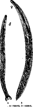
Figure 1

Figure 1. Head end -
buccal capsule (according to Looss), with larvae.
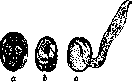
Figure 3. a and b - eggs in various stages of development; c - larva hatching from the egg (according to Manson).
organism, A. duodenale, for the most part, does not perform this complex path, but more often directly stops in the small intestines, where it continues its development (Fülleborn), reaching sexual maturity in 3-5 weeks, after another 2 molts. It is already clear from the developmental cycle of A. duodenale that the sources of invasion can be, on the one hand, infected soil and water and, on the other hand, food, drink, tobacco, etc., contaminated with infective stages. The circumstance that larvae undergo their development in the ground at a certain degree of warmth and moisture and that they are capable of penetrating into man through intact skin explains the fact that ancylostomiasis is especially widespread in the tropical and subtropical zones of the globe, and in the temperate zone among miners, ditchdiggers, brickyard workers, plantation workers, etc., acquiring the significance of an occupational disease here. In hot countries, ancylostomiasis therefore predominantly affects the poorest strata of the population, whose anti-hygienic living conditions create wide opportunities for "household" infection of man. Ancylostomiasis. Under the name ancylostomiasis (ancylostomiasis), one now understands a disease of humans and certain other mammals caused by roundworms (see Nematodes) of the genus Ankylostoma Dubini (1843). Two species of this genus can parasitize humans - A. duodenale Dubini (1843) and A. brasiliense de Faria (1910). The greatest distribution and significance in human pathology belongs to Ancylostoma duodenale, first discovered by Dubini in 1838 in Italy.
ma duodenale, first discovered by Dubini in 1838 in Italy.
Figure 4. Rhabditiform form.
discovery (by Leich- later a causal connection was established by tenstern-y). between the parasitism of this nematode (in the small intestines) and the severe diseases that were rampant in many places on earth and went by various names, such as: Cachexia africana (s. america-na), Cachexie aqueuse, Anemie des pays chauds and others. The true etiology of these diseases was clarified only in 1854 by Griesinger, who pointed out that the cause of the so-called Egyptian chlorosis, which afflicted more than a quarter of Egypt's population, is A. duodenale. In Europe, serious attention was first paid to this disease during the excavation of the Saint Gotthard tunnel, when it was discovered that the mass cases of severe anemia among the workers were of parasitic origin. Finally, it was established that the diseases of brick workers and the 'Anaemia montana' of coal miners, which until then had been associated with working conditions without light and breathable air, were of the same etiology.-To fully assess the pathogenic significance of A., the following points should be taken into account: 1. A., when penetrating through the skin, undergoes a complex migratory path in the host's body, accompanied by certain tissue trauma (skin, blood, lungs). 2. When attaching to the intestinal wall, A. penetrate deeply into its tissues (up to the submucosa), repeatedly change their attachment points in the process, leaving traces in the form of bleeding defects in the mucosa. 3. Being hematophages, A. at the same time secrete a special secretion that prevents blood clotting and toxins in which hemolytically acting substances predominate. In connection with this, the pathogenic effect of A. consists of 1) mechanical damage to the host's tissues, 2) intoxications and 3) inoculation of microbial flora in the areas of mechanical damage. According to the clinical picture of the disease, French clinicians distinguish the following (sometimes difficult to differentiate) forms: 1) acute gastrointestinal forms (sudden onset, disturbance of gastrointestinal tract function, subsequently-anemia, edema), 2) chronic gastrointestinal forms (slower course with the same symptoms), 3) cachectic form (gastrointestinal symptoms recede into the background, severe anemia, emaciation), 4) mild form (anemia, dizziness, palpitations, etc.), 5) atypical form (skin rashes, catarrhal condition of the upper respiratory tract). With a small number of A. in the body, no painful manifestations are often noted. Changes in the blood do not provide clear indications for diagnosis. Eosinophilia is not always noted, mainly in mass infestations. The significance of ankylostomiasis as a factor contributing to other diseases is illustrated by the research of Kofoid and Tucker, who, based on the examination of 22,842 American soldiers, came to the following conclusions: in those areas where the prevalence of ankylostomiasis exceeds 10%, mortality is 87.5% higher than in areas where the infestation rate is below 10%; in units where the prevalence of ankylostomiasis is 30%, mortality from pneumonia is three times greater than in units with a low percentage of ankylostomiasis (10%); among persons infected with ankylostomiasis, there is an increased tendency to suffer from tonsillitis, bronchitis, pneumonia and measles; mental abilities in ankylostomiasis patients are reduced by 25%. The diagnosis is made with certainty by finding A. eggs in the feces. Since cases of ankylostomiasis infection without clear clinical manifestations (with a small number of parasites) are not uncommon, it is necessary to assess the intensity of invasion by the method of 'quantitative helminthological coprological analysis'. Assessment of invasion intensity is especially necessary in mass examinations for monitoring health improvement measures. The prognosis in uncomplicated cases, with the possibility of therapeutic intervention, is favorable: expulsion of the parasites leads to complete restoration of health.-The implementation of preventive measures is complicated by the fact that A. larvae, living in the external environment, enter the body both through the skin and per os. In connection with this, it is not sufficient to observe basic hygiene rules regarding food and drink, and main attention should be directed to protecting against the invasive agent in the external environment. This is especially true for those places that, on the one hand, provide a favorable environment for the development of larvae, and on the other, where working conditions promote the most complete contact of a person with this infected environment (mines, rice fields, plantations, etc.). It must be remembered that here the source of spread of the invasion is people infected with ankylostomiasis, resp. their feces. Therefore, the most radical measure is the mandatory and universal deworming of ankylostomiasis carriers with subsequent disinfection of their feces. For endemic foci of ankylostomiasis, the Rockefeller organization (International Health Board, Rockefeller Foundation U. S. A.), which conducts a significant, especially in colonies, anti-ankylostomiasis campaign, recommends universal deworming of the entire population, as this measure is practically easier to implement and cheaper than deworming only known carriers after their preliminary examination. Along with broad sanitary education of the population, the following measures are recommended: the provision of sanitary toilets (an unsanitary toilet is a breeding ground for ankylostomiasis); in houses-the installation of wooden or cement floors; wearing shoes; in enterprises, especially those unfavorable for ankylostomiasis, periodic medical examination of workers for ankylostomiasis, examination of new employees for ankylostomiasis. In mines-provision of portable toilets and supply of good drinking water. For disinfection of soil, a 3% solution of lysol is proposed. As anthelmintics for ankylostomiasis, the most effective are Thymol (4.0-10.0 pro die, 2.0 pro dosi, after 1-2 hours with subsequent laxative), Ol. Chenopodii (1.5 in one or 2 doses, after 2 hours-laxative), carbon tetrachloride (Carboneum tetrachloratum, CC14) in a single dose of 3 cubic cm (1 cubic cm=1.6g).-Ankylostomiasis is one of the most widespread diseases, being endemic for almost half of the world's population. The entire belt of the earth from 36° N. latitude to 30° S. latitude is completely infected with ankylostomiasis; it affects over 500 million people, i.e., more than 25% of the world's population. In many places, the infection rate reaches 100%. Of European countries, Italy is most affected; in other states, ankylostomiasis is mostly concentrated in coal mining enterprises (France, Germany, Great Britain, Spain, Belgium, Holland, Austria, etc.)-Until recent years, the USSR was considered free of ankylostomiasis. There were assumptions (Skryabin) about the infection of Donbass mines, but an organized expedition (25th Union Helminthological Expedition in 1925) to investigate this question found no evidence of this disease in Donbass. Since 1925, the first reports of ankylostomiasis in various places in the USSR have appeared, and at present there are already data on the significant spread of this disease in the form of endemic foci in Transcaucasia (Azerbaijan, Georgia, Abkhazia, Armenia) and Central Asia (Turkestan). The English, under the name 'hookworm disease' (German 'Hakenwurmkrankheit'), understand not only ankylostomiasis but also necatorosis (see). Since the causative agents of these helminthiases are A. and Necator, which are representatives of the same family A., according to the nomenclature proposed by Skryabin and Schulz, the name combining these helminthiases will be 'ankylostomidoses'. In carnivores in the USSR, A. caninum (Ercolani, 1859) is quite common, which is unable to parasitize in humans.
Related articles
Mentioned in
Cite this page
“Ancylostoma.” Soviet Medical Encyclopedia. English translation of Bolshaya Meditsinskaya Entsiklopediya, 1st ed. (Moscow, 1928–1936), ed. N. A. Semashko. https://sovietmedicalencyclopedia.pages.dev/article/ancylostoma/