Trichinellosis
Historical document, translated for reference. It reflects medical knowledge of the 1920s–30s and is not medical advice.
Summary
Trichinellosis is a parasitic disease of humans, pigs, and carnivores caused by the nematode Trichinella spiralis. The parasite infects hosts through consumption of contaminated meat and forms cysts in muscle tissue.
Encyclopedia article (1928–1936)
Trichinellosis, a helminthic disease of humans, pigs, and carnivores, caused by the parasitization in the bodies of these animals of nematodes Trichinella spiralis (Owen, 1835)(figs. 1 and 2), belonging to the family Trichinellidae (Ward, 1907). Extremely small, barely visible to the naked eye adult parasites are localized in the small, and with intense infestations also in the large intestine, penetrating (females) for fixation deep into the mucous membrane. The size of the male is 1.4-1.6 mm X 0.04 mm. The posterior end is provided with two large processes on either side of the cloaca, located ventrally, and two pairs of papillae between them. The size of the female is 3-4 mm X 0.060 mm. The vulvar opening is located at the anterior end of the body approximately at the boundary of the first and one-fifth of its length. The female is viviparous. The size of the larvae laid is 0.09-0.16 mm X 0.006-0.009 mm. Infection with T. occurs from eating pork or other animals' meat containing viable trichinae larvae, which are in the encapsulated state in the muscles. From the meat digested in the intestinal tract, encapsulated larvae are released; the capsules dissolve and the larvae reach sexual maturity within 28-40 hours. The laying of numerous larvae by females begins 6-7 days after infection, previously the females burrow deep into the intestinal wall and with the anterior end penetrate into the central lymphatic vessel; in some cases the entire female is located in the dilated lymphatic vessel of the mucous or submucous membrane, more rarely penetrates deep into the mesenteric glands. Entering the lymphatic system, the larvae along with the lymph pass into the bloodstream, by which they are distributed throughout the body. The most intensely infested by trichinae larvae are the diaphragm legs, intercostal muscles, tongue muscles, pharyngeal muscles, eye muscles. Upon reaching the site of localization, the larvae penetrate through the sarcolemma into the muscle cell, where they grow somewhat, coil spirally and become encapsulated due to the reaction of the surrounding tissue. The capsule, which in the vast majority of cases contains one larva, has a lemon-like shape. Over time, the larvae may die and the capsule may become calcified. However, this process occurs at different times in different individuals. In humans, calcification of trichinae
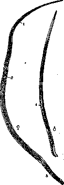
Figure 1. Trichinella spiralis. Left adult female; right adult male: 1- genital opening; 2- papillae; embryoincapsule; 3- the same embryo after 15 days (encapsulated).
capsules usually occurs from several months to several years after infection. However, there are known cases when larvae retained viability for 20, 24 years, etc. The presence of trichinae in human muscles was established as early as 1828 by Piko (Paget). Later, a student (Paget), finding numerous trichinae capsules in the muscles of a human corpse, drew attention to the worm coiled inside them. The structures he found were given to Owen, who in 1835 described them and named them-Trichina spiralis, later changed by Railliet to Trichinella, since the name Trichina already existed in insects. The life cycle of this parasite was deciphered through experiments by Leuckart (1859), Virchow (1859), Zenker (1860), and others. In the infection of T. in humans, infected pork plays a role, and various methods of its processing in most cases are insufficient for the death of trichinae larvae. The latter resist low temperatures well. Thus, according to Leuckart, keeping trichinellosis-infected pork for 3 days at t°-2°-25° is insufficient for the death of larvae. The viability of the latter is preserved for 57 days at t°-12.2° (Ransom). Salting kills larvae only in the superficial layers of meat, and in salted ham, for example, they can retain viability for more than one year. Smoking is also insufficient for the death of larvae. Even when boiling and frying, due to the poor thermal conductivity of meat, larvae in the depth of the piece may remain viable. Finally, in rotting meat, the viability of larvae can be preserved for 2-3 months.
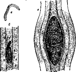
Figure 2. Trichinella spiralis: a-embryo; b-embryo just having penetrated the muscle; c-the same embryo after 15 days (encapsulated).
Trichinae are cosmopolitan, and human trichinosis can be found wherever pork is consumed. Abroad, trichinosis in pigs is especially widespread in America (Boston 4-5%; Chicago 8%). In European countries in the 19th century, trichinosis was extremely widespread in Germany, where there were several trichinosis epidemics; some of them had a high mortality rate. Within the USSR, trichinosis as an endemic disease of humans is more common in Belarus. Trichinosis is also found in the North Caucasus and in Ukraine; there are cases of locally acquired human trichinosis in the Ryazan region, in the Volga German Republic, and in Siberia. Undoubtedly, human trichinosis is more widespread than is known to us, since in most cases physicians are unable to diagnose it. This applies not only to the USSR but also to other countries (Brumpt, 1927 also points to physicians' inability to diagnose trichinosis). In the spread of human trichinosis, besides the pig, whose meat infects humans, rats play a major role, from which pigs can become infected. In areas where trichinosis in pigs is prevalent, rats are always significantly infested with trichinae. Dogs and cats may also have considerable importance in the trichinae cycle. The symptomatology of trichinosis can vary depending on the number of viable trichinae in the meat consumed. Mild cases may be completely asymptomatic or be mistaken for mild influenza. In moderate and severe cases, several periods of the disease can be noted, which however are not always sharply defined. The earliest symptoms of the disease are gastrointestinal disorders, which however may sometimes be completely absent. Nausea, belching, and vomiting are most commonly observed. There may be diarrhea, sometimes very severe, or constipation. After consuming a large amount of heavily infested meat, death may occur within 1-2 days from extensive hemorrhagic inflammations of the intestinal mucosa. The rise in temperature begins on the 3rd-4th day; it increases until the 9th-11th day to 40-41°C, after which a prolonged febrile period sets in, lasting 4-7 weeks, in milder cases 2-3 weeks. The period of maximum temperature coincides with the penetration of the new generation into the muscles. At this time, facial edema, especially of the eyelids, appears. These edemas are extremely characteristic symptoms of trichinosis and are usually observed even in mild cases. In Belarus, for a long period, they sought to elucidate the etiology of a disease known as 'odutlovatka' which turned out to be trichinosis (Alisov). In more severe cases, edema is also observed on the extremities. During this same period of the disease, muscles begin to swell and harden; pain appears (in mild cases of the disease, swelling and sometimes muscle pain may be absent). Pain in the tongue makes speech difficult; hoarseness and even aphonia, painful sensations during breathing, pain when moving the eyeball, etc., are observed. Death most often occurs between the 4th and 7th week of the disease. Recovery in mild cases can occur in 2-3 weeks, while in moderate cases it is prolonged from 6 weeks to 3-4 months. The mortality rate in different epidemics ranges from insignificant to 25-30%. The diagnosis of trichinosis in clear-cut cases presents little difficulty; the acute onset with a temperature that increases for several days, edema of the eyelids and face in the absence of protein in the urine, and pronounced eosinophilia constitute a characteristic symptom complex of trichinosis. Eosinophilia appears by the 8th day after infection and can reach very high figures, usually 15-30% and even 78% (Fuchs) and 85% (Pevzner), and persists for quite a long time. Trichinosis often leads to confusion with typhoid or typhus fever, in which case the diazo reaction and the Weil-Felix reaction, which are negative in trichinosis, assist in the differential diagnosis. Examination of the urine makes it easy to rule out acute inflammation of the kidneys. Milder trichinosis sometimes extremely resembles influenza, for which it is undoubtedly often mistaken without the appropriate blood analysis (eosinophilia). In doubtful cases, biopsy can resolve the question, however, its application is not possible before the 3rd week of the disease. Furthermore, in weak infestations in trichinosis, biopsy may also give a negative result. Finding adult parasites in the feces, although possible, presents considerable difficulties and has no diagnostic value if the parasites are not found. Attempts at serodiagnosis of trichinosis do not yet have diagnostic value. Therapy for trichinosis in the early stages of the disease should be directed toward removing adult trichinae from the intestine. Therefore, laxatives are indicated in cases of constipation and normal stool. In cases of diarrhea, the latter should not be stopped. Anthelmintics are also recommended: santonin, thymol, benzene. The latter, according to Mosler, is prescribed as follows: Benzoli 6.0; Mucilag. Gummi arab. 25.0; Succi Liquir. 8.0; Aq. Menthae pip. 120.0. Shake well. Give a tablespoonful every 1-2 hours. Even after the first period of the disease, anthelmintic treatment is not superfluous, since females sometimes remain in the intestinal tract for several weeks, continuing to lay larvae. At present, we do not have reliable means that could affect the young larvae, whether migrating or encapsulated, and thereby cure the disease. Therefore, the physician's measures should be directed toward supporting the patient's strength and helping the body in the process of encapsulating the larvae. For the latter purpose, calcium preparations and a diet rich in calcium salts are indicated. Prevention should be directed along the lines of veterinary-sanitary institutions. Mandatory trichinoscopy of all pig carcasses on slaughterhouses and veterinary-sanitary points with subsequent rational rejection and disinfection of infested material is necessary. The fight against rats as a reservoir of infested material, as well as against stray dogs, is of great importance. It is necessary to carry out extensive sanitary-educational work among the population, explaining the factors in the spread of trichinosis infestation, its significance, and also promoting personal prevention (thorough thermal processing of pork). It is equally necessary for physicians to become more familiar with trichinosis. Rational zoohygienic maintenance of pigs in farms and the elimination of stray pigs wandering through village streets and squares are essential elements in the prevention of trichinosis.
Related articles
Cite this page
“Trichinellosis.” Soviet Medical Encyclopedia. English translation of Bolshaya Meditsinskaya Entsiklopediya, 1st ed. (Moscow, 1928–1936), ed. N. A. Semashko. https://sovietmedicalencyclopedia.pages.dev/article/trichinellosis/