Dipylidium Caninum L
Historical document, translated for reference. It reflects medical knowledge of the 1920s–30s and is not medical advice.
Summary
Dipylidium caninum is a tapeworm parasite that infects the intestines of dogs, cats, and occasionally humans. The article describes its morphology, life cycle, clinical manifestations, diagnosis, treatment, and prevention.
Encyclopedia article (1928–1936)
DIPYLIDIUM CANINUM L, 1767, syn.- Taenia cucumerina, cucumber-shaped or cucumber tapeworm. A tapeworm parasite in the small intestines of dogs and cats, also found in the same organ in humans. The strobila reaches up to 350 mm in length with a maximum width of mature segments of about 3 mm (Fig. 1 and 2). The width of the scolex is 0.35-0.86 mm. On the club-shaped rostellum there are 3-4 rows of hooks (numbering 48-60), of which the anterior ones reach 0.011-0.015 mm, while the posterior ones are about 0.006 mm. Mature segments (6-7 mm long and 2-3 mm wide) are shaped like cucumber seeds and are often colored pinkish in life. The genital openings are located somewhat behind the middle of the segment's length. Each egg capsule contains several eggs (8-15-21). The eggs are round, reaching 0.034-0.04 mm in diameter; the diameter of the oncosphere is 0.025-0.036 mm, and the length of the embryonic hooks is 0.011-0.014 mm. The egg capsules are oval in shape. The definitive hosts are domestic carnivores (cats and dogs) and some predatory mammals (wolves, foxes, jackals). Man is an accidental host in which, however, the parasite is able to reach sexual maturity. The intermediate host is the dog and cat flea (Ctenocephalus canis et felis), the human flea (Pulex irritans), and the dog biting louse (Trichodectes canis), in the body cavity of which the larva of the worm lives in the cysticercoid stage.
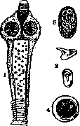
Figure 1. 1 - scolex; 2 - hooks of the scolex; 3 - cocoon with eggs; 4 - egg with oncosphere.
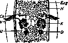
Figure 2. Hermaphroditic segment: Cb - bursa cirri; V - vagina; U - uterus; D - vitelline duct; K - ovary; Я - testes; Exd - excretory canal.
ta (oncospheres) are ingested by larvae of fleas feeding on various organic substances, and the oncosphere first settles in the body cavity of the larva; as the larva transforms into an adult insect, the embryo of the parasite passes into the body cavity of the latter and transforms into a cysticercoid. When the flea is swallowed by the definitive host (carnivore, human), the cysticercoid transforms into a sexually mature worm. Man can thus become infected only if he accidentally (while eating together with cats and dogs) swallows a flea infested with a cysticercoid with food. Regarding the age distribution of D., Blan-chard's statistics are interesting: in infants aged 5 to 6 months, 33.9% of all described cases were noted; from 7 months to 3 years - 42.09%; from 4 to 8 years - 10.16%; from 8 to 20 years - 3.39%; over 20 years - 10.16%. Later literature reports cases of even earlier infection: specifically at 40 days of age and even 20 days (Vacca; 1909). More than one hundred cases of D. parasitism in humans have been described. In the USSR, D. caninum was found in humans 13 times: 2 cases by Brandt, 1 by Krüger, 1 case in Uzbekistan (1926), 4 cases in Armenia (1926), 1 case in Tashkent (1927), 2 cases in Moscow (1927), 1 case in Cherepovets, 1 case in the North Caucasus (1927). The rare finding of this parasite is probably due to the poor familiarity of physicians with this form, which is why it is sometimes confused with better-known cestodes of the family Taeniidae. In humans, single specimens were most commonly encountered - from two to ten; however, cases of very intense invasion have also been described, when the number of parasites reached more than 200 specimens. The pathogenic effect of the parasite consists of the same factors as in most other cestodes. Calami-da (1901) experimentally established that extracts of D. have a hemolytic effect. In invasion by this parasite, cases can occur without visible symptoms; in other cases, severe disturbances of the intestine or nervous system are observed, which can be eliminated only by expelling the parasites. Diagnosis is made by finding oncospheres, parasite eggs, or cocoons in fecal masses. Treatment is carried out with extract of male fern. Prevention: avoid close contact (mainly for children, especially during meals) with dogs and cats. With respect to these animals, especially when kept indoors, it is necessary to take the following measures: 1) periodically deworm them (at least once every 3 months) to eliminate parasitism, 2) keep clean the place allocated for the animal's lodging, in particular to disinfect with boiling water (to destroy flea larvae; see Fleas), 3) comb out ectoparasites.
Related articles
Mentioned in
Cite this page
“Dipylidium Caninum L.” Soviet Medical Encyclopedia. English translation of Bolshaya Meditsinskaya Entsiklopediya, 1st ed. (Moscow, 1928–1936), ed. N. A. Semashko. https://sovietmedicalencyclopedia.pages.dev/article/dipylidium-caninum-l/