Inflammation
Historical document, translated for reference. It reflects medical knowledge of the 1920s–30s and is not medical advice.
Summary
Inflammation is a complex of local pathological processes involving vascular changes, exudation, increased temperature, pain, and functional impairment. The article explores its historical understanding, morphological characteristics, and physiological mechanisms.
Encyclopedia article (1928–1936)
Inflammation. Contents: Morphology and pathological physiology of I. . .626 Experimental study of I........631 Causes of I...................632 Character of I...................633 Comparative pathology of I..........636 Mechanism of development of I.............636 Course of I. and influence of factors.......637 Spread of I...............642 Outcomes of I.....................642 Terminology of I................644 Principles of treatment of I..............644 Inflammation (Lat. inflammatio, Gr. phlogosis), such a complex of local pathological processes of morphological, functional, and physicochemical character, which develops along the course of the vascular (and generally mesenchymal) apparatus under the influence of various irritants on the body tissues. The most essential components of the inflammatory symptom-complex are: 1. Acute (local) circulatory disorders in the form of active hyperemia of vessels; clinically - redness (rubor). 2. Exit from vessels of liquid and formed elements of blood - so-called exudation; clinically - edema, tension, swelling (tumor). 3. Increase in temperature of the area of I. (calor). 4. Pain (dolor). 5. Impairment of function (functio laesa) from the side of the inflamed tissue, as a symptom of damage (alteration) of it, in particular - acute disorders of tissue metabolism in the form of its sharp increase, with development of tissue acidosis and increase in their molecular concentration.-The main symptoms of I. (calor, rubor, tumor, dolor) were established already by Hippocrates, Celsus, Galen. Over a series of centuries there was a dispute, which of these symptoms is cardinal, and at first calor, then rubor was taken as such; in the formula ubi stimulus, ibi affluxus was expressed also the connection of I. with the influence of external factors. The very concept of I. arose only in the times of Greek medicine, although already in the ancient Egyptians such designations of I., as "blood brother", "companion of pus", "father of tumor" testify to familiarity with some sides of this process. The ancient doctrine of I. about the abundant influx of warm blood, accompanied by exudation from dilated vessels, pains and impairment of function, has preserved its basis even to the present time. Galen's doctrine of I. as a local fever, his indications of the curative nature of I. are also not without deep interest for modernity. During the XVI-XIX centuries I. was interpreted from different points of view: chemical (v. Helmont), mechanical (Bellini), animistic (Stahl); while the treatment of I. followed Hippocrates. The creator of the modern doctrine of I. is Hunter (end of XVIII century), who first pointed out the significance of endogenous factors in I. (location of I., peculiarities of the organism); in particular, his terms: "healthy and unhealthy I." should be placed in parallel with modern terms - normergic and allergic. I. With the appearance of the microscope pathology only refined the old doctrine in histological respect, and even later, with the flourishing of the so-called molecular pathology, broad possibilities opened up for studying the physicochemical processes in I. It should be noted, however, that with the discovery of new technical possibilities the disputes about I. have by no means become less acute; on the contrary, new difficulties have arisen in connection with attempts to delimit I. from other pathological processes. As could be expected, a complete delimitation has not been achieved to this day: I. is not an isolated chapter of pathology; the problem of I. closely borders on the problems of degeneration, regeneration, hyperplasia and even neoplasia; it is incorrect from this to draw the conclusion (Andral, Thoma) that the very concept of inflammation must be abolished, since in a similar position is also the question of many other pathological processes, for example, tumors. It is essentially important in defining the concept of I. (to this day there is no generally accepted definition of I.) to consider above all its complexity, i.e., to consider this process as an integrating sum of individual (intractable) symptoms, even if the latter are represented weakly individually. The existing even at present great disagreements on the question of the essence of I. are to a large extent conditioned by the one-sidedness of approach, and in particular also by the fact that they want to find an absolute criterion of I., for example, taking as such this or that histological, physicochemical reactions, ignoring I. as a complex clinical-anatomical, rich in variations symptom-complex. A similar error was also made by Metchnikoff, who centered all the essence of I. in phagocytosis, although the main idea of his doctrine of I. as a process analogous to digestion can still be considered correct today. Morphology and pathological physiology of inflammation. The inflamed surface of the skin, serous, mucous membranes is always more or less brightly red in color, while in parenchymatous organs the redness is blurred by the presence of significant degenerative phenomena, and therefore may be completely absent and even replaced by anemia, for example, in the so-called interstitial I. The consistency of tissues in I. is either sharply increased (e.g., carbuncle) or decreased (e.g., brain in encephalitis), depending on the structure of the organ (tissue) and the very character of I. Usually the consistency changes even along the course of the same process, and in acute inflammation this change goes in the direction of increase or decrease of turgor, or even appearance of fluctuation (for example, in purulent inflammation); in chronic forms usually the reverse phenomenon takes place due to the occurrence of scleroses. The elasticity of tissues in I. is always decreased; their elasticity becomes less perfect, although it may be increased. On microscopic examination, strong hyperemia of vessels is found, especially of arterioles, capillaries and small veins, with characteristic pulsation (the latter is often felt by the patient himself). The blood circulation, being at the very beginning of inflammation somewhat accelerated (effect of irritation of dilators), is later inhibited depending, in part, on the thickening of the blood (discharge of plasma - exudation - into the surrounding tissues), but mainly from the discrepancy between the amount of blood flowing and the width of the vascular bed (too great dilatation of the latter). In I. in stagnant organs, as well as under the action of strong irritants, the phase of acceleration may be absent. The highest degree of circulatory disorders in I. can be considered the development of blood stasis, thrombi, significant hemorrhages. From the ordinary neuroparalytic or neurotonic hyperemia, inflammatory hyperemia differs, first, by its duration-, secondly, by the fact that inflammatory hyperemia is caused, mainly, by the loss of elasticity of the connective tissue surrounding the capillaries, and finally, by the fact that at the height of I. the vascular system is not excitable either directly or through the nervous system. Every I. is accompanied by exudation, i.e., exit from vessels of liquid and formed elements of blood. The very oozing, or exudation of parts of blood plasma represents a process of selective and sequential excretion by vessels of various protein bodies, starting with the smallest (according to the size of their molecules) - type of albumin, then euglobulin, pseudoglobulin and finally fibrinogen (Bechhold, Oswald); oozing of fibrinogen presupposes the greatest degree of porosity of the vascular wall, after which follows the emigration of whole cells; the increased porosity of the vascular "ultrafilter" is a consequence of the sharp dilatation of vessels (formation of stomata, see Stomata), as well as of the disturbance of physicochemical properties of their walls. The blood pressure in the vessels of the inflamed area is increased, which favors exudation; this also increases the amount of outgoing lymph, which has significance for the whole organism in the sense of pyrogenic action from the inflammatory focus, intensification of blood coagulation processes, etc. The process of exudation, or emigration of formed elements may concern both white and red blood corpuscles; the greatest significance has the emigration of polymorphonuclear leukocytes, occurring according to the following scheme. Before emigration proper, there occurs a change in the character of the blood column in the vessel in the sense that the normal differentiation of this column into the axial part, where the specifically heavier formed elements of blood are located, and into the peripheral (plasmatic) part disappears under the influence of slowing of blood flow, and the leukocytes (as specifically lighter) recede into the plasmatic layer and accumulate near the very wall of the vessel (see figure 1), giving the phenomenon of so-called "marginal standing" (for the first time a physical explanation of this phenomenon was given by Shklyarevsky in 1865); however Fahraeus (1928) denies such an explanation, pointing out the significance of the size of the cell (see Blood Circulation). More and more losing in the speed of progression, the leukocytes finally become firmly fixed at the walls; after the moment of fixation follows a change in the shape of the leukocyte and precisely on the side turned to the vessel wall (see figure 2); these changes reduce to the formation of so-called pseudopodia (see), which penetrate through the stomata of the vascular wall, stretching and acquiring more and more bizarre outlines (see figure 2); at the height of emigration the whole body of the leukocyte has the appearance of a thread with two club-shaped ends (in the lumen of the vessel and outside it); on
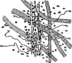
Figure 1. Marginal standing and emigration of white
balls. Frog tongue. Semi-schematic (according to Aschoff). At the end, there is a complete exit of the cell beyond the vessel—infiltration of the surrounding tissue (see Figure 3). Emigration, or diapedesis (see), of erythrocytes is a frequent but not constant phenomenon; it is sharply expressed in hemorrhagic I. and then seems to be inversely proportional to the strength of leukocyte emigration.-The next group of cellular processes in I. are the processes of proliferation, or production. They consist in the fact that the connective tissue elements that build
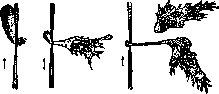
Figure 2. Process of leukocyte emigration through
the vessel wall; the formation of pseudopodia. Arrows indicate the direction of blood flow (according to Aschoff). The vascular walls (endothelium, cells of the adventitia), and other mesenchymal cells (so-called wandering cells at rest, fibroblasts) swell, round off, separate from their inherent connections (the process of complexation, see.), and multiply vigorously, predominantly mitotically, ultimately creating entire colonies of young connective tissue cells (so-called histiocytes), which together with leukocytes form the inflammatory infiltrate (see Figure 3). Due to such a reaction of local cells, the boundaries of the vessels become significantly blurred, a phenomenon particularly facilitated by intravascular proliferation and subsequent emigration of endothelial derivatives. As a result of productive processes, the complete disappearance of vascular contours is sometimes observed (see Figure 9) with the formation of so-called granulomas in place of the vessel; thus are formed tuberculous nodules, gummas, typhoid, malarial granulomas, etc. The distinction between proliferation and emigration processes encounters certain difficulties due to the proven possibility of leukocyte formation in situ through the same proliferative process, for example, from elements of the vascular wall, and according to Mollendorf, who denies the evidential value of emigration in Cohnheim's experiment—even from fibroblasts; all this forces us to consider the very division of inflammation into exudative and productive (see below) to a certain extent conditional. At the beginning of inflammation, emigration phenomena are usually predominant; subsequently, productive processes quickly superimpose and then predominate, imperceptibly transitioning into the regeneration phase (see).-The third group of morphological processes in inflammation consists of alterative changes affecting the entire complex of degenerative, destructive, and necrobiotic phenomena in the focus of inflammation (see Alteration). Along with cellular changes in inflammation, changes in so-called paraplastic substances are observed: disintegration, dissolution of collagen and elastic fibers, processes of resorption of the ground substance of bone, cartilage, etc. (tissue amorphization phenomena). Exudation, proliferation, and alteration constitute the morphological triad, which is mandatory for any inflammatory process. The predominance of one or another group of phenomena in this triad allows the classification of inflammation as exudative, productive, and alterative (see below). Functional changes in inflammation are collectively designated as functio laesa. Under this designation are hidden phenomena of different orders; thus, it may be a decrease or complete loss of function of a given organ (for example, in nephritis, neuritis, myositis); on the other hand, phenomena of the opposite order are observed: a state of hyperfunction, for example, increased sweating in some dermatitis, enhanced spermatogenesis in the inflamed testicle, etc.; however, phenomena of the latter kind are transient and quickly give way to weakening or paralysis of function. The conventional nature of the designation functio laesa also follows from the fact that every inflammation, since it is necessarily accompanied by a sharp increase in tissue metabolism (see below), is a hyperfunction of the mesenchymal apparatus; but since we will analyze inflammation from a particular, resp. organopathological point of view, we will inevitably come to recognizing functio laesa as predominantly a negative factor. Thus, the mesenchyme in inflammation is in a state of hyperfunction; the specific carriers of the mesenchyme (organs, differentiated tissues) most often exhibit all clinical signs of hypofunction—either from the very beginning of inflammation or after a temporary phase of their hyperfunction. There are no specific functional disturbances characteristic only of inflammation, although this does not exclude the possibility of basing the diagnosis of inflammation on certain most frequently encountered functional signs.-The rule that inflammation is accompanied by pain has many exceptions depending on the location of the lesion. Thus, inflammation of most parenchymatous organs (liver, kidneys, lungs) is usually devoid of this symptom. Pain is most significant in inflammation of serous membranes, skin, periosteum, and it varies in character: stabbing, burning, dull, aching, drilling, pulsating, etc. The intensity of pain in inflammation depends not only on the number of nerve branches but also on the nature of the tissue, for example, in terms of its distensibility; low distensibility usually gives the greatest pain (pulpitis, paronychia). Pain in inflammation can be of a radiating nature, sometimes it is projected at considerable distances from the inflamed area (so-called eccentric projection): this phenomenon is not without practical significance, as it can lead to false conclusions about the location of inflammation (see Symptomatic pain).-The increase in temperature in the area of inflammation is explained, mainly, by the increased influx of blood, which usually maintains body temperature. Some authors see the center of gravity of this symptom in the enhancement of local metabolism, i.e., in increased heat production. Physicochemical changes in tissues during inflammation are significant and are expressed in the violation of the normal setting ('isotonicity') of tissue colloidal systems, in terms of changes in the ratio of H- and OH-ions on the one hand, and Na-K-Ca-ions on the other; these changes proceed in the direction of increasing H-ions (H-hyperionia), i.e., tissue acidosis, and an increase in K and Na ions. Parallel to the violation of isotonicity, there is also a violation of isotonicity, i.e., molecular concentration, in terms of a sharp increase in osmotic pressure (up to triple the normal, for example, D=1.4°). Both phenomena—H-hyperionia and osmotic hypertension have one common cause—the extremely enhanced tissue metabolism in the area of inflammation, i.e., increased breakdown of tissue protein molecules, which in turn depends on the increase in enzymatic autolytic, particularly oxidative, processes; an increase in CO2 (by 3-4 times) is always observed in the inflamed tissue. This 'metabolic fire', 'hyperplety' of tissue juices (Schade), with violation of colloidal systems, entails a series of functional and morphological changes in the focus of inflammation. Thus, tissue acidosis (according to Schade, it can increase 50-fold!) by itself causes vasodilation, and subsequently—swelling of the vessel walls with an increase in their porosity. Acidosis is possibly associated with tissue turbidity (turbid swelling, see.), fat decomposition (see.), basophilia (see.), loss of tissue elasticity, and phenomena of hemagglutination (stasis).-The increase in molecular concentration causes a decrease in the surface tension of the colloids of the protoplasm of erythrocytes, leukocytes, histiocytes, making the cells more sticky and, in particular, apparently contributing to the development of the 'marginal standing' phenomenon. The further action of the same factor leads (according to Schade's theory) to a change in cell configuration, the formation of pseudopodia, preceding the phenomenon of emigration. The formation of inflammatory edema finds its explanation in the increase of adsorption processes, i.e., the binding of water, proteins, and salts by tissues as a result of the increase in their osmotic concentration, as well as due to the change in viscosity of the exuded proteins, predominantly globulins, and the phenomena of their clotting (fibrinogen) upon contact with denatured tissue surfaces. The binding of water itself in inflammation has its characteristic features; it is based on endosmotic and molecular imbibition (Hofmeister) of structural elements with swelling, homogenization (amorphization) of the latter; circulation in tissue clefts ceases at this time. All the above allows one to compare the inflammatory focus with a concentrated salt solution in a dialyzing sheath. Modern data obtained by physicochemical methods, to a certain extent, restore and deepen the so-called attraction theory of inflammation of the late 18th and early 19th centuries (Boerhaave, Magendie, Poiseuille, Virchow), which saw the essence of this process precisely in the increased tissue activity and in the attraction of nutrients by damaged tissues. Supporters of this theory (in particular Virchow until the end of his life) stubbornly adhered to the position that circulatory disturbances, diapedesis, emigration have only secondary and minor importance; a return to this theory is also encountered now in the views of some modern clinicians and researchers (such as Bier, Roux). The experimental study of inflammation is carried out on various cold-blooded and warm-blooded animals, with frogs, guinea pigs, and rabbits being the most suitable. The classic experiment on frogs is Cohnheim's experiment: in a curarized frog, the intestine is pulled out through a lateral abdominal wound, the mesentery of which is carefully spread on the microscope stage; acting on the mesentery with various irritants, the development of inflammation is observed; thus Cohnheim discovered the phenomenon of emigration; however, this phenomenon had been described earlier by Dutrochet (1842) and Waller (1846). Ricker and his school conducted similar experiments on the omentum, pancreas of rabbits, guinea pigs (see Ricker's law). Changes in the vessels can also be studied using the capilloscopy method.
The morphology of I. is studied by the usual methods accepted in histology. The most important aid for this is experiments with tissue cultures (Maximov); however, this idea was first implemented by Ziegler (1876). The study of physicochemical processes is carried out using methods accepted in this field (Schade, Hoeber). Causes of I. are very diverse, but ultimately come down to the effect of various physical, chemical, and biological irritants in the broadest sense of the word. It is difficult to imagine such an exogenous factor that could not, under certain conditions, be a cause of I.; if we add to this the group of endogenous factors, then it becomes understandable the extraordinary prevalence of inflammatory processes in the animal kingdom. Among external factors, injuries in the direct and conditional sense of the word (violation of integrity, heat, cold, light, X-rays, chemical substances) have the greatest significance; then microorganisms, animal parasites, where on the one hand, the active principle is the very presence of bacteria or parasites (as a foreign body), and on the other hand, and mainly, the products of their life activity and decay (bacterial proteins, endotoxins, etc.). The significance of the latter moment (decay) is especially clearly demonstrated in cases of cysticercosis, when a mild reaction around living parasites clearly sharpens upon their death, creating only at this moment the most distinct anat. and clin. syndrome of the disease. The same is indirectly emphasized by the facts of pronounced microbism of tissues (for example, in spirochetoses) without any morphological reaction. Among endogenous moments, one should point to necrotic tissue masses formed in the body itself (e.g., cancerous decay, infarcts), as well as deposits of organic and inorganic salts; here we see a close relationship of I. to the so-called organization. Finally, I. can be neurogenic; classical examples of this are herpes zoster and apparently pemphigus, as well as the scar phenomenon (acute I.) at the site of a healed wound after being bitten by a rabid animal at the beginning of rabies disease. A bright example of I. as a psychosensitive reflex act can be the experiments of Doswald, Kreibich, Heller, and Schultz (Doswald, Kreibich, Heller, Schultz) in obtaining I. as a result of a hypnotic session, e.g., by suggesting to the subject that a red-hot object is touching him. The question of the significance of the nervous system as an etiological factor should not be confused with the even larger question of the significance of the nervous connection in the mechanism of development of I. in general, i.e., independently of causal factors (about this below). Of great theoretical importance is the fact that I. can be a manifestation of purely physiological or formative processes; it is known, e.g., that the digestive tract in the midst of digestion (especially with a protein-rich diet) can present typical pictures of I., such as: hyperemia, emigration of leukocytes, desquamation of the epithelium, etc.; a similar reaction was observed in the mucous membrane of the rabbit's uterus when it was irrigated with spermatozoa. The processes of formation of some organs, for example, mammary, thyroid
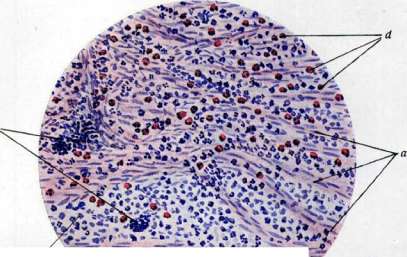
Figure 3, Acute appendicitis [medium magnification, staining - hematoxylin-eosin]: d - bundles of the muscular layer of the appendix; below they are strongly separated by inflammatory infiltrate (it), consisting mainly of leukocytes with an admixture of eosinophils (rf); c - groups of lymphoid cells [proliferation) along the course of small vessels (according to the preparation of I. V. Davydovsky). v (9; ' Re? of Figure 4, Chronic inflammation (area of sclerosis): a - infiltration with plasma cells; wheel-shaped cell nuclei (^adkern); a light perinuclear zone is visible; b - connective tissue (fibrous) layers (according to the preparation of I. V. Davydovsky). TO ST. IhKrttLWX'.SC. glands, the entire process of regression of the gill apparatus, the dropping off of the tail in the tadpole, the transition from the larval stage (higher insects, echinoderms) to the final form, etc., also proceed under the banner of inflammation, sometimes accompanied even by bright alterative phenomena, hemorrhages, etc. These examples indicate that inflammation, remaining for us the most significant pathological process, has its physiological prototypes, and sometimes is even one of the principles of building new tissue. The character of V. varies depending on the producing factor, on the location of V., i.e., the anatomical and physiological peculiarities of tissues or organs, as well as the peculiarities of the organism itself, its immunobiological reactivity. The following main forms of V. are distinguished: exudative, productive, and alterative. 1. V. is called exudative when the phenomena of exudation of parts of plasma, formed elements of blood, come to the fore. Depending on the properties of the exudate, they speak of serous V. with predominant seepage of fluid rich in protein but not coagulating, of fibrinous V. - when masses of fibrin (as a result of the exudation of plasma fibrinogen) appear in the tissues or on their surface, of purulent V. - with abundant emigration of leukocytes, of hemorrhagic - with predominance of diapedesis of erythrocytes in the entire picture. More often, however, mixed forms of exudate are observed, e.g., sero-fibrinous, fibrino-purulent, etc. Very often in the same case, exudative V. passes through a series of stages: e.g., starting as serous, it then passes into purulent. Exudative V. belongs to the number of most acute and typically occurring forms; the free inflammatory exudate (the so-called effusion) in the body cavities, for example, in pleurisy, peritonitis, represents a fluid, always more or less cloudy depending on the amount of suspended formed elements, yellowish (or reddish in hemorrhagic V.) in color and with a high protein content, usually over 4%; sometimes the amount of protein is almost equal to the percentage of plasma proteins (7-8%). Some difficulties may be presented by the recognition of pure forms of serous V., namely - in the sense of distinguishing it from simple edema or dropsy (i.e., transudate), especially in those not infrequent cases when V. is superimposed on an undoubtedly edematous substrate; thus, cases of ascitic-peritonitis are observed with heart defects, with liver cirrhosis. Measurement of the percentage of protein, cytological study of the centrifugate, histological study of the cavity walls clarify the nature of the process. To more or less pure forms of serous V. can be attributed second-degree burns, erysipelas, malignant edema, the initial periods of some pneumonias (see figure 7).- Hemorrhagic V. often very much resembles vulgar bleeding; such, for example, is hemorrhagic meningitis in anthrax sepsis; at present, all cases are considered when microscopically only bleeding is confirmed and there is neither serous impregnation nor cellular reaction, they are simply classified as hemorrhages. Classical representatives of hemorrhagic V. are cases of anthrax, plague, 'Spanish' flu, often tuberculosis, streptococcal infections, as well as aseptic forms of V. of body cavities, associated with their growth by malignant tumors, e.g., in cancer of the pleura. - Fibrinous V. consists either in fibrinous impregnation of the inflamed tissue or, which has greater practical significance, in the formation on the surface of the affected organ, especially of serous and mucous membranes, of membranous grayish deposits, the so-called false membranes, for which this form is also called false-membranous (or pseudomembranous) V. There are several varieties of fibrinous V. Thus, they speak of croupous V. when the fibrinous exudate in the form of a gray film 0.5 mm thick or more lies almost freely on the surface of the mucous membrane and can be easily removed from it, e.g., in the so-called croup of the larynx (see figure 8); the mucous membrane itself and the epithelium covering it retain their anatomical integrity in this case. In those cases when the fibrinous exudate is tightly adjacent to the surface of the mucous membrane and the latter bears more or less deep necrobiotic changes, while being impregnated with fibrin, it is customary to speak of diphtheritic V. (see figure 6). Intermediate forms are also called croupo-diphtheritic, or diphtheroid V. Instead of 'diphtheritic' V., Aschoff proposed the term 'scab-forming' (verschorfende) V. in order, on the one hand, to emphasize the most important moment here of necrosis (formation of a scab) and then of an ulcer (diphtheritic V. - is always ulcerative V.), and on the other hand, to avoid the use of the word 'diphtheria', i.e., the acute infectious disease, for which, by the way, diphtheritic V. is not obligatory. Diphtheritic V. is most often observed in the urinary bladder, uterus (e.g., in septic endometritis), intestine (e.g., in dysentery, uremic colitis). All other conditions being equal, e.g., with the same infection, fibrinous V. of mucous membranes lined with stratified epithelium more often has a diphtheritic character; on mucous membranes lined with multi-row cylindrical epithelium, it has a croupous character; in other words, not only the nature of the infection but also the anatomical substrate determine this or that variety of fibrinous V. This position, however, has many exceptions. Along with the generally accepted view that fibrin in the said deposits and in the depth of the tissue is of hematogenous origin, i.e., represents plasma fibrinogen, there are indications of local formation of fibrin from collagen fibers of connective tissue, from cell protoplasm [the theory of the so-called 'fibrinoid transformation' (Virchow, Grawitz, Neumann)]. The causes of fibrinous forms of V. are various infections (diphtheria, scarlet fever, diplococcus, streptococcus) and intoxications (mercury poisoning, uremia, etc.).- Purulent V. is characterized by abundant emigration of leukocytes and the formation of pus (see), i.e., exudate consisting, morphologically, almost exclusively of leukocytes; the latter abundantly infiltrate the tissue; often complete melting of the tissue and the formation of an abscess, phlegmon occur. Opening on the surface of an organ, for example, the skin or mucous membrane, purulent V. gives rise to the formation of ulcers (see); spreading along the lymphatic spaces, or along the line of least resistance, usually from top to bottom, purulent deposits make very winding paths (the so-called fistulas), opening somewhere on the surface, or form freely lying in the tissues new deposits (migratory abscesses). Among the causes of purulent V., the so-called pus-forming bacteria most often appear. Often, however, purulent V. is sterile; such can be obtained by the introduction into tissues of certain chemical substances, e.g., turpentine. -.G n i l o s t-n o b (or ichorous) V. is a variety of other forms of exudative V., most often fibrinous and purulent; it is caused by the entry of putrefactive bacteria into the focus, which leads to the decomposition of the exudate and the formation of foul-smelling gases. To the group of exudative V. is also attributed the so-called catarrh, or catarrhal V., observed only on mucous membranes. In essence, it is not an independent form of V., and is characterized only by a number of additional circumstances, such as the admixture of mucus to this or that exudate, as well as of shed, usually mucoid-degenerated epithelial cells (see figure 9). However, the admixture of mucus depends, mainly, on the enhanced activity of the mucous glands on the basis of inflammatory hyperemia; the mucous membranes show all the usual signs of V. They distinguish serous, mucous, purulent, and desquamative catarrh. 2. In p r o d u c t i v e V. the phenomena of proliferation, i.e., processes of inflammatory tissue neoplasia, come to the fore. This type of V. can have a vulgar character, i.e., not have an etiological characteristic; in other cases, the product of cellular proliferation are the so-called granulomas, i.e., more or less clearly outlined, most often round foci of granulation tissue, sometimes distinguished by special morphological features, on the basis of which they are sometimes designated as specific granulomas (see).
A classic example of productive inflammation are tuberculous tubercles and syphilitic gummas; among acute infections, typhus, typhoid fever, rabies, rheumatism, and others occupy the most prominent place. Along with various infections, causes of productive inflammation include difficult-to-resorb foreign bodies, irregularities in the blood supply to the inflamed area, and other factors. 3. Finally, the concept of alterative inflammation follows from the term alteration itself (see). This type of inflammation is observed almost exclusively in parenchymatous organs (heart, liver, kidneys, brain), in nerves, and in muscles. At present, the term 'alterative inflammation' has replaced the so-called parenchymatous inflammation, under which, however, ordinary degenerative changes of organs were often described, which today (since they exist independently) are not classified as inflammation. Among the conditions for the development of alterative inflammation, the anatomical substrate and immunobiological reactive peculiarities of the organism are of particular importance (see below). Comparative pathology of inflammation, developed by Mechnikov, considers that inflammation in its primitive manifestations can occur in organisms standing at a very low level of development, for example, in those without blood vessels. A prick in a sponge gives the effect of accumulation of wandering cells, which then merge into a plasmodial mass. Productive processes as a rudimentary form of inflammation, its prototype, are also observed in jellyfish, annelids, and earthworms. In general, the productive element in inflammation is the oldest in phylogenesis; exudation, emigration of leukocytes arise even later than the phylogenetic date of development of the vascular system with leukocytes in the blood; thus, in the earthworm, axolotl, and triton (in their young larvae), the existing vessels hardly participate in inflammation; the same is true in mollusks, snails; in the larvae of axolotl and triton and in closely related Cephalopoda, clear accumulations (emigration) of leukocytes are already observed. The very ability to suppuration coincides with the appearance of a well-developed endothelial lining in the vessels. Subsequently, in warm-blooded animals and humans, there are no fundamentally new 'instruments of inflammation'; only the rate of development of inflammation, 'inflammatory readiness,' changes in the sense of increasing the speed of development of the entire picture, its greater brightness, which is undoubtedly facilitated by the participation in the mechanism of development of inflammation of the highly developed nervous connection. The latter, along with the highly developed vascular system, also makes inflammation in mammals more dependent on the general condition of the organism. In plants, with the exception of some lower forms (for example, plasmodia of myxomycetes), inflammation as such does not exist; various tissue injuries here lead only to regeneration processes. Mechanism of development of inflammation in highly developed organisms, in particular in humans, is described as follows. One irritant or another, acting on tissue, causes in it the above-mentioned complex of physicochemical phenomena that exist for some time independently as a mandatory pre-stage of inflammation, still without a clear cellular and vascular reaction. Rössle considers this pre-stage as Vorverdauung, i.e., as some prelude to proper digestion (Verdauung), in which he, following Mechnikov, sees the essence of inflammation. Cellular and vascular processes follow, however, as soon as these humoral processes reach the proper degree of development (increase in acidosis, accumulation of tissue autolysates, etc.). Further development proceeds automatically, with the fading of some and the intensification of other symptoms of both cellular and humoral character. A particular difficulty in the pathogenesis of inflammation is the question of the role of the nervous system here. That the nervous connection is not necessary for the development of inflammation has now been proven experimentally; comparative pathological observations also speak of this; on the other hand, the same experiments and observations show that the role of the nervous system in the mechanism of development of inflammation is significant. Thus, in favor of this speaks the very possibility of neurogenic inflammation, the possibility of influencing the course of inflammation by severing the nervous connection and the presence of centripetal reflexes going from the focus of inflammation along the corresponding reflex arc and returning (centrifugally) to the site of inflammation. These reflexes coming from the focus of inflammation give a new influx of blood-the so-called secondary congestion. This also includes the so-called intracutaneous vasomotor reflex, expressed, for example, in increased sweating of the skin at the site of a burn or in the appearance of the Pirquet reaction on the side opposite to the site of inoculation, but symmetrically to it, etc. Undoubtedly, therefore, that the inflammatory process not only 'self-regulates' at the site but also receives a certain regulation through the nervous system, which must be emphasized especially in the so-called allergic forms of inflammation. Proponents of the neuropathological theory of inflammation (Nepe, Rokitansky, at present Ricker), who claim that every inflammation arises as a reflex act following the irritation of sensory nerves, that inflammation is always neurogenic hyperemia with subsequent tissue processes dependent on it, have not received substantial support in recent years in such categorical formulations. Course of inflammation and influence of factors. By course, inflammation is distinguished as acute and chronic; the course of inflammation, as well as its morphological character, depends on many factors, such as: the active agent (e.g., virulence of the microbe), local conditions for the development of inflammation and peculiarities of the organism as a whole. Thus, tuberculosis, leprosy, syphilis, difficult-to-resorb foreign bodies more often combine with chronic productive forms of inflammation; various coccal infections, strongly irritating, e.g., caustic substances, more often give acute exudative forms. It should, however, be borne in mind that the same factor can give different courses and forms of inflammation, and conversely, different factors can cause identical clinical and anatomical pictures of inflammation. Streptococcus can be a pus-forming microbe; but no less often it gives serofibrinous, hemorrhagic, and sometimes purely productive and moreover chronic forms of inflammation (e.g., in sepsis lenta); tuberculosis, along with pictures of productive inflammation, often in the same case gives pictures of acute exudative inflammation. Various modifications of inflammation can be obtained in animals with the anthrax bacillus, typhoid bacillus, in connection with fluctuations in factors of susceptibility and virulence. The above makes it understandable why any strict specification of microbes according to the character of inflammation they cause is impossible. On the contrary, starting from the clinical-anatomical peculiarities of inflammation, it is possible to speak with greater probability about its etiology; thus, seeing a serohemorrhagic (sanguineous) exudate or its impregnation of the bottom and edges of a wound or cellular tissue, one can correctly suspect fresh streptococcal infection; observing the soft meninges impregnated with yellowish exudate, one can think of diplococcus; when seeing an abundant flow of turbid mucous exudate from an incision in a diseased lung, suspicion of Friedländer's pneumonia arises; blue-green pus, a fibrinous coating in the larynx is involuntarily associated in our minds with Bacillus pyocyaneus, Bacillus diphtheriae (of course, all this will only be more or less probable assumptions). The explanation for the circumstance that the same causative factor can give different pictures of inflammation lies in the following local and general factors. 1. Duration of action of the factor causing inflammation; thus, by changing the time of immersion of a rabbit's ear in water at t° 50°, one can obtain all possible variants of inflammation, from hyperemia to severe necrotic forms. 2. Importance of anatomical-physiological peculiarities of tissues and organs; this factor is particularly clearly evident when comparing inflammation in vascular and avascular tissues, e.g., in the myocardium and heart valves or in the cornea and conjunctiva of the eye; significant quantitative and qualitative differences are always observed when comparing inflammation in the so-called parenchymatous organs, on mucous membranes, serous sheets. Diphtheria in the same case can cause catarrhal inflammation of the pharynx, diphtheritic inflammation of the larynx, and croupous inflammation of the trachea. 3. Importance of blood circulation. Intensification of arterial hyperemia leads to intensification of exudation; thus, a moderate burn of the rabbit's ear on the sympathectomized side causes strong inflammatory phenomena compared to the control side; thus, arterial hyperemia is not only a symptom of inflammation but also an important causative factor. Anemia of the inflamed area weakens exudation, on which the effect of the sometimes observed beneficial action of ice, local bloodletting is based; it is possible that remote bloodlettings (e.g., from the veins of the elbow in croupous pneumonia) have a similar action; this probably also includes the beneficial effect of some aero- and hydrotherapeutic measures in inflammation of internal organs. However, in those cases where exudate is already abundant, anemia of the inflamed area can give the opposite effect-intensification of alterative changes, even complete necrosis, as was observed in Samuel's experiments when an anemicized rabbit's ear was smeared with croton oil.
The explanation for the phenomena mentioned should be sought in the fact that arterial hyperemia (like plethora and hydremia in general), by increasing the mass of incoming blood, also increases the mass of exudate and the intensity of exchange in the area of inflammation. Indeed, the predisposition of edematous tissues to inflammation is known, and on the other hand, the beneficial effect of a dry diet is noted, which, by smoothing out the moment of hydremia, as it were, counteracts this predisposition. Venous hyperemia has various effects on inflammation. Thus, leg ulcers with varicose veins heal faster when the limb is elevated and properly bandaged; the frequency of hypostatic pneumonias in severely ill, immobile patients is well known, which is why preventive measures are taken in the form of frequent repositioning of these patients to prevent the development of stagnant phenomena in the lungs. True, Wir pointed to the possibility of treating inflammation with venous hyperemia, but this apparent contradiction is explained by the fact that Wir's hyperemia is not vulgar venous stasis: the speed of blood flow and tissue exchange processes are disturbed here to a much lesser extent. 4. The state of nervous connections; the complete absence of such connections makes the entire picture of inflammation less vivid: individual symptoms of inflammation are delayed in development, the course of inflammation loses its pace; only in exceptional cases does denervation stop the development of inflammation. The severe pictures of inflammation often observed with significant disturbances of innervation are caused not only by accompanying trophic disorders of tissues but also by indirect circumstances such as the greater susceptibility of such parts to external factors (for example, due to sensory disorders in syringomyelia), as well as the admixture of vascular disorders, etc. 5. The significance of the reactive capabilities of the organism itself; these capabilities do not represent something unchanging. Even in the growing fetus and newborn, we encounter a significant difference in the picture of inflammation, for example, in congenital syphilis, and moreover, the inflammatory capability itself represents one of the functions of growth and development. Age, nutritional conditions, hereditary-constitutional factors, the entire sum of external factors, past diseases, especially infectious ones—all this leaves its imprint on the immunobiological properties and reactive capabilities of the organism, ultimately creating all the diversity of inflammatory processes both qualitatively and quantitatively. Let us recall the peculiarities of the tuberculous process in children and adults, during pregnancy, diabetes, etc. Kaufmann investigated the exudate from Spanish fly in people with various diseases and showed how these diseases influence the character of the exudate obtained from the fly. In the process of artificial immunization of an animal, the most acute form of hemorrhagic inflammation in anthrax (with septic phenomena) can be transformed into a sluggish and no longer fatal form of vulgar purulent inflammation. In a similar experiment, the acute exudative reaction in streptococcal infection can turn into a purely productive process. It is well known the absence of suppuration processes in rheumatics, in the so-called sepsis lenta; on the other hand, they speak of an exudative diathesis, or an asthenic, anergic inflammation, when they want to emphasize the weakness of the developing symptoms of inflammation, the sluggishness of its course and healing. Exhausted starving subjects show a comparatively sluggish, as if cinematically stretched picture of inflammation; conversely, strong, robust people have a more lively in pace and more pronounced in quantitative reaction, which apparently lies at the basis of the paradoxical fact of the relative severity of the course of infectious diseases (e.g., influenza, typhoid, typhus, etc.) in people of 'good build and nutrition'. It is very probable that in the case of strong therapeutic interventions (such as seroproteinotherapy, chemotherapy) we are dealing with an artificial modification of old and even the acquisition of new inflammatory capabilities and possibilities in a given organism for a given infection at a given moment. The significance of immunobiological factors is particularly evident in the experimental study of inflammation, when the factor of sensitization of the animal is introduced. Inflammation is designated as normergic if the sum of reactive processes in inflammation does not go beyond the usual phenomena dual for a given animal species for a given virus. With appropriate sensitization of the animal, it turns out that upon introduction of the same virus (which may also be of non-organized nature, e.g., serum protein), inflammation already proceeds allergically, i.e., differently in clinical and morphological terms (allergic inflammation). This tissue allergy is expressed either in the weakening of the most important symptoms of inflammation, especially alteration and exudation, less so proliferation (so-called allergy with a negative sign, bordering on the phenomenon of local immunity), or, conversely, in the strengthening of some symptoms with a change in their interrelationship (for example, strengthening of proliferation at the expense of exudation). This allergy with a positive sign can take the form of an extremely rapidly developing process called hyperergic (or anaphylactic) inflammation; it is characterized by rapidity of pace, expressed alterative phenomena (necroses), serohemorrhagic exudate (serous apoplexy of tissue), and suppression of cellular (productive and exudative) phenomena. A typical representative of hyperergic inflammation is the Arthus phenomenon (see Anaphylaxis), rare cases of acute gangrenous inflammation upon introduction of antitoxin serum into the skin (see Figure 5); this probably also includes the so-called Spanish influenza, and according to Lauche, also lobar croupous pneumonia. However, every infectious disease
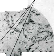
Figure 7. Serous pneumonia (medium magnification): a - swollen desquamated epithelium of alveoli; in places - desquamated in layers (l); b - leukocytes in the alveolar spaces; c - the same barely noticeable serous fluid (according to Aschoff).
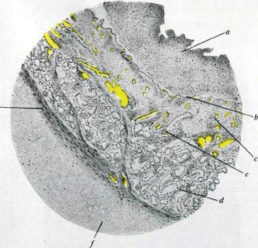
Figure 8. Croupous-diphtheritic inflammation of the larynx (weak magnification): a - fibrinous membrane with leukocytes and disintegration of epithelium. Under it is seen a deep layer of preserved epithelial cells (£). Even deeper is seen the swollen mucous membrane (g) with marked hyperemia of vessels and cellular infiltration; d - glands of the mucous membrane; e - musculature; f - cartilage (according to Aschoff).
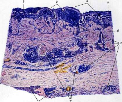
Fig. 3. Chronic inflammation of the gallbladder (weak magnification, staining: hematoxylin-eosin): a - preserved cylindrical epithelium of the bladder; b - catarrhal exudate (mucus, leukocytes, desquamated epithelial cells); on the surface of the mucosa; c - small-cell infiltration of the mucous membrane; d - muscular membrane of the bladder; e - infiltrates in the depth of the wall and on the serous surface; f - blood vessels (according to I. V. Davydovsky's preparation)
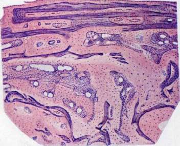
Fig. 15. Galisterele in osteomalacia. Section through a rib without decalcification of bone. Staining: hematoxylin-eosin. The blue color corresponds to normal lime-containing bone; the pink color corresponds to osteoid tissue, i.e., bone that has lost its lime in the process of galisterele (according to D. P. Pogozhevsky). In some diseases, the process may be accompanied by pictures of allergic, in particular hyperergic, inflammation; clinically, the latter gives the greatest severity of the course. The ability to hyperergic inflammation is not the same in different tissues; the skin occupies first place here; it is interesting, for example, that the Pirquet reaction, which is an allergic reaction, cannot be obtained in its typical form in internal organs (Klopstock). From all that has been stated, it is clear how closely the problems of inflammation and immunity are connected. He goes even further, pointing out that in essence all so-called immune reactions demonstrate to us the perfection of the inflammatory capabilities of mesoderm and precisely in the sense of enhanced and accelerated (or weakened and delayed) digestive ability of it with respect to one or another repeated irritant; and this author considers inflammation as an act of intratissue (cellular) and humoral digestion. The course of chronic inflammation is distinguished by a number of features. Hyperemia here is not so pronounced, and moreover, it is mainly of a venous nature, so that the inflamed area has a bluish tint. Pain and elevation of temperature are also weakly expressed, sometimes absent; swelling and hardening, however, have other bases here and depend on the strong development of both young granulation tissue and denser, later scar connective tissue (induration phenomena). In general, the main sign of chronic inflammation is the predominance of proliferative processes of both purely inflammatory and regenerative order; the separation of the latter presents here insurmountable difficulties. In addition to the proliferation of connective tissue elements, atypical proliferation of epithelium is often noted in chronic inflammation, and cases are observed of the transition of the process into a neoplastic one, i.e., into cancer. Such are essentially all tar cancers obtained experimentally by prolonged irritation of the skin with tar; these include stomach cancers that arise on the sites of ulcers, etc. The infiltrate in chronic inflammation consists, for the most part, of small lymphoid cells (see Fig. 9), which are, for the most part, a product of the multiplication of local connective tissue elements, as well as larger cells of the "polyblast" type of Maximov or "histiocytes" of Aschoff. Often, infiltrates consist mainly of so-called plasma cells (Plasmazellen, see Fig. 4). Periodic exacerbations (Lat.-exacerbatio) should be considered an essential attribute of chronic inflammation, which are expressed externally in an intensification of redness, swelling, pain, and microscopically—in the admixture to the infiltrate of exudate cells, mainly leukocytes. One should not think that every round- or small-celled infiltrate signifies chronic inflammation; such accumulations of round cells in tissues are also observed without inflammation, e.g., in connection with an intensification of the fermentative or hormonal activity of the organ, in connection with the appearance of hematopoietic function in the stroma. Chronic inflammation, like acute, can be exudative, productive, and alterative. The closest causes of chronic inflammation are most often an unremoved irritant within the focus of inflammation, the duration of the action of external factors, local circulatory disorders, e.g., venous stasis, sharp changes in the tissue of the organ after a previous acute inflammation, so that even physiological irritants can maintain or exacerbate inflammation, for example, in nephritis, some colitis. From an immunobiological point of view, chronic inflammation can sometimes be explained by the fact that the corresponding antigen, acting on the tissues allergically, is slowly absorbed by them, since one of the properties of the allergic (in the sense of hyperergic) tissue reaction is the tendency to rapid isolation, binding of the antigen and the tendency to encapsulation, i.e., processes that are by their nature long-lasting, chronic. Inflammation rarely from the very beginning proceeds as chronic. Spread of inflammation occurs either along the length of a given tissue (per continuitatem), or by contact (per contiguitem), or per metastasis; in the latter case, it is more correct to speak not of the spread of inflammation, but of the dissemination, or metastases, of one or another irritant, since inflammation as such, being a local process, does not metastasize. Aschoff's view that there is also a general inflammation of the body (e.g., in febrile diseases) is not shared by the majority of pathologists; the very pyrogenicity of inflammation, i.e., the appearance of fever in inflammation somewhere, does not yet mean the spread of specifically inflammatory factors. Outcomes of inflammation. Elimination of inflammation is possible only if the corresponding irritant is removed, all products of tissue decay are removed, and the violations of tissue metabolism itself (tissue energetics) are smoothed out. Inflammation inevitably passes into the period of regeneration (see), and especially in chronic inflammations, regenerative and inflammatory processes can exist in parallel for a long time, weakening and strengthening one at the expense of the other. As a result of regeneration, either the former relationships are restored (restitutio) or scleroses, scars arise. The different outcome is determined by the form of inflammation, its duration. Thus, among exudative forms, serous, croupous, all types of acute catarrhal inflammation end in absorption or mechanical removal of the exudate and complete restoration of the former relationships. Purulent inflammations with the formation of abscesses, ulcers, fistulas always lead to healing with scarring; the same applies to diphtheritic forms, bright alterative ones, and to all chronic forms. Productive inflammation is most often accompanied by scleroses. At present, the view on the essence of "complete" recovery has somewhat changed: such is understood to a certain extent conditionally, since very often at the site of such complete healing a locus minoris resistentiae forms (recurrence of erysipelas of the face, croupous inflammation of the lungs, increased sensitivity of the bladder, conjunctiva after their former inflammation, etc.). This decrease in resistance is more correctly, apparently, to be considered as increased sensitivity, i.e., as allergy or hyperergy of the tissue (see above). The opposite state of decreased sensitivity is also observed; for example, Samuel after recovery from inflammation caused by croton oil observed a sharp decrease in sensitivity of the same place to other irritants, for example, to burning. That anatomical healing is not always accompanied by physiological recovery follows, for example, from Ricker's experiments with suprarenin, which on the conjunctiva after a former conjunctivitis gave a clear effect of vasodilation. The significance of inflammation for the organism, on the one hand, follows as if from the very definition of inflammation as a reaction of the organism to one or another irritation, thus representing a certain regular act. As for the expediency of this reaction, it is undoubtedly that this expediency is very conditional; in particular, it is completely wrong to assign to it an explanatory significance, i.e., to consider inflammation as a protective reaction (defensive inflammation of Aschoff), and to interpret all phenomena of inflammation as elements or stages of the struggle of two beginnings, all the more so that the harmful beginning (for example, a burn) can act momentarily and cause inflammation without being in the future one of the objects of the supposed struggle. To consider inflammation as always an expedient reaction is out of the question because it is extremely often accompanied by such significant changes in organs vital for life that the continuation of the life of the individual in the order of such "protection" becomes impossible. It was also pointed out above that it is precisely hyperergic inflammation, representing the highest manifestation of inflammatory capability, that carries the greatest damage to tissues and the greatest danger to the whole organism. The very hyperemia in inflammation, insofar as it is accompanied by the influx of bactericidal substances, the emigration of leukocytes with their phagocytic activity, can be called expedient, but the same hyperemia contains the danger of dissemination of the virus with general infection of the organism. In experiments with diplococci (Lubarsch), obtained from exudate in croupous pneumonia, it was possible to show that the transitions of the stages of hepatization from red to red-gray and gray are accompanied by a clear decrease in the virulence of the diplococcus, so that the latter, obtained from exudate at the end of gray hepatization, loses the ability to give a general septic process (in a rabbit), and the matter is limited to a local reaction in the form of purulent inflammation. This experiment as if shows that the protective action of the "juices" of the organism takes place—the diplococcus loses its virulence during the inflammatory process; but, on the other hand, the very inflammation of the lungs as a reaction, neither in its volume nor in its possible consequences, of course, cannot be recognized as an expedient or protective reaction.
Inflammation is first and foremost a regularly cyclical, blindly automatic complex process, individual components of which can be simultaneously harmful, beneficial, and indifferent to the organism. Terminology of Inflammation. All inflammatory processes are denoted by the prefix "itis" at the end of the corresponding Latin or Greek word, i.e., the name of a tissue or organ (myocarditis, nephritis, etc.); in Russian, "itis" is translated as "it" (miokardit, nefrit). Inflammation of certain organs has long received special designations (angina, pneumonia, carbuncle). If one wishes to indicate inflammation of a membrane or capsule of an organ, the prefix "peri" is placed before the word (pericarditis, perigastritis), and if inflammation affects the surrounding cellular tissue, the prefix "para" is used (parametritis, paronephritis). Qualitative characteristics of inflammation, spatial, and topographical relations are also commonly denoted: for example, purulent limited peritonitis (peritonitis purulenta circumscripta), superficial catarrhal appendicitis (appendicitis catarrhalis superficialis), lobar pneumonia, acinous tuberculosis. It is often necessary to introduce etiological and pathogenetic factors into the designation: tuberculous pleurisy, hypostatic pneumonia. One should avoid calling such pathological conditions (pathos) inflammation, which, differing in a certain stability and staticity, are actually a consequence of inflammation rather than an inflammatory (i.e., dynamic) process (nosos); thus, fibrous adhesions of the pleura, pericardium after former pleurisy, pericarditis cannot be designated as pleurisy, pericarditis, even with the adjective "fibrous." It is incorrect to affix the prefix "itis" to such ailments where inflammation as such is not assumed; abuses of the term "myocarditis" are particularly frequent. Reparative inflammation is called an independent form of inflammation that arises after some other process, e.g., inflammation around an infarct. Since in such cases inflammation plays the role of a demarcating process in relation to adjacent normal tissues, this form is also called demarcation inflammation, and the zone of inflammation, which has the appearance of a red line, is called the demarcation line. The term "defensive" inflammation, also "independent" inflammation (Lyubarsch), associated mainly with infection, has not become widespread. Principles of treating Inflammation. are reduced to either 1) eliminating the irritating factor, or 2) changing the rate of development of inflammation, its character, or 3) influencing individual symptoms (e.g., pain). The first requires no special explanation. In the second case, such means as heat, compresses are used, which increases the influx of blood, exudation, proliferation processes and achieves completion of the process in a shorter period of time. The application of cold aims to cause the opposite effect—denervation of the area of inflammation and mitigation of the quantitative aspect of the process, mainly exudation, with which the analgesic effect of cold is probably also associated. It is often necessary to influence the very character of the process—for example, one strives to give a greater acuity to the chronic course of inflammation (important in sluggish courses of some parasitic dermatitis, avitaminotic wounds, etc.); methods for refreshing wound surfaces also belong here. In recent years, the question of preventing hyperergic forms of inflammation, which are the most acute in course and prognostically least favorable, has theoretically been resolved; thus, if a sensitized animal before the introduction of an antigen, which should cause hyperergic inflammation, is injected into tissues a sufficient amount of a colloidal solution, e.g., dyes, then the effect of the antigen disappears. Along with local effects on the focus of inflammation, there are also methods of general effect on the organism (vaccination, protein therapy, physiotherapy, chemotherapy, etc.). In these cases, the aim is to influence the general and local tissue metabolism processes, the physico-chemical and enzymatic processes in tissues, and also to try to act directly on the causative agent of inflammation; this includes, for example, salvarsan therapy in spirochetoses. However, in these cases, the physician's efforts are directed not so much at inflammation as such as at its outcomes, i.e., at regeneration processes (see Wounds). In recent years, the method of treating inflammation with X-rays has been proposed. Doses of 20% HED are used (see Dosimetry). Most authors indicate in this case an analgesic effect, as well as acceleration of the rate of inflammation. In chronic inflammation, the results of X-ray therapy are doubtful, sometimes negative. The beneficial effect of calcium salts (both at the site of inflammation and when administered internally) is also noted. Experimentally, it has been possible to show that these salts weaken adsorption processes, in particular the binding of water by tissues. Of great theoretical interest is the successful application of alkalis, in particular as an analgesic; this indirectly emphasizes the importance of the acidosis factor in the symptomatology of inflammation.
Related articles
Mentioned in
Cite this page
“Inflammation.” Soviet Medical Encyclopedia. English translation of Bolshaya Meditsinskaya Entsiklopediya, 1st ed. (Moscow, 1928–1936), ed. N. A. Semashko. https://sovietmedicalencyclopedia.pages.dev/article/inflammation/